


 biology
Similar presentations:


")


Interstitial Lung Disease
1.
Interstitial Lung DiseaseThe Pleura and Chest Wall
2.
Objectives• Interstitium
• Pleural disease
• Chest wall disease
3.
Interstitial disease• What is the interstitium?
• What does the interstitium do?
• What are the pathophysiological effects of
interstitial disease?
• What are the clinical manifestations?
4.
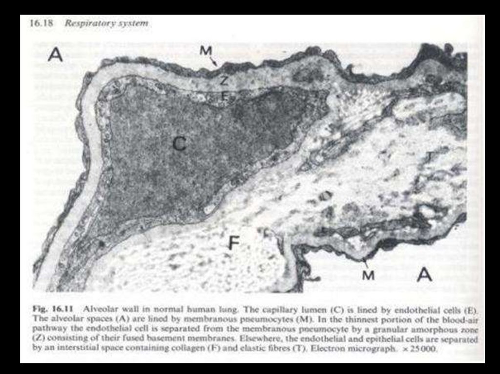
What is the interstitium?5.
6.
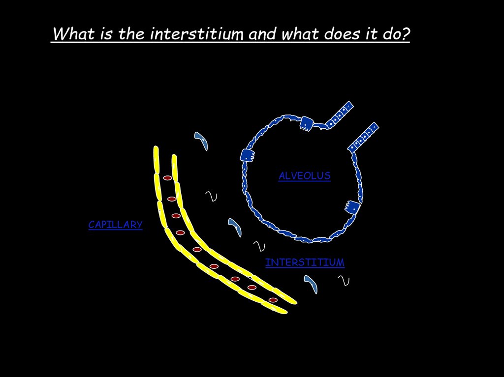
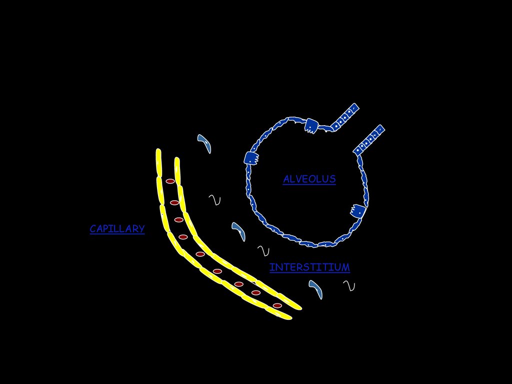
What is the interstitium and what does it do?.
.
.
.
.
CAPILLARY
. . .
.
.
.
.
ALVEOLUS
.
..
. . . ..
INTERSTITIUM
7.
Does interstitial disease effect just the interstitium?NO !
Structures affected:
Cells involved:
Acini
Alveoli lumen
Bronchiolar lumen
Bronchioles
Epithelial
Endothelial
Mesenchymal
Macrophages
Recruited inflammatory cells
Chronic Diffuse parenchymal lung disease’…
8.
VentilationDiffusion
Perfusion
O2
CO2
9.
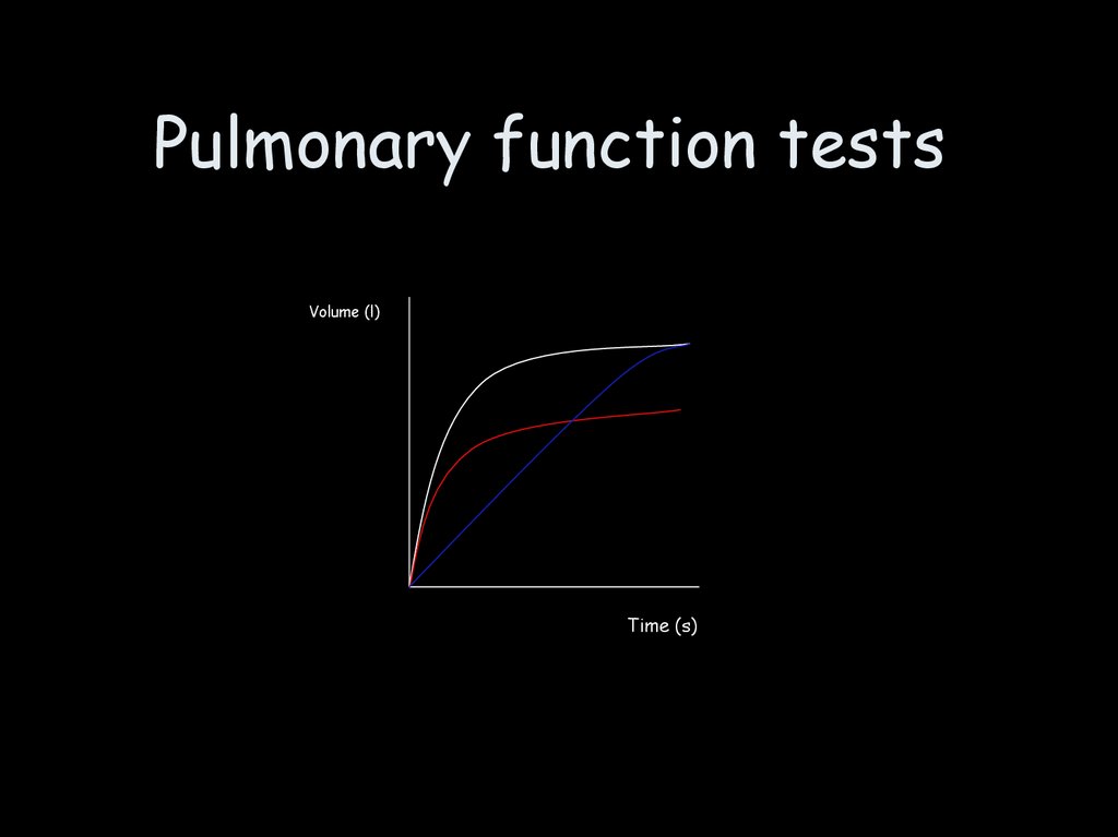
Pulmonary function testsVolume (l)
Time (s)
10.
Patient 159 year old male
Respiratory rate 24/min, HR 106,
Oxygen saturations 87%
Shortness of breath & dry cough,
increasing 1 year - breathless with
dressing
Chest examination - diffuse
bilateral crackles, reduced air
entry
Bilateral pitting ankle oedema
Rheumatoid arthritis (on
methotrexate) x 15 years
Current smoker 40 years.
Pigeon fancier
11.
12.
13.
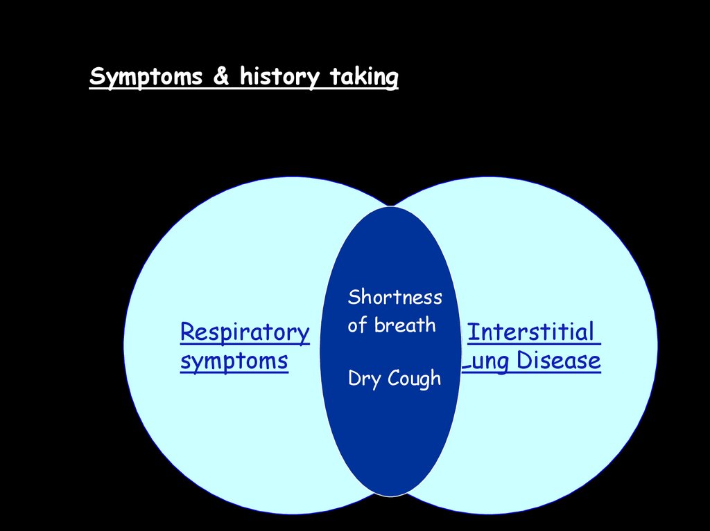
Symptoms & history takingRespiratory
symptoms
Shortness
of breath
Dry Cough
Interstitial
Lung Disease
14.
..
.
.
.
CAPILLARY
. . .
.
.
.
.
ALVEOLUS
.
..
. . . ..
INTERSTITIUM
15.
• Common clinical features• Symptoms 1-Chronic dry cough
2-Exertional dyspnea
• Signs
1-Clubbing
2-Basal inspiratory crepitations
• Laboratory 1-High ESR
2-Pulmonary infeltrate and reduced
lung size
3-Restrictive pattern of pulmonary
function tests
16.
Pulmonary function tests• Spirometry 1-Decreased FEV1,FEV
(Normal FEV1/FVC)
2-Decreased TLC
3-Mildly Decreased PEF
4-Markedly Decreased DLCO
• Blood gasses 1-Hypoxia
2-Hypocapnea
• (Type 1 respiratory failure)
17.
ExaminationSigns of underlying disease
Cyanosis
Clubbing
Tachycardia
Tachypnoea
chest movement
Course crackles
Signs of right heart failure
18.
Bloodtests
Interstitial Lung Disease
Occupational
•Asbestosis
•Silicosis
•Coal Workers
pneumoconiosis
Treatment
related
Connective
tissue disease
•Radiation
•Methotrexate
•Nitrofurantoin
•Amiodarone
•Chemotherapy
•Rh. Arthritis
•SLE
•Polymyositis
•Schleroderma
•Sjogren’s
Immunological
•Sarcoidosis
•Hypersensitivity
pneumonitis
Idiopathic
•CFA/IPF
•UIP/NSIP
•DIP
•LIP
•RB-ILD
•BOOP
19.
Idiopathic interstitial pneumonitis (IIP)A variety of histological
descriptions (UIP,NSIP,DIP,RBILD, BOOP)
Histological descriptions - high
inter and intra observer
variability
Often poor correlation with CT
chest & clinician
Biopsy may not help with
management
More cellular - more steroid
responsive
Presents 60-70 years old
Cough/ Breathlessness
CXR/Chest - basilar, bilateral,
subpleural fibrosis +/- ground
glass
Restrictive PFT’s
Biopsy - variable findings
Treatment- observe/steroids
Prognosis - depends on cause
20.
Asbestos21.
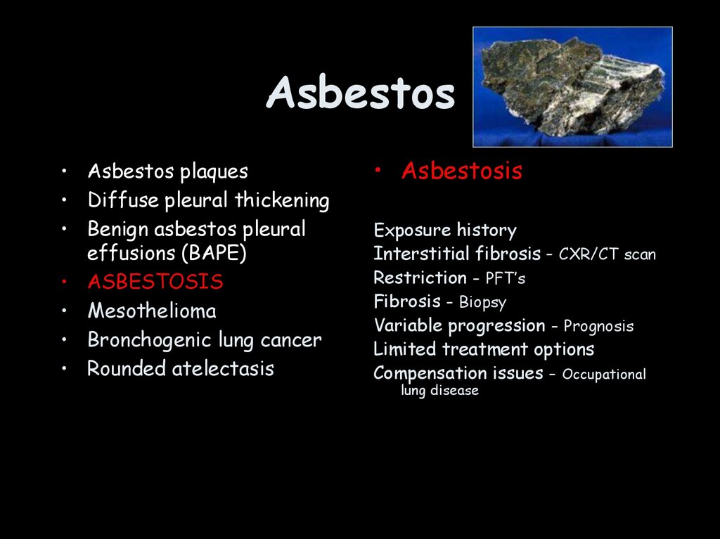
Asbestos• Asbestos plaques
• Diffuse pleural thickening
• Benign asbestos pleural
effusions (BAPE)
• ASBESTOSIS
• Mesothelioma
• Bronchogenic lung cancer
• Rounded atelectasis
• Asbestosis
Exposure history
Interstitial fibrosis - CXR/CT scan
Restriction - PFT’s
Fibrosis - Biopsy
Variable progression - Prognosis
Limited treatment options
Compensation issues - Occupational
lung disease
22.
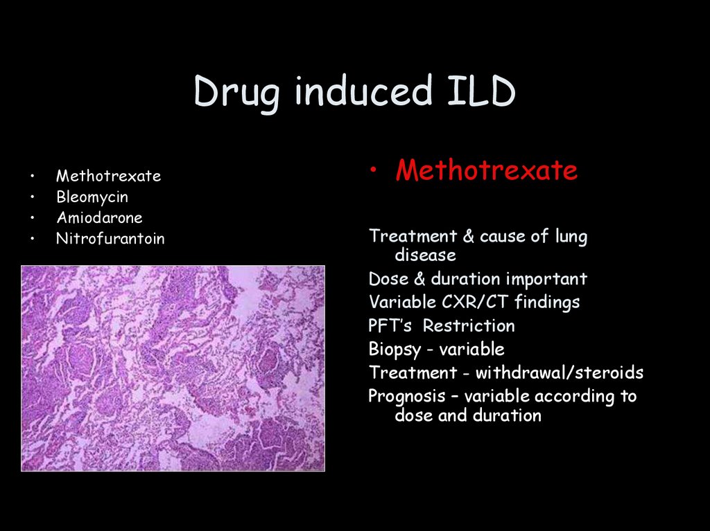
Drug induced ILDMethotrexate
Bleomycin
Amiodarone
Nitrofurantoin
• Methotrexate
Treatment & cause of lung
disease
Dose & duration important
Variable CXR/CT findings
PFT’s Restriction
Biopsy - variable
Treatment - withdrawal/steroids
Prognosis – variable according to
dose and duration
23.
Rheumatoidlung disease
24.
Connective tissue diseaseDermatomyositis/ Polymyositis
Rheumatoid lung disease
Sjogren’s Syndrome
Systemic Lupus erythematosis
Schleroderma
Rheumatoid arthritis
May predate arthritic symptoms
Disease or treatment may be cause
Male > female
Variable CXR/CT findings
PFT’s Restriction/normal
Biopsy- variable findings
Treatment - rheumatoid
drugs/observation
Prognosis - variable
25.
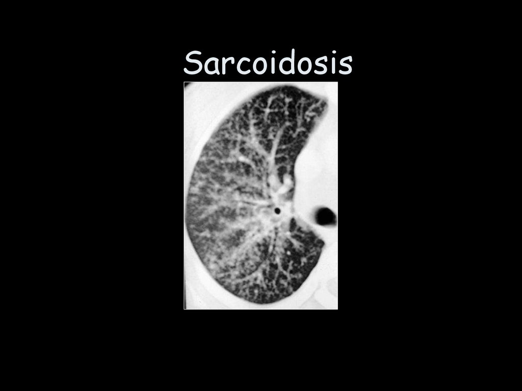
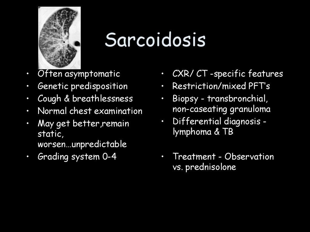
Sarcoidosis26.
SarcoidosisOften asymptomatic
Genetic predisposition
Cough & breathlessness
Normal chest examination
May get better,remain
static,
worsen…unpredictable
• Grading system 0-4
• CXR/ CT -specific features
• Restriction/mixed PFT’s
• Biopsy - transbronchial,
non-caseating granuloma
• Differential diagnosis lymphoma & TB
• Treatment - Observation
vs. prednisolone
27.
Interstitial disease• What is the interstitium?
• What does the interstitium do?
• What are the pathophysiological effects of
interstitial disease?
• What are the clinical manifestations?
28.
Objectives• Interstitium
• Pleural disease
• Chest wall disease
29.
Pleural Disease• Anatomy
• Effusions
• Malignancy
30.
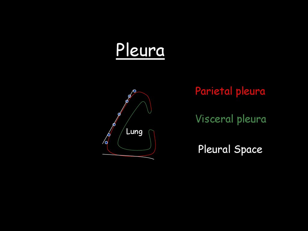
PleuraParietal pleura
Visceral pleura
Lung
Pleural Space
31.
LUNGVisceral pleura
Fat pad
Parietal pleura
Endothoracic fascia
Innermost intercostal
Intercostal fat
& vessels
Intercostal muscles
32.
Functions of the pleural space• Allow movement of lung and chest wall
• Coupling of chest wall and lung - inward lung recoil,
outward chest wall recoil
• Pleural fluid circulation
Lung
33.
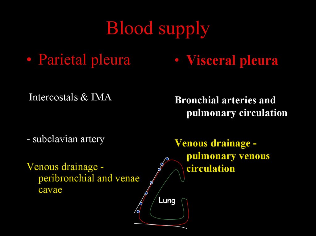
Blood supply• Parietal pleura
• Visceral pleura
Intercostals & IMA
Bronchial arteries and
pulmonary circulation
- subclavian artery
Venous drainage pulmonary venous
circulation
Venous drainage peribronchial and venae
cavae
Lung
34.
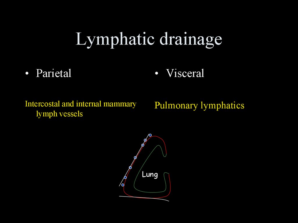
Lymphatic drainage• Parietal
• Visceral
Intercostal and internal mammary
lymph vessels
Pulmonary lymphatics
Lung
35.
Pleura - innervationParietal pleura - somatic,
sympathetic & parasympathetic
Phrenic & intercostal nerves
Lung
Visceral pleura - devoid of
somatic innervation
36.
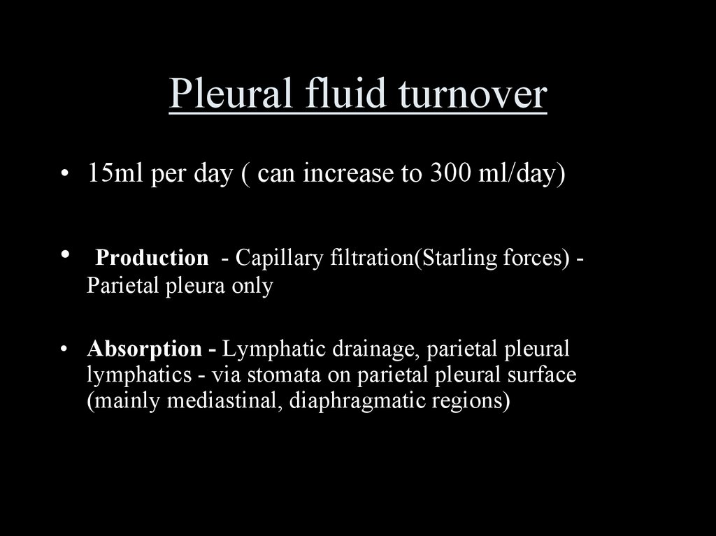
Pleural fluid turnover• 15ml per day ( can increase to 300 ml/day)
Production - Capillary filtration(Starling forces) Parietal pleura only
• Absorption - Lymphatic drainage, parietal pleural
lymphatics - via stomata on parietal pleural surface
(mainly mediastinal, diaphragmatic regions)
37.
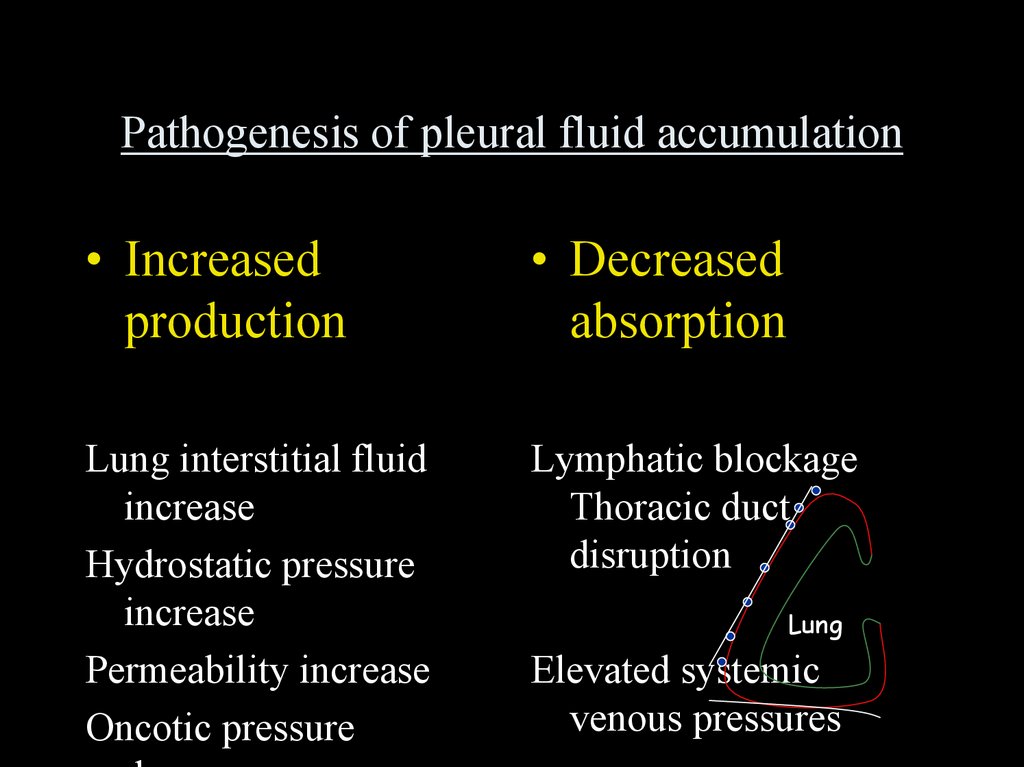
Pathogenesis of pleural fluid accumulation• Increased
production
• Decreased
absorption
Lung interstitial fluid
increase
Hydrostatic pressure
increase
Permeability increase
Oncotic pressure
Lymphatic blockage
Thoracic duct
disruption
Lung
Elevated systemic
venous pressures
38.
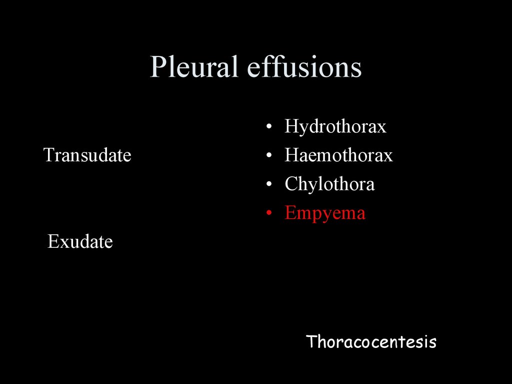
Pleural effusionsTransudate
Hydrothorax
Haemothorax
Chylothora
Empyema
Exudate
Thoracocentesis
39.
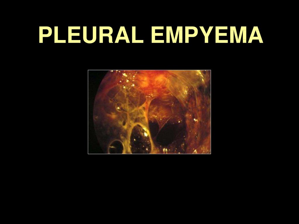
PLEURAL EMPYEMA40.
PLEURAL EMPYEMADefinition
90
82
Collection of pus in
the pleural cavity
commonly secondary
to a pneumonia
80
70
60
50
40
30
20
8
10
0
PNEUMONIA
TUMOR
6
SURGERY
3
TB
1
FOREIGN BODY
41.
EMPYEMA: complicationsfistula
fibrothorax
chronic
empyema
trapped
lung
empyema
necessitatis
functional
restriction
42.
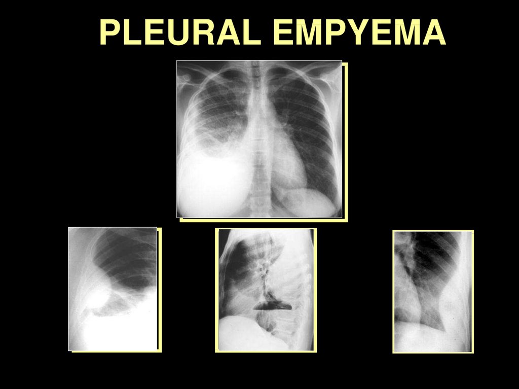
PLEURAL EMPYEMA43.
PLEURAL EMPYEMA44.
Pleural malignancy• Metastatic
• Primary - mesothelioma
• Mesothelioma
Asbestos exposure
Pain, breathlessness
Effusion, mediastinal pleural
enhancement
Chemotherapy, palliative &
radical surgery
Poor prognosis
45.
Pleural Disease• Anatomy
• Effusions
• Malignancy
46.
Objectives• Interstitium
• Pleural disease
• Chest wall disease
47.
Chest wall disease• Congenital
• Acquired
Pectus deformities
Scoliosis
Kyphosis
Muscular dystrophy
Trauma
Iatrogenic
Ankylosing spondylitis
Motor neurone disease
48.
Chest wall diseaseVentilation
Volume (l)
Time (s)
49.
Chest wall diseaseVentilation
Sleep disordered breathing
Poor clearance of secretions
Atelectasis
Pneumonia