


 biology
Similar presentations:


Anatomical Basis of Breathing
1.
Dr. Monqith Mazin2.
Objectivesa.
b.
c.
d.
Describe thoracic wall: bones and muscles
Define the muscles of respiration
Define the mediastinum and its contents
Describe the pleura, pleural cavity and
pleural reflections
e. Recognize the mechanism of breathing
3.
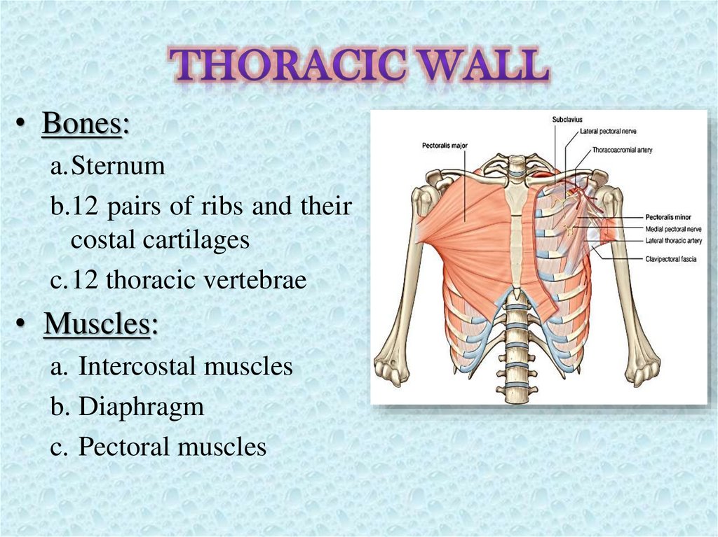
• Bones:a.Sternum
b.12 pairs of ribs and their
costal cartilages
c.12 thoracic vertebrae
• Muscles:
a. Intercostal muscles
b. Diaphragm
c. Pectoral muscles
4.
• Sternum:flat bone consists of three
parts:
1. Manubrium
2. Body
3. Xiphoid process
5.
• Ribs:12 pairs of flat bones. Divided into 3
types:
1. True ribs (1-7)
2. False ribs (8-10)
3. Floating ribs (11-12)
Also the ribs divided into:
a. Typical ribs (3-9)
b. Atypical ribs (1, 2, 10 ,11, 12)
6.
• Head with two articularsurfaces
• Neck
• Tubercle with two parts
• Shaft with an angle and
costal groove
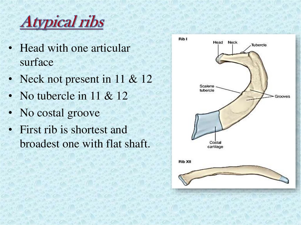
7.
• Head with one articularsurface
• Neck not present in 11 & 12
• No tubercle in 11 & 12
• No costal groove
• First rib is shortest and
broadest one with flat shaft.
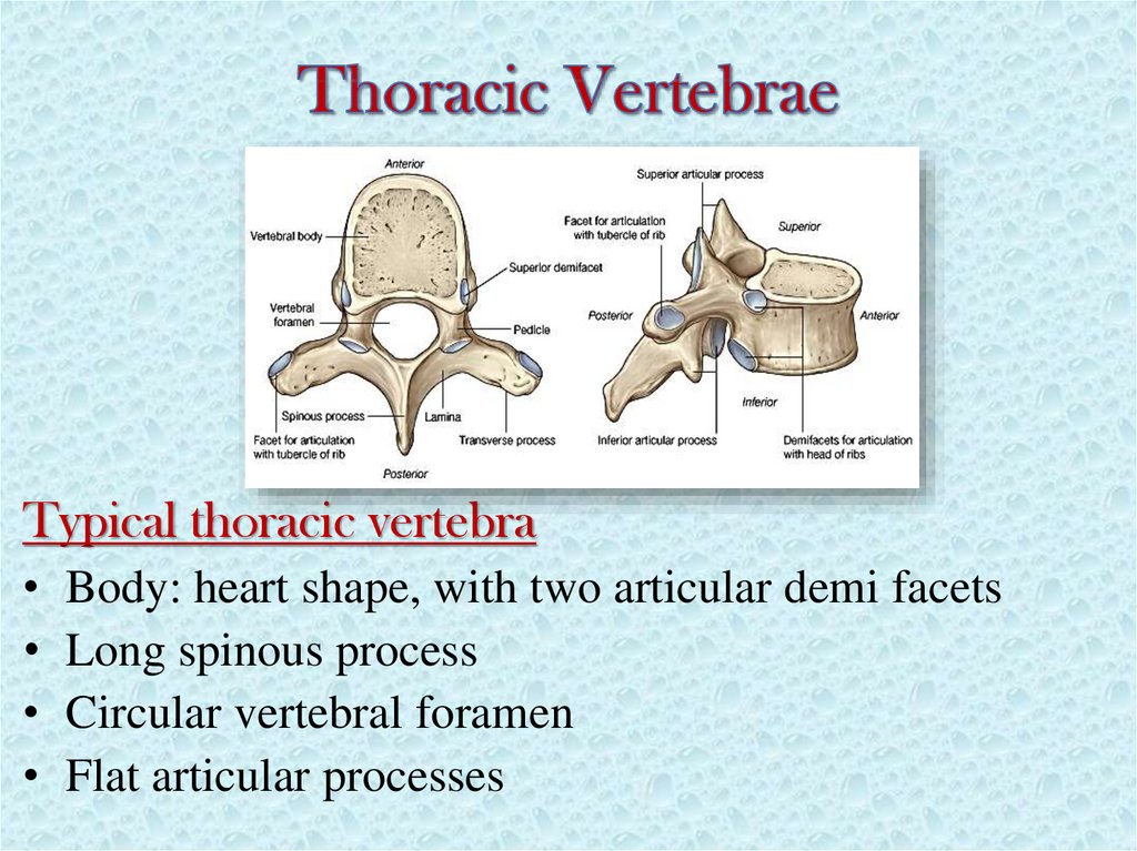
8.
Typical thoracic vertebraBody: heart shape, with two articular demi facets
Long spinous process
Circular vertebral foramen
Flat articular processes
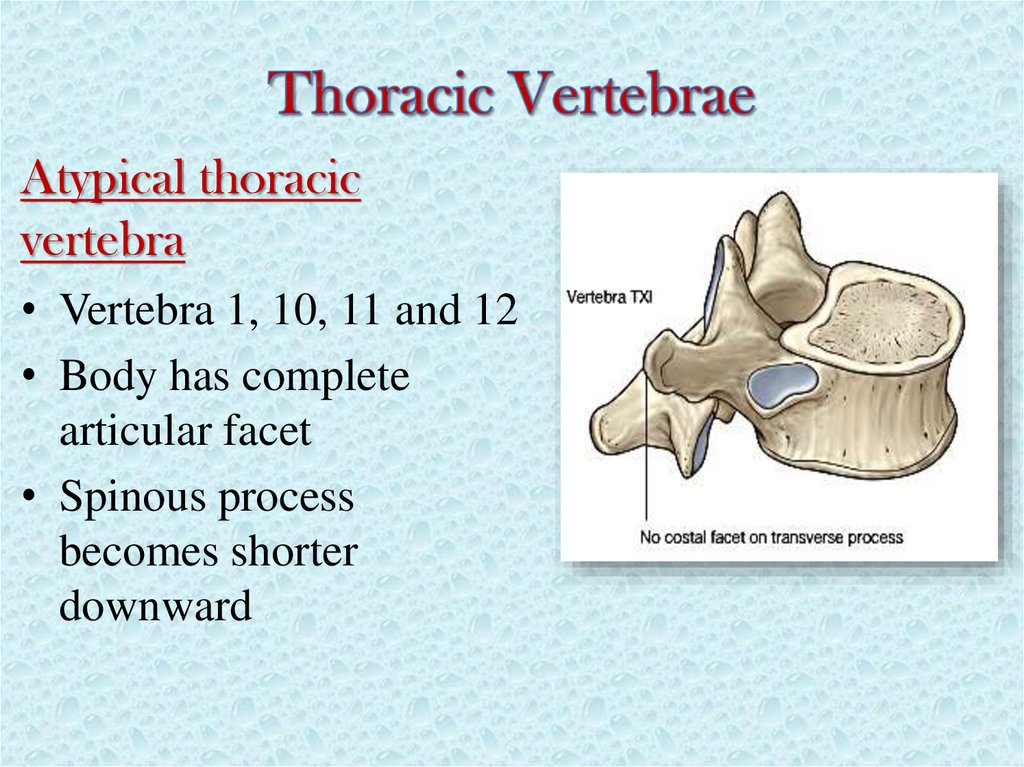
9.
Atypical thoracicvertebra
• Vertebra 1, 10, 11 and 12
• Body has complete
articular facet
• Spinous process
becomes shorter
downward
10.
Intervertebral joints:a. Symphyses: vertebral bodies;
b. synovial joints: articular
processes
Costovertebral joints:
synovial
Sterno-costal joints:
a. First
rib:
Primary
cartilaginous
b. 2nd- 7th ribs: synovial
Inter-chondral joints:
synovial
11.
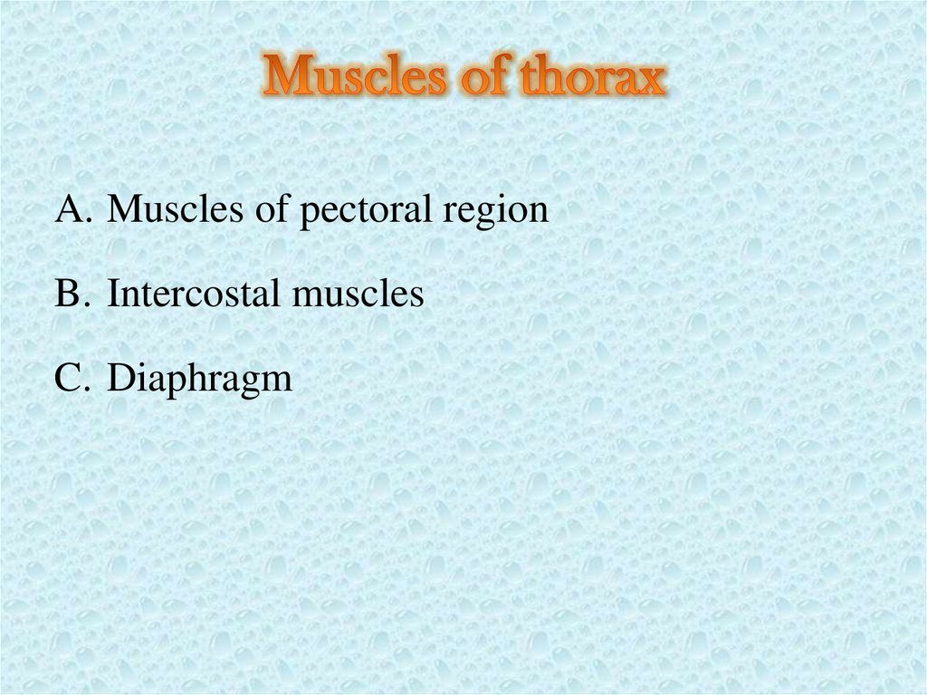
A. Muscles of pectoral regionB. Intercostal muscles
C. Diaphragm
12.
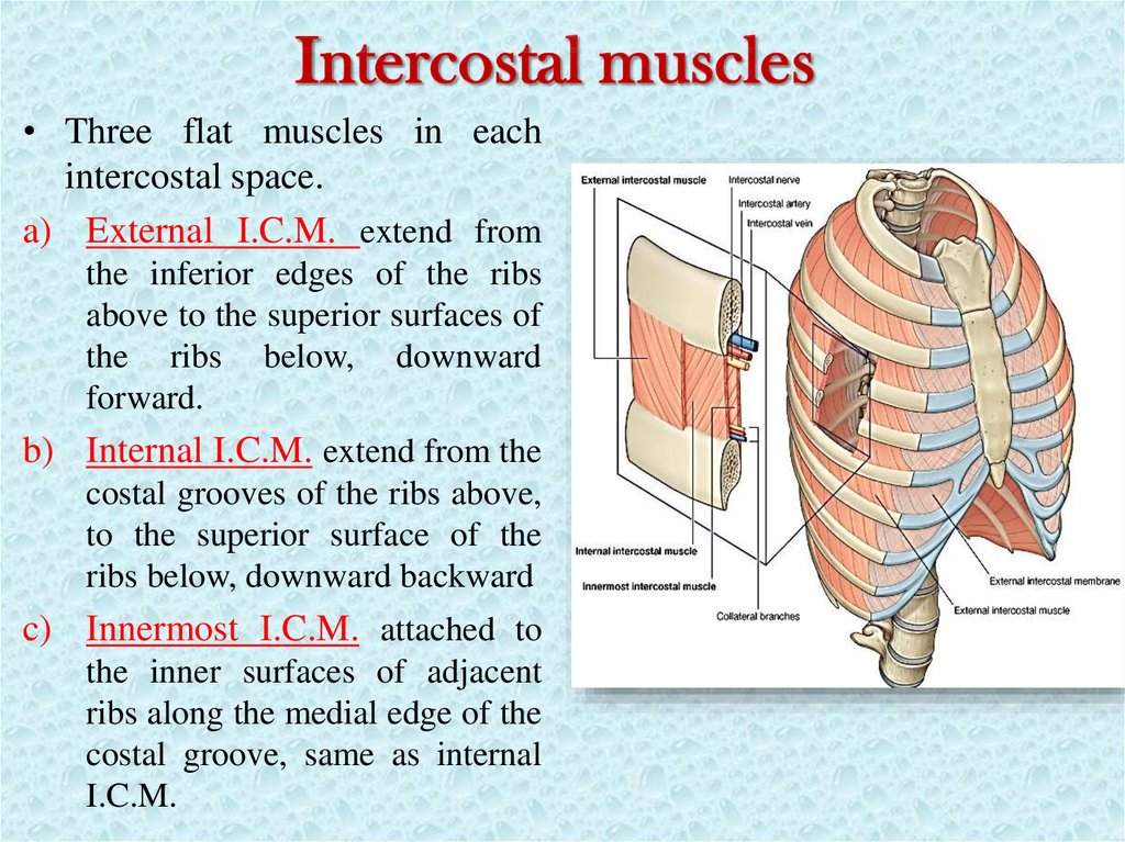
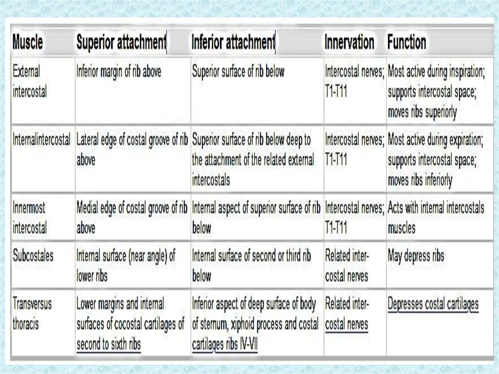
• Three flat muscles in eachintercostal space.
a) External I.C.M. extend from
the inferior edges of the ribs
above to the superior surfaces of
the ribs below, downward
forward.
b) Internal I.C.M. extend from the
costal grooves of the ribs above,
to the superior surface of the
ribs below, downward backward
c) Innermost I.C.M. attached to
the inner surfaces of adjacent
ribs along the medial edge of the
costal groove, same as internal
I.C.M.
13.
Transversus thoracisfrom the posterior aspect of the
xiphoid process, the inferior part
of the body of the sternum, and
the adjacent costal cartilages of
the lower true ribs. They pass
superiorly and laterally to insert
into the lower borders of the
costal cartilages of ribs III to VI.
Subcostales
They extend from the internal
surfaces of one rib to the internal
surface of the second or third rib
below. Their fibers parallel the
course of the internal intercostal
muscles and extend from the
angle of the ribs to more medial
positions on the ribs below.
14.
15.
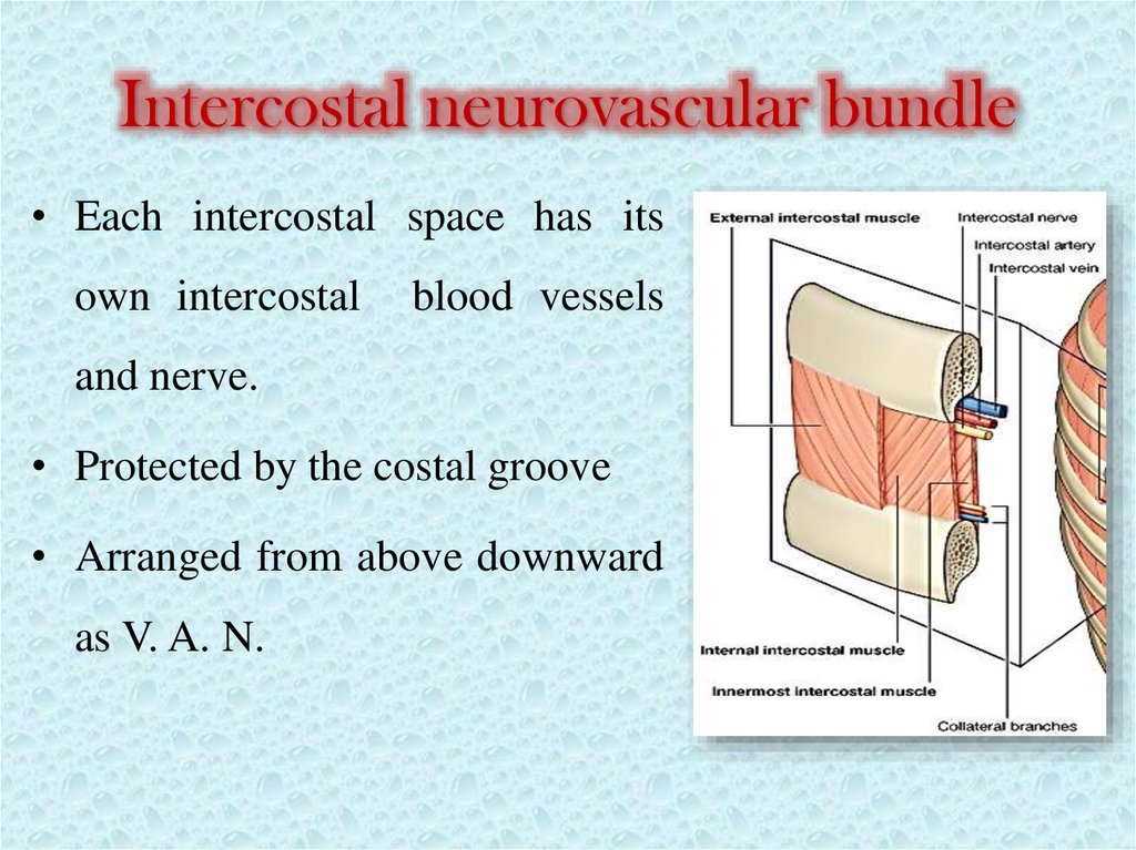
Intercostal neurovascular bundle• Each intercostal space has its
own intercostal
blood vessels
and nerve.
• Protected by the costal groove
• Arranged from above downward
as V. A. N.
16.
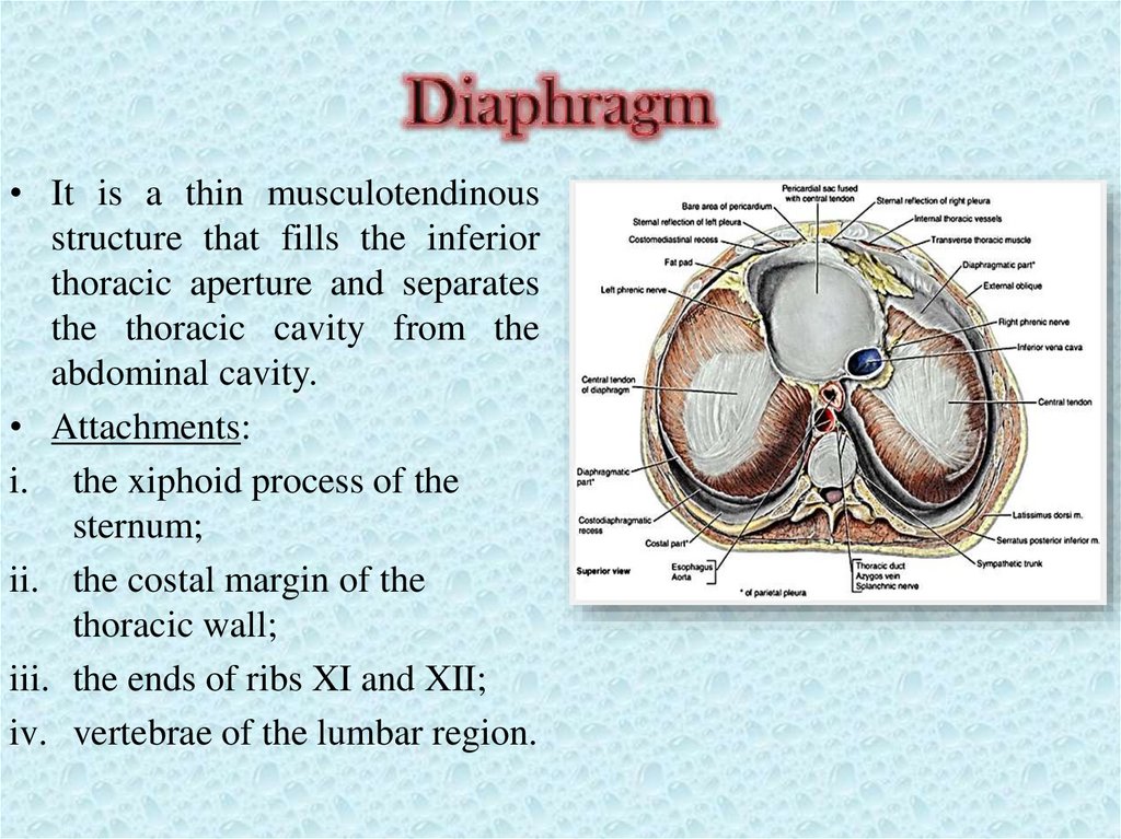
• It is a thin musculotendinousstructure that fills the inferior
thoracic aperture and separates
the thoracic cavity from the
abdominal cavity.
• Attachments:
i. the xiphoid process of the
sternum;
ii. the costal margin of the
thoracic wall;
iii. the ends of ribs XI and XII;
iv. vertebrae of the lumbar region.
17.
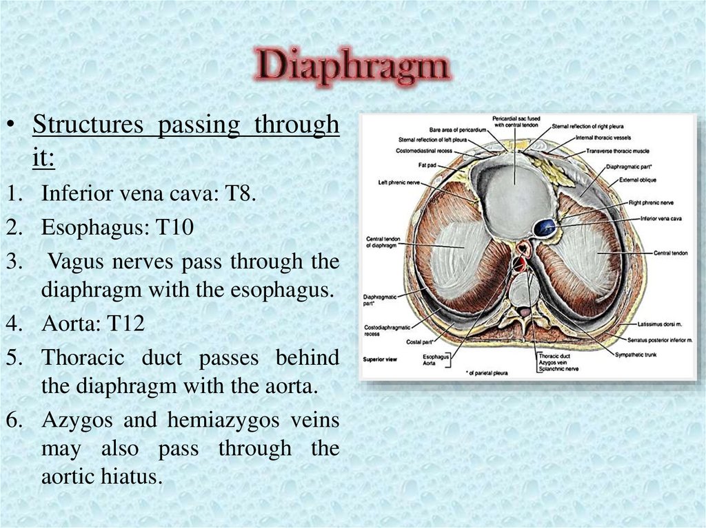
• Structures passing throughit:
1. Inferior vena cava: T8.
2. Esophagus: T10
3. Vagus nerves pass through the
diaphragm with the esophagus.
4. Aorta: T12
5. Thoracic duct passes behind
the diaphragm with the aorta.
6. Azygos and hemiazygos veins
may also pass through the
aortic hiatus.
18.
• Blood supply:i. From above, Pericardiacophrenic and Musculophrenic
arteries; branches of the internal thoracic artery.
ii. From below, inferior phrenic arteries, which branch
directly from the abdominal aorta.
• Nerve supply:
Phrenic nerves (C3 to C5)
19.
• The cavity of thorax extends fromsuperior to inferior thoracic apertures.
• Superior thoracic aperture is bounded
by T1 vertebra, 1st ribs and manubrium.
• Inferior thoracic aperture is bounded by
attachments of diaphragm.
• It is divided into bilateral pleural
cavities and a central mediastinum
20.
• Each pleural cavity is lined by asingle layer of flat mesothelial
cells, and an associated layer of
supporting
connective
tissue;
together, they form the pleura.
• It is divided into two major types,
based on location:
a) Parietal pleura; lines pleural
cavity
b) Visceral pleura; adheres to and
covers the lung.
• Plural space between the two
layers contain thin film of serous
fluid.
21.
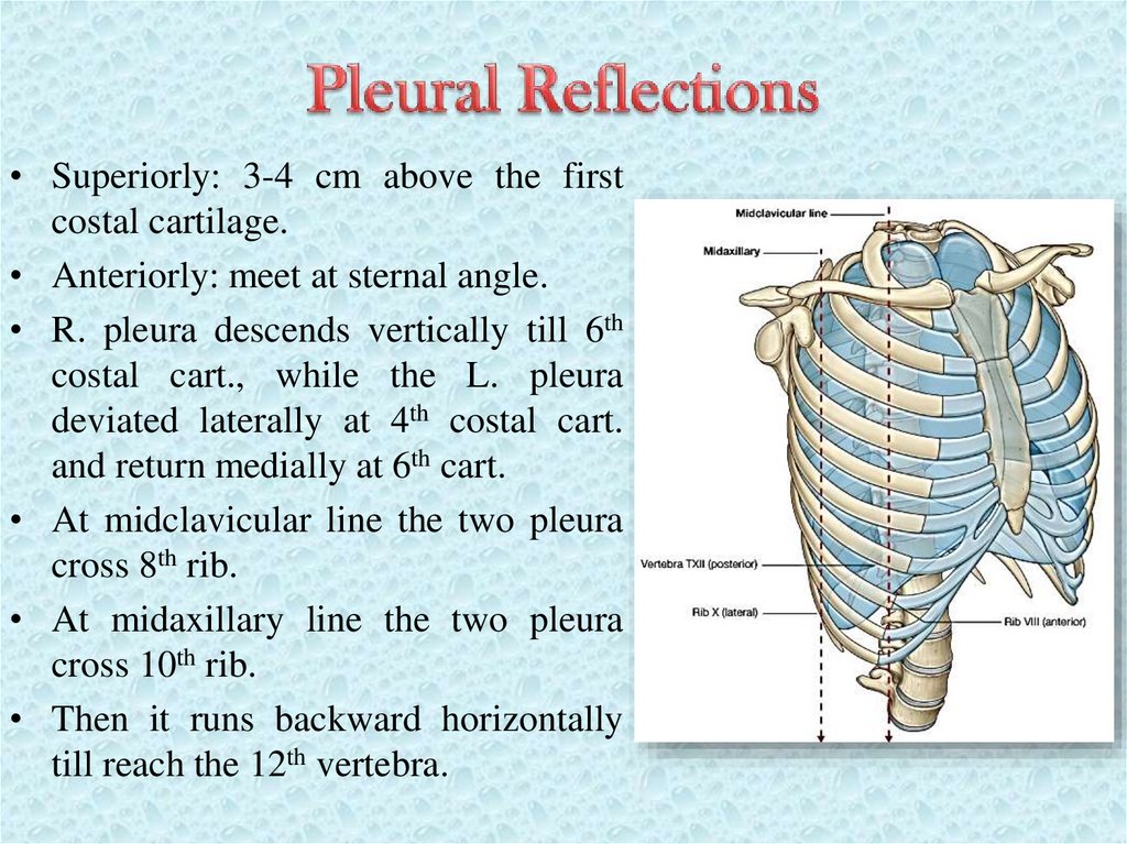
• Superiorly: 3-4 cm above the firstcostal cartilage.
• Anteriorly: meet at sternal angle.
• R. pleura descends vertically till 6th
costal cart., while the L. pleura
deviated laterally at 4th costal cart.
and return medially at 6th cart.
• At midclavicular line the two pleura
cross 8th rib.
• At midaxillary line the two pleura
cross 10th rib.
• Then it runs backward horizontally
till reach the 12th vertebra.
22.
• Spaces where the twolayers of pleura become
opposed as the lung do not
fill the pleural cavity.
a) Costomediastinal
recesses
b) Costodiaphragmatic
recesses
23.
• One of the principal functions of the thoracic wall and thediaphragm is to alter the volume of the thorax and thereby
move air in and out of the lungs.
• During breathing, the dimensions of the thorax change in the
vertical, lateral, and anteroposterior directions.
• The primary muscle of respiration is the diaphragm.
• Accessory muscles of respiration assist the diaphragm include:
I.C.M., Pectoral muscles, neck muscles and abdominal
muscles.
24.
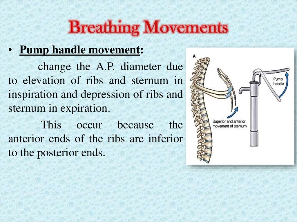
• Pump handle movement:change the A.P. diameter due
to elevation of ribs and sternum in
inspiration and depression of ribs and
sternum in expiration.
This occur because the
anterior ends of the ribs are inferior
to the posterior ends.
25.
• Bucket handle movementIncreases
dimensions
of
the
lateral
the
thorax,
because the middles of the shafts
tend to be lower than the two
ends.
When
the shafts
are
elevated, the middles of the
shafts move laterally.
26.
So,• In Inspiration:
i. Diaphragm contracts and depressed that increases
vertical diameter of thoracic cavity.
ii. Elevation of anterior parts of ribs with the sternum by
pump handle mechanism increases the anteroposterior
diameter of thoracic cavity.
iii. Elevation of middle parts of ribs by bucket handle
mechanism increases the lateral diameter of thoracic
cavity.
• In expiration:
vice versa