


")


 biology
Similar presentations:


")


Circulation and Gas Exchange
1. Chapter 42
Circulation andGas Exchange
PowerPoint® Lecture Presentations for
Biology
Eighth Edition
Neil Campbell and Jane Reece
Lectures by Chris Romero, updated by Erin Barley with contributions from Joan Sharp
Copyright © 2008 Pearson Education, Inc., publishing as Pearson Benjamin Cummings
2. Overview: Trading Places
• Every organism must exchange materials withits environment.
• Exchanges ultimately occur at the cellular level.
• In unicellular organisms, these exchanges
occur directly with the environment.
Copyright © 2008 Pearson Education, Inc., publishing as Pearson Benjamin Cummings
3.
• For most cells making up multicellularorganisms, direct exchange with the
environment is not possible.
• Gills are an example of a specialized exchange
system in animals.
• Internal transport and gas exchange are
functionally related in most animals.
Copyright © 2008 Pearson Education, Inc., publishing as Pearson Benjamin Cummings
4.
How does a feathery fringe help this animal survive?5. Circulatory systems link exchange surfaces with cells throughout the body
• In small and/or thin animals, cells canexchange materials directly with the
surrounding medium.
• In most animals, transport systems connect
the organs of exchange with the body cells.
• Most complex animals have internal transport
systems that circulate fluid.
Copyright © 2008 Pearson Education, Inc., publishing as Pearson Benjamin Cummings
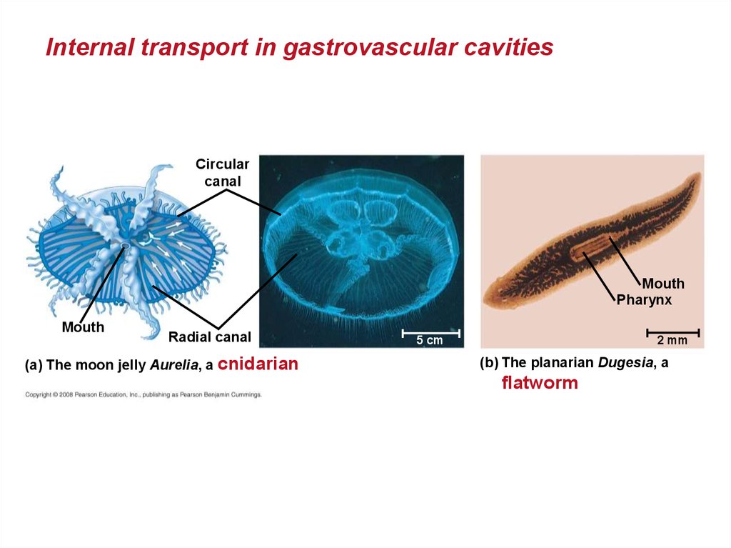
6. Gastrovascular Cavities
• Simple animals, such as cnidarians, have abody wall that is only two cells thick and that
encloses a gastrovascular cavity.
• This cavity functions in both digestion and
distribution of substances throughout the body.
• Some cnidarians, such as jellies, have
elaborate gastrovascular cavities.
• Flatworms have a gastrovascular cavity and a
large surface area to volume ratio.
Copyright © 2008 Pearson Education, Inc., publishing as Pearson Benjamin Cummings
7.
Internal transport in gastrovascular cavitiesCircular
canal
Mouth
Pharynx
Mouth
Radial canal
(a) The moon jelly Aurelia, a cnidarian
5 cm
2 mm
(b) The planarian Dugesia, a
flatworm
8. Open and Closed Circulatory Systems
• More complex animals have either open orclosed circulatory systems.
• Both systems have three basic components:
– A circulatory fluid = blood or hemolymph.
– A set of tubes = blood vessels.
– A muscular pump = the heart.
Copyright © 2008 Pearson Education, Inc., publishing as Pearson Benjamin Cummings
9.
• In insects, other arthropods, and mostmolluscs, blood bathes the organs directly in
an open circulatory system.
• In an open circulatory system, there is no
distinction between blood and interstitial fluid,
and this general body fluid is more correctly
called hemolymph.
Copyright © 2008 Pearson Education, Inc., publishing as Pearson Benjamin Cummings
10.
• In a closed circulatory system, the blood isconfined to vessels and is distinct from the
interstitial fluid.
• Closed systems are more efficient at
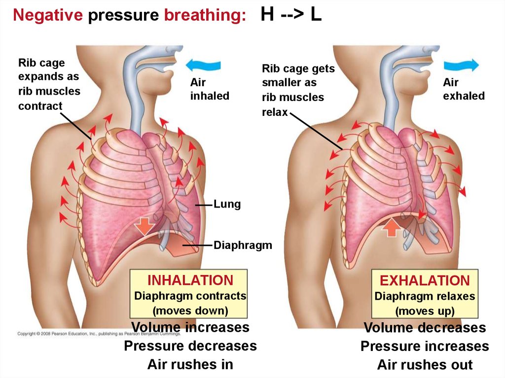
transporting circulatory fluids to tissues and
cells.
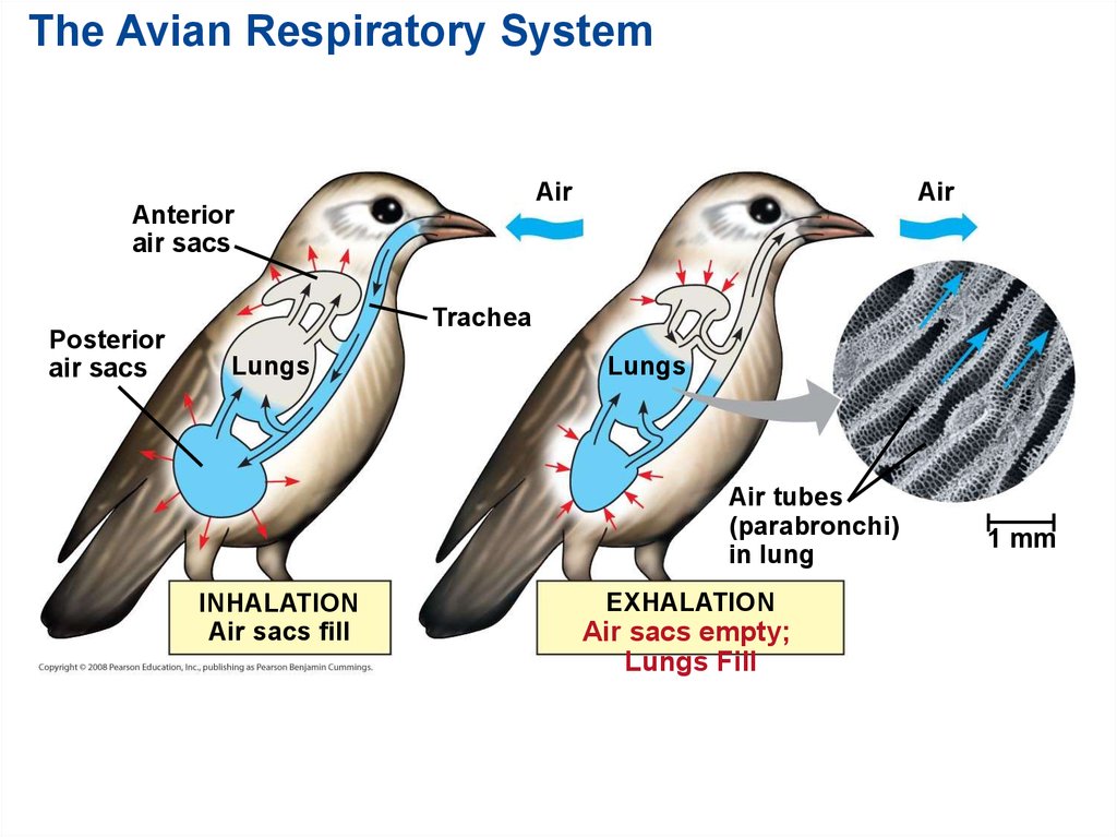
Copyright © 2008 Pearson Education, Inc., publishing as Pearson Benjamin Cummings
11.
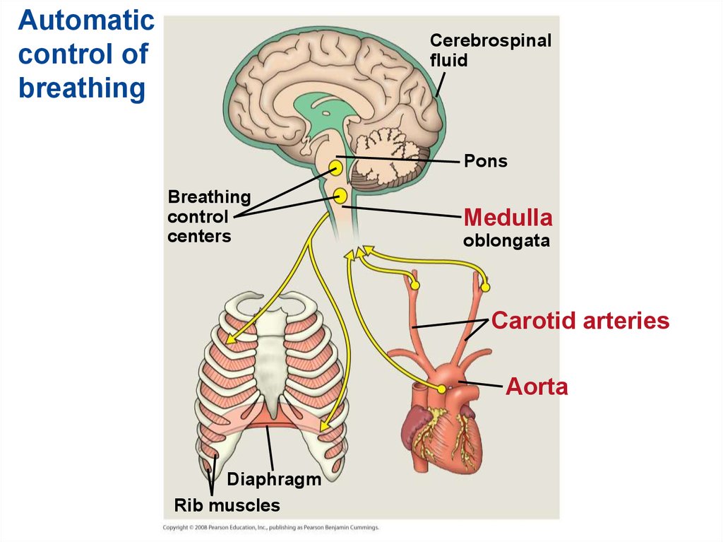
Open and closed circulatory systemsHeart
Hemolymph in
sinuses
surrounding organs
Pores
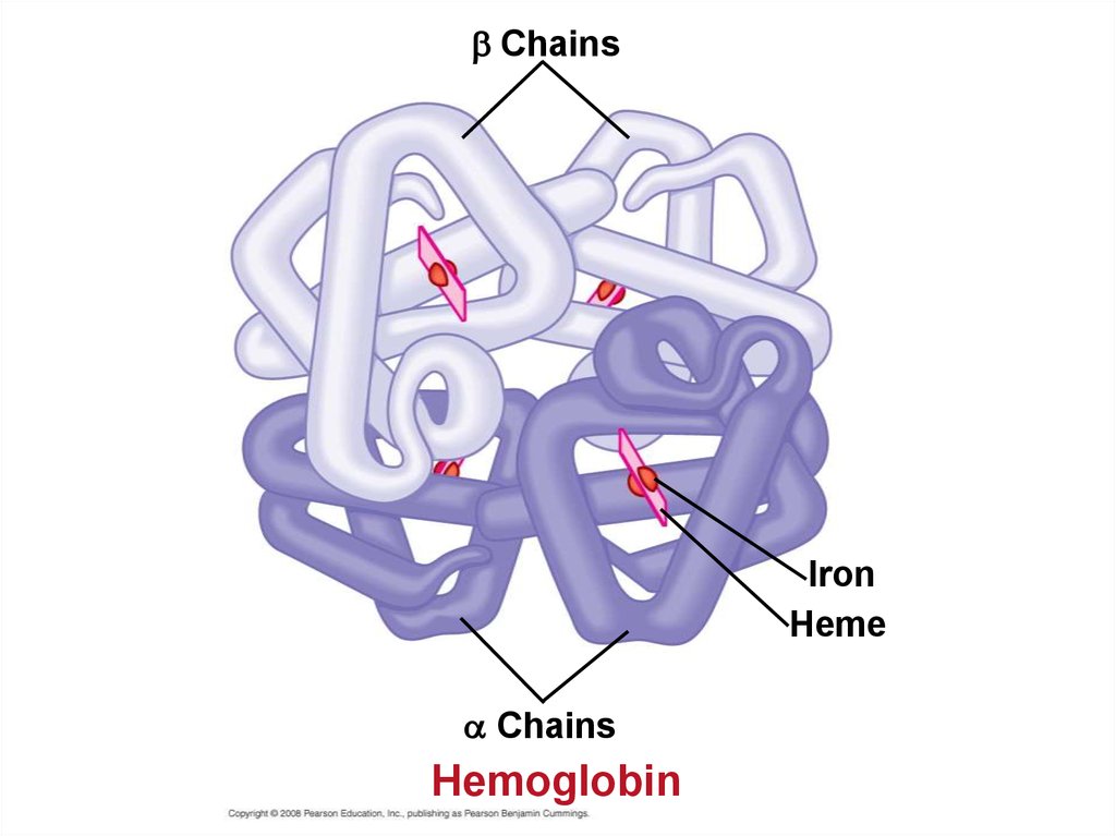
Blood
Small branch vessels
In each organ
Interstitial
fluid
Dorsal vessel
(main heart)
Tubular heart
(a) An open
Heart
circulatory system
Auxiliary hearts
(b) A closed
Ventral vessels
circulatory system
12. Organization of Vertebrate Closed Circulatory Systems
• Humans and other vertebrates have a closedcirculatory system, often called the
cardiovascular system.
• The three main types of blood vessels are:
arteries - away from the heart.
veins - toward the heart.
capillaries - exchange with body cells.
Copyright © 2008 Pearson Education, Inc., publishing as Pearson Benjamin Cummings
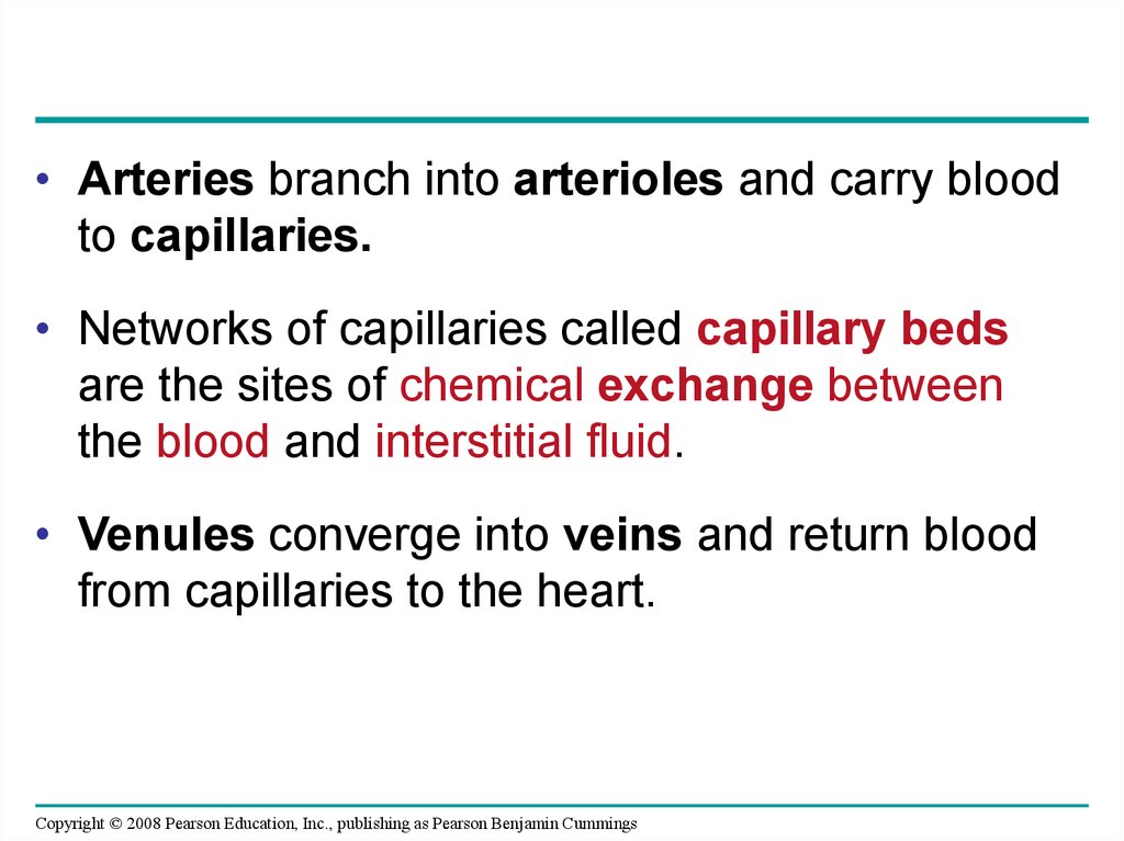
13.
• Arteries branch into arterioles and carry bloodto capillaries.
• Networks of capillaries called capillary beds
are the sites of chemical exchange between
the blood and interstitial fluid.
• Venules converge into veins and return blood
from capillaries to the heart.
Copyright © 2008 Pearson Education, Inc., publishing as Pearson Benjamin Cummings
14.
• Vertebrate hearts contain two or morechambers.
• Blood enters through an atrium and is pumped
out through a ventricle.
Atria - receive blood
Ventricles - pump blood
Copyright © 2008 Pearson Education, Inc., publishing as Pearson Benjamin Cummings
15. Single Circulation
• Bony fishes, rays, and sharks have singlecirculation with a two-chambered heart.
• In single circulation, blood leaving the heart
passes through two capillary beds before
returning.
Copyright © 2008 Pearson Education, Inc., publishing as Pearson Benjamin Cummings
16.
Singlecirculation
in fishes
Gill capillaries
Artery
Gill
circulation
Ventricle
Heart
Atrium
Vein
Systemic
circulation
Systemic capillaries
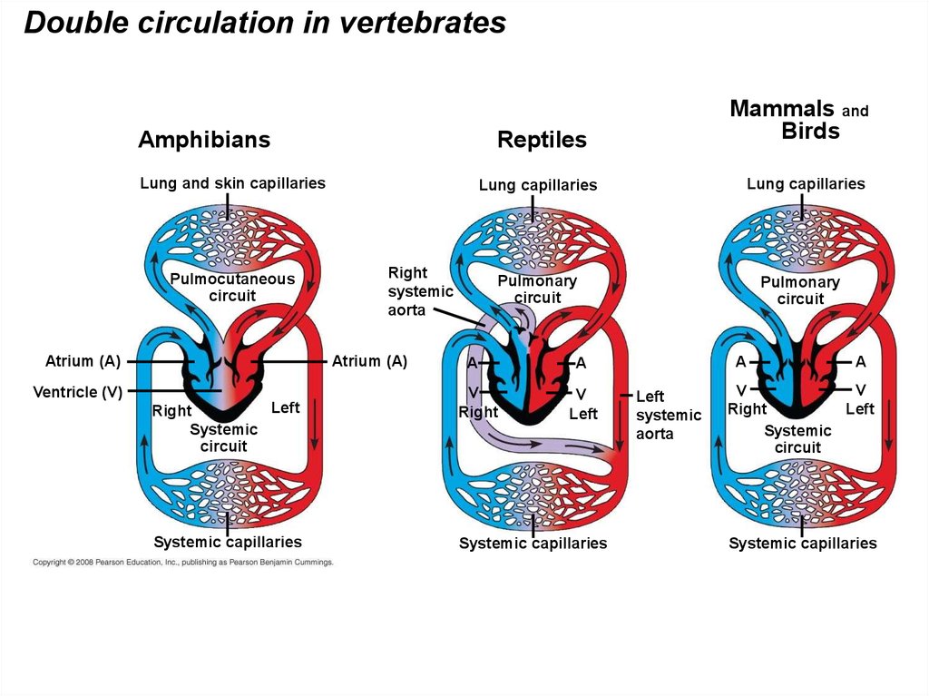
17. Double Circulation
• Amphibian, reptiles, and mammals havedouble circulation.
• Oxygen-poor and oxygen-rich blood are
pumped separately from the right and left sides
of the heart.
Copyright © 2008 Pearson Education, Inc., publishing as Pearson Benjamin Cummings
18.
Double circulation in vertebratesAmphibians
Reptiles
Lung and skin capillaries
Pulmocutaneous
circuit
Atrium (A)
Left
Right
Systemic
circuit
Systemic capillaries
Lung capillaries
Lung capillaries
Right
systemic
aorta
Atrium (A)
Ventricle (V)
Mammals and
Birds
Pulmonary
circuit
A
V
Right
Pulmonary
circuit
A
A
V
Left
Systemic capillaries
Left
systemic
aorta
A
V
V
Right
Left
Systemic
circuit
Systemic capillaries
19.
• In reptiles and mammals, oxygen-poor bloodflows through the pulmonary circuit to pick up
oxygen through the lungs.
• In amphibians, oxygen-poor blood flows
through a pulmocutaneous circuit to pick up
oxygen through the lungs and skin.
• Oxygen-rich blood delivers oxygen through the
systemic circuit.
• Double circulation maintains higher blood
pressure in the organs than does single
circulation.
Copyright © 2008 Pearson Education, Inc., publishing as Pearson Benjamin Cummings
20. Adaptations of Double Circulatory Systems
Amphibians:• Frogs / amphibians have a three-chambered
heart: 2 atria and 1 ventricle.
• The ventricle pumps blood into a forked artery
that splits the ventricle’s output into the
pulmocutaneous circuit and the systemic
circuit.
• Underwater, blood flow to the lungs is nearly
shut off.
Copyright © 2008 Pearson Education, Inc., publishing as Pearson Benjamin Cummings
21. Reptiles (Except Birds)
• Turtles, snakes, and lizards have a threechambered heart: two atria and one ventricle.• In alligators, caimans, and other crocodilians a
septum - partially or fully divides the ventricle.
• Reptiles have double circulation, with a
pulmonary circuit - lungs and a systemic circuit.
Copyright © 2008 Pearson Education, Inc., publishing as Pearson Benjamin Cummings
22. Mammals
RA --> RV --> LUNGS --> LA --> LV --> BodyMammals
• Mammals and birds have a four-chambered
heart with two atria and two ventricles.
• The left side of the heart pumps and receives
only oxygen-rich blood, while the right side
receives and pumps only oxygen-poor blood.
• Mammals and birds are endotherms and
require more O2 than ectotherms.
Copyright © 2008 Pearson Education, Inc., publishing as Pearson Benjamin Cummings
23. Coordinated cycles of heart contraction drive double circulation in mammals
• Blood begins its flow with the right ventriclepumping blood to the lungs.
• In the lungs, the blood loads O2 and unloads
CO2
• Oxygen-rich blood from the lungs enters the
heart at the left atrium and is pumped through
the aorta to the body tissues by the left
ventricle.
• The aorta provides blood to the heart through
the coronary arteries.
Copyright © 2008 Pearson Education, Inc., publishing as Pearson Benjamin Cummings
24.
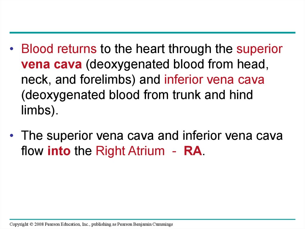
• Blood returns to the heart through the superiorvena cava (deoxygenated blood from head,
neck, and forelimbs) and inferior vena cava
(deoxygenated blood from trunk and hind
limbs).
• The superior vena cava and inferior vena cava
flow into the Right Atrium - RA.
Copyright © 2008 Pearson Education, Inc., publishing as Pearson Benjamin Cummings
25.
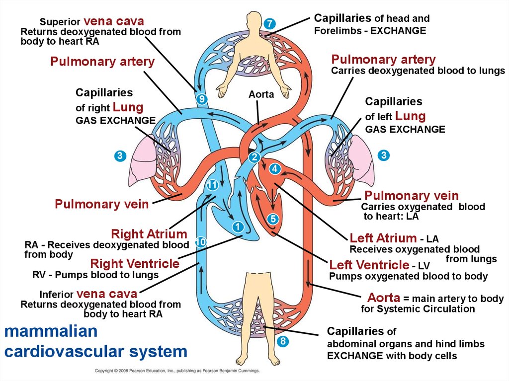
Superior vena cavaReturns deoxygenated blood from
body to heart RA
Capillaries of head and
7
Forelimbs - EXCHANGE
Pulmonary artery
Pulmonary artery
Capillaries
of right Lung
GAS EXCHANGE
Carries deoxygenated blood to lungs
Aorta
9
Capillaries
of left Lung
GAS EXCHANGE
3
3
2
4
11
Pulmonary vein
Pulmonary vein
Right Atrium
1
Carries oxygenated blood
to heart: LA
5
Left Atrium - LA
RA - Receives deoxygenated blood 10
from body
Receives oxygenated blood
from lungs
Left Ventricle - LV
Pumps oxygenated blood to body
Right Ventricle
RV - Pumps blood to lungs
Inferior vena cava
Returns deoxygenated blood from
body to heart RA
mammalian
cardiovascular system
Aorta = main artery to body
for Systemic Circulation
Capillaries of
8
abdominal organs and hind limbs
EXCHANGE with body cells
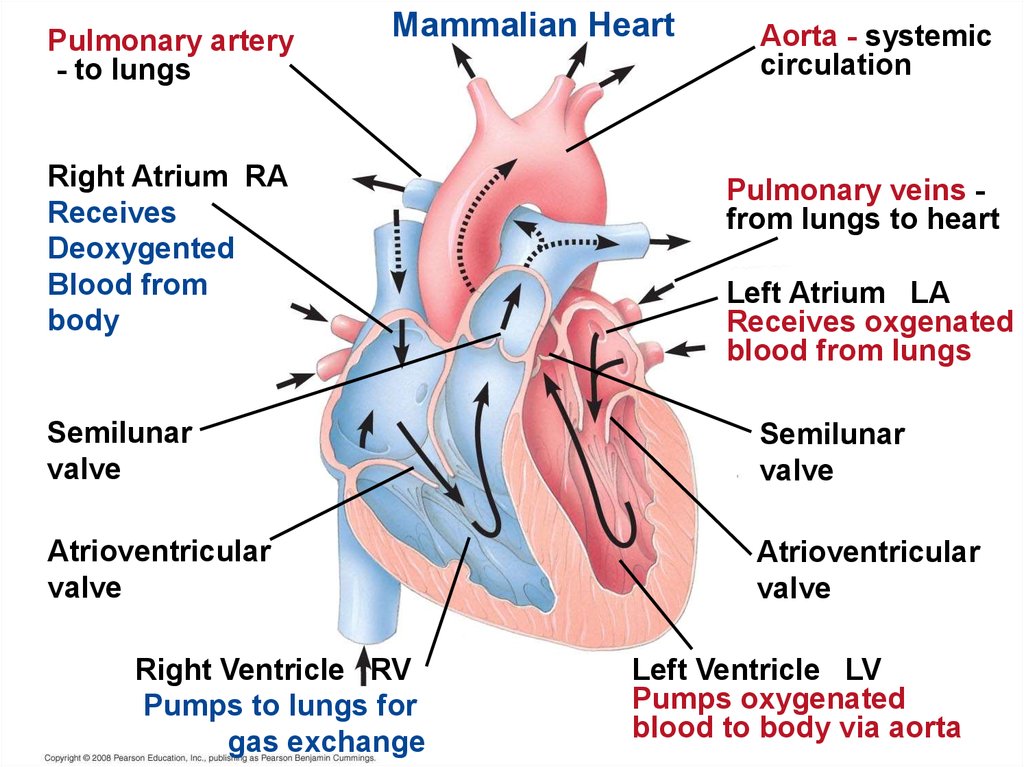
26. The Mammalian Heart: A Closer Look
• A closer look at the mammalian heart providesa better understanding of double circulation.
• RIGHT side = deoxygenated blood from body
pumped to lungs.
• LUNGS = gas exchange.
• LEFT side = oxygenated blood from lungs
pumped to body.
Copyright © 2008 Pearson Education, Inc., publishing as Pearson Benjamin Cummings
27.
Pulmonary artery- to lungs
Mammalian Heart
Right Atrium RA
Receives
Deoxygented
Blood from
body
Aorta - systemic
circulation
Pulmonary veins from lungs to heart
Left Atrium LA
Receives oxgenated
blood from lungs
Semilunar
valve
Semilunar
valve
Atrioventricular
valve
Atrioventricular
valve
Right Ventricle RV
Pumps to lungs for
gas exchange
Left Ventricle LV
Pumps oxygenated
blood to body via aorta
28.
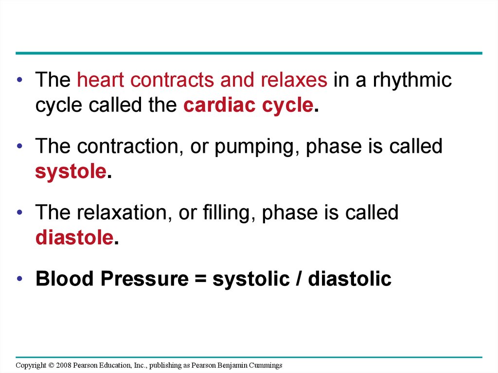
• The heart contracts and relaxes in a rhythmiccycle called the cardiac cycle.
• The contraction, or pumping, phase is called
systole.
• The relaxation, or filling, phase is called
diastole.
• Blood Pressure = systolic / diastolic
Copyright © 2008 Pearson Education, Inc., publishing as Pearson Benjamin Cummings
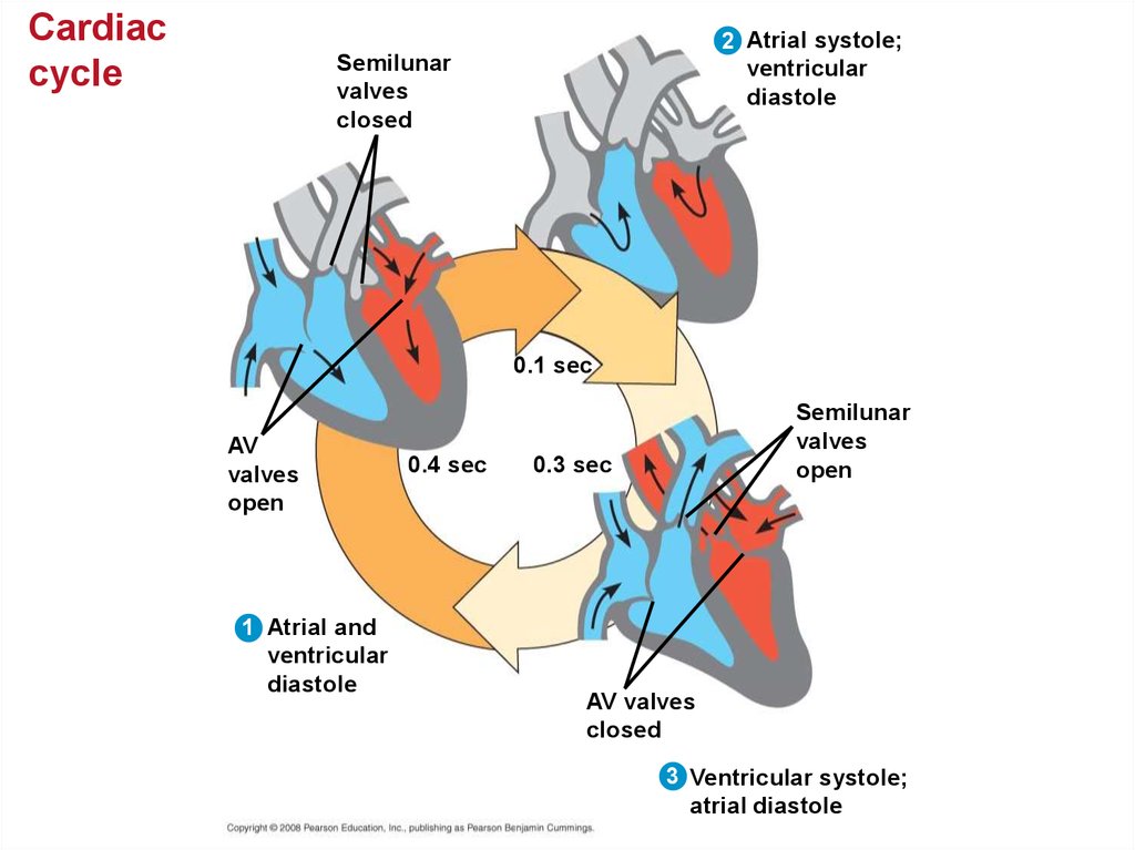
29.
Cardiaccycle
2 Atrial systole;
ventricular
diastole
Semilunar
valves
closed
0.1 sec
AV
valves
open
1 Atrial and
ventricular
diastole
0.4 sec
Semilunar
valves
open
0.3 sec
AV valves
closed
3 Ventricular systole;
atrial diastole
30.
• The heart rate, also called the pulse, is thenumber of beats per minute.
• The stroke volume is the amount of blood
pumped in a single contraction.
• The cardiac output is the volume of blood
pumped into the systemic circulation per
minute and depends on both the heart rate
and stroke volume.
Copyright © 2008 Pearson Education, Inc., publishing as Pearson Benjamin Cummings
31.
Four valves prevent backflow of blood in the heart:• The atrioventricular (AV) valves separate each
atrium and ventricle.
• The semilunar valves control blood flow to the aorta
and the pulmonary artery.
• The “lub-dup” sound of a heart beat is caused by the
recoil of blood against the AV valves (lub) then against
the semilunar (dup) valves.
• Backflow of blood through a defective valve causes a
heart murmur.
Copyright © 2008 Pearson Education, Inc., publishing as Pearson Benjamin Cummings
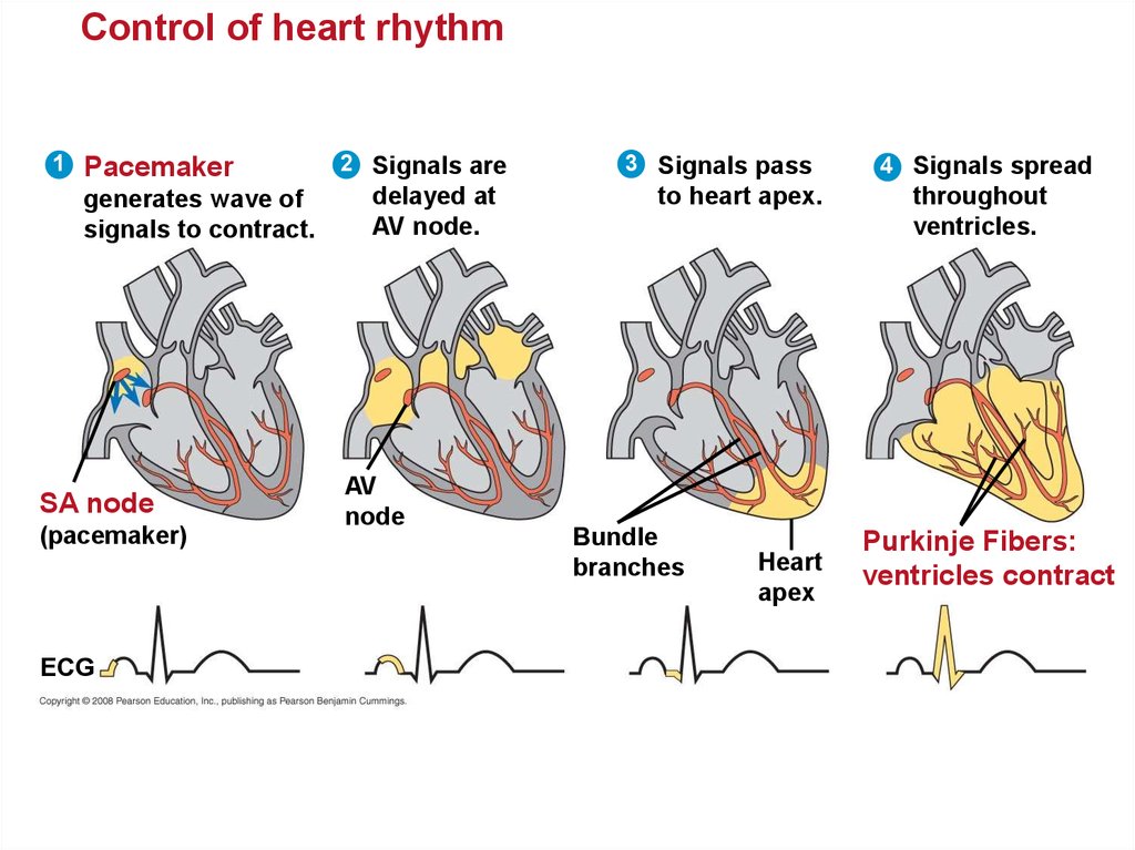
32. Maintaining the Heart’s Rhythmic Beat
• Some cardiac muscle cells are self-excitable = theycontract without any signal from the nervous system.
• The sinoatrial (SA) node, or pacemaker, sets the rate
and timing at which cardiac muscle cells contract.
• Impulses from the SA node travel to the
atrioventricular (AV) node. At the AV node, the
impulses are delayed and then travel to the Purkinje
fibers that make the ventricles contract.
• Impulses that travel during the cardiac cycle can be
recorded as an electrocardiogram (ECG or EKG).
The pacemaker is influenced by nerves, hormones,
body temperature, and exercise.
Copyright © 2008 Pearson Education, Inc., publishing as Pearson Benjamin Cummings
33.
Control of heart rhythm1
Pacemaker
generates wave of
signals to contract.
SA node
(pacemaker)
ECG
2 Signals are
delayed at
AV node.
AV
node
3 Signals pass
to heart apex.
Bundle
branches
Heart
apex
4 Signals spread
throughout
ventricles.
Purkinje Fibers:
ventricles contract
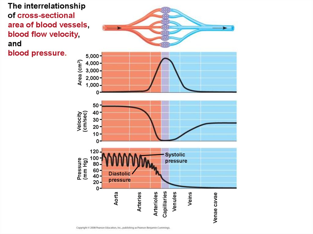
34. Patterns of blood pressure and flow reflect the structure and arrangement of blood vessels
• The physical principles that govern movementof water in plumbing systems also influence the
functioning of animal circulatory systems.
• The epithelial layer that lines blood vessels is
called the endothelium.
Copyright © 2008 Pearson Education, Inc., publishing as Pearson Benjamin Cummings
35.
Structureof
blood
vessels
Artery
Vein
SEM
100 µm
Valve
Basal lamina
Endothelium
Smooth
muscle
Connective
tissue
Endothelium
Capillary
Smooth
muscle
Connective
tissue
Artery
Vein
Capillary
15 µm
Red blood cell
Venule
LM
Arteriole
36.
• Capillaries have thin walls, the endotheliumplus its basement membrane, to facilitate the
exchange of materials.
• Arteries and veins have an endothelium,
smooth muscle, and connective tissue.
• Arteries have thicker walls than veins to
accommodate the high pressure of blood
pumped from the heart.
• In the thinner-walled veins, blood flows back to
the heart mainly as a result of muscle action.
Copyright © 2008 Pearson Education, Inc., publishing as Pearson Benjamin Cummings
37. Blood Flow Velocity
• Physical laws governing movement of fluidsthrough pipes affect blood flow and blood
pressure.
• Velocity of blood flow is slowest in the capillary
beds, as a result of the high resistance and
large total cross-sectional area.
• Blood flow in capillaries is necessarily slow for
exchange of materials.
Copyright © 2008 Pearson Education, Inc., publishing as Pearson Benjamin Cummings
38.
4,0003,000
2,000
1,000
0
50
40
30
20
10
0
Systolic
pressure
Venae cavae
Veins
Venules
Capillaries
Arterioles
Diastolic
pressure
Arteries
120
100
80
60
40
20
0
Aorta
Pressure
(mm Hg)
Velocity
(cm/sec)
Area (cm2)
The interrelationship
of cross-sectional
area of blood vessels,
blood flow velocity,
and
blood pressure.
5,000
39. Blood Pressure
• Blood pressure is the hydrostatic pressure thatblood exerts against the wall of a vessel.
• In rigid vessels blood pressure is maintained;
less rigid vessels deform and blood pressure is
lost.
Copyright © 2008 Pearson Education, Inc., publishing as Pearson Benjamin Cummings
40. Changes in Blood Pressure During the Cardiac Cycle
• Systolic pressure is the pressure in thearteries during ventricle contraction /systole; it
is the highest pressure in the arteries.
• Diastolic pressure is the pressure in the
arteries during relaxation /diastole; it is lower
than systolic pressure.
• A pulse is the rhythmic bulging of artery walls
with each heartbeat.
Copyright © 2008 Pearson Education, Inc., publishing as Pearson Benjamin Cummings
41. Regulation of Blood Pressure
• Blood pressure is determined by cardiac outputand peripheral resistance due to constriction of
arterioles.
• Vasoconstriction is the contraction of smooth
muscle in arteriole walls; it increases blood
pressure.
• Vasodilation is the relaxation of smooth
muscles in the arterioles; it causes blood
pressure to fall.
Copyright © 2008 Pearson Education, Inc., publishing as Pearson Benjamin Cummings
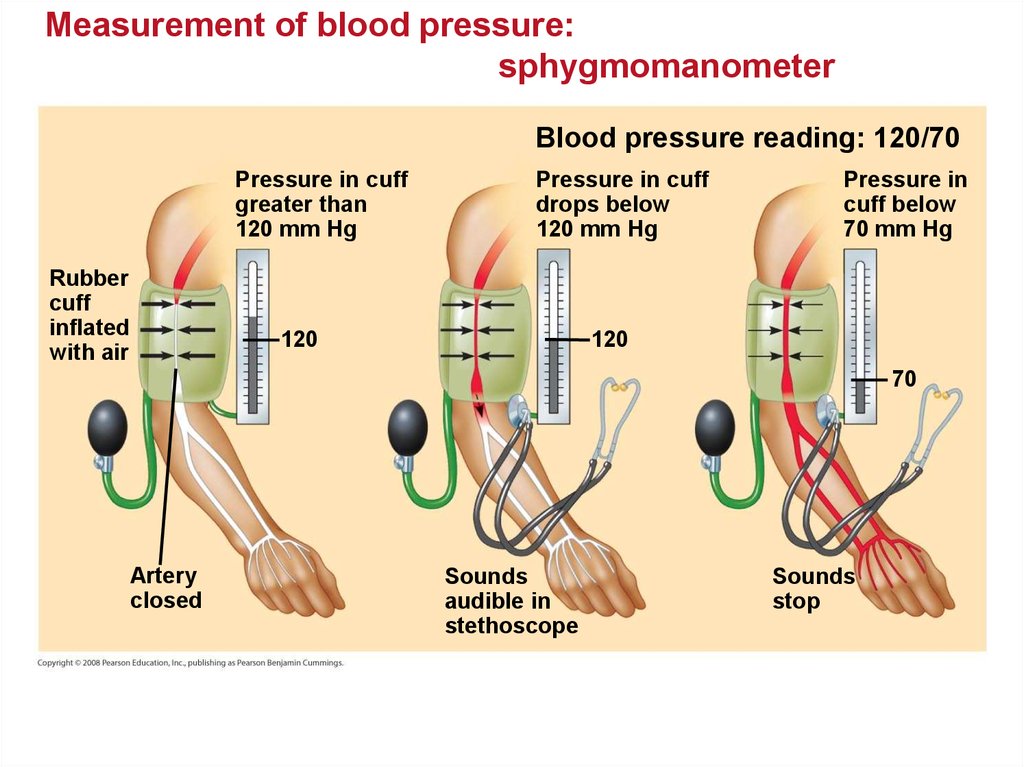
42.
• Vasoconstriction and vasodilation help maintainadequate blood flow as the body’s demands change.
• The peptide endothelin is an important inducer of
vasoconstriction.
• Blood pressure is generally measured for an artery in
the arm at the same height as the heart.
• Blood pressure for a healthy 20 year old at rest is
120 mm Hg at systole / 70 mm Hg at diastole.
Copyright © 2008 Pearson Education, Inc., publishing as Pearson Benjamin Cummings
43.
Question: How do endothelial cells control vasoconstriction?RESULTS
Leu
Met
Ser
Endothelin
Ser
Cys Ser Cys —NH +
3
Asp
Lys
Glu Cys Val Tyr Phe Cys His Leu Asp Ile
Cys
Ile Trp
—COO–
Trp
Parent polypeptide
1
53
73
Endothelin
203
44.
Measurement of blood pressure:sphygmomanometer
Blood pressure reading: 120/70
Pressure in cuff
greater than
120 mm Hg
Rubber
cuff
inflated
with air
Pressure in cuff
drops below
120 mm Hg
120
Pressure in
cuff below
70 mm Hg
120
70
Artery
closed
Sounds
audible in
stethoscope
Sounds
stop
45.
• Fainting is caused by inadequate blood flow tothe head.
• Animals with longer necks require a higher
systolic pressure to pump blood a greater
distance against gravity.
• Blood is moved through veins by smooth
muscle contraction, skeletal muscle
contraction, and expansion of the vena cava
with inhalation.
• One-way valves in veins / heart prevent
backflow of blood.
Copyright © 2008 Pearson Education, Inc., publishing as Pearson Benjamin Cummings
46.
Blood flow in veinsDirection of blood flow
in vein (toward heart)
Valve (open)
Skeletal muscle
Valve (closed)
Copyright © 2008 Pearson Education, Inc., publishing as Pearson Benjamin Cummings
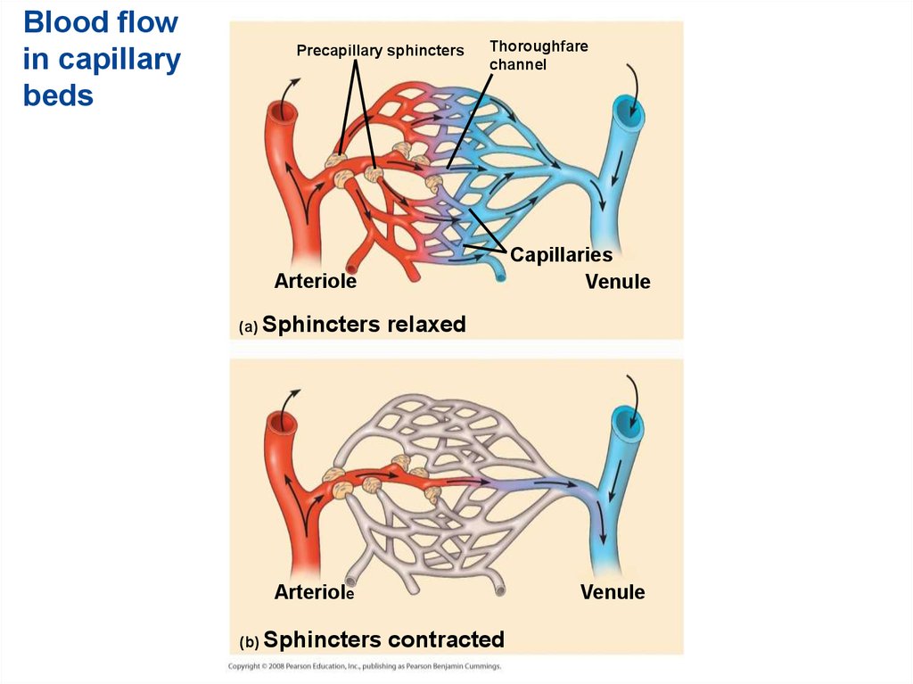
47. Capillary Function
• Capillaries in major organs are usually filled tocapacity. Blood supply varies in many other
sites.
• Two mechanisms regulate distribution of blood
in capillary beds:
– Contraction of the smooth muscle layer in the
wall of an arteriole constricts the vessel.
– Precapillary sphincters control flow of blood
between arterioles and venules.
Copyright © 2008 Pearson Education, Inc., publishing as Pearson Benjamin Cummings
48.
Blood flowin capillary
beds
Precapillary sphincters
Thoroughfare
channel
Capillaries
Venule
Arteriole
(a) Sphincters
relaxed
Arteriole
(b) Sphincters
Venule
contracted
49.
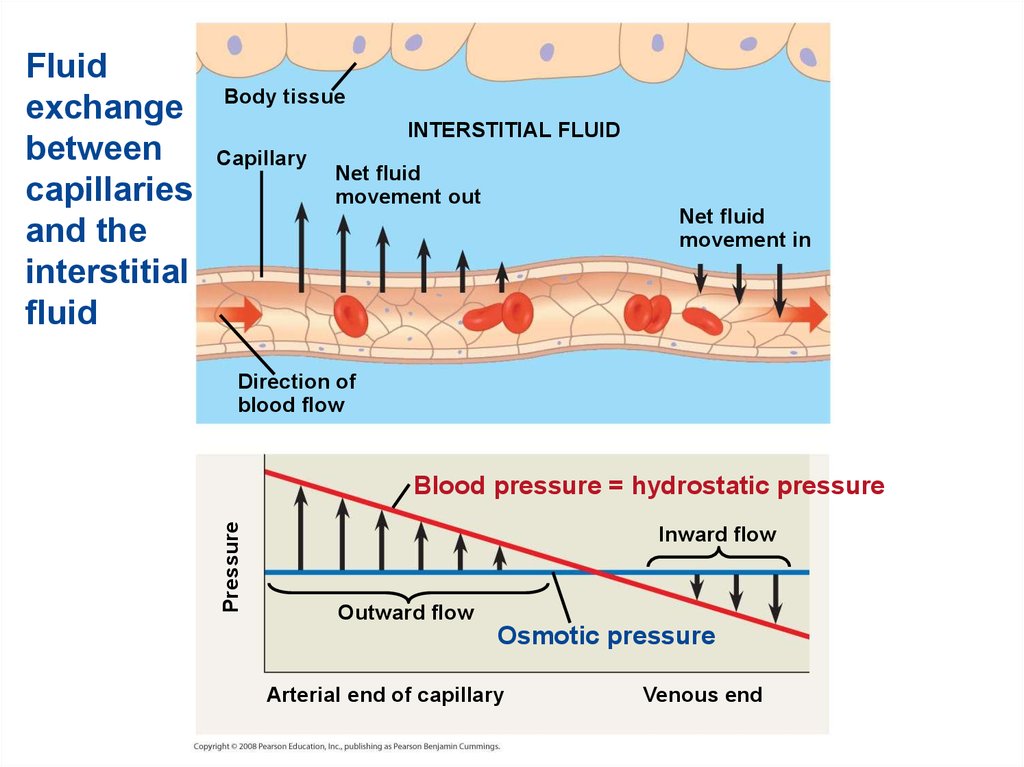
• The critical exchange of substances betweenthe blood and interstitial fluid takes place
across the thin endothelial walls of the
capillaries.
• The difference between blood pressure and
osmotic pressure drives fluids out of
capillaries at the arteriole end and into
capillaries at the venule end.
Copyright © 2008 Pearson Education, Inc., publishing as Pearson Benjamin Cummings
50.
Body tissueINTERSTITIAL FLUID
Capillary
Net fluid
movement out
Net fluid
movement in
Direction of
blood flow
Blood pressure = hydrostatic pressure
Pressure
Fluid
exchange
between
capillaries
and the
interstitial
fluid
Inward flow
Outward flow
Osmotic pressure
Arterial end of capillary
Venous end
51. Fluid Return by the Lymphatic System
• The lymphatic system - returns fluid thatleaks out in the capillary beds … restoring
filtered fluid to blood maintains homeostasis.
• This system aids in body defense.
• Fluid, called lymph, reenters the circulation
directly at the venous end of the capillary bed
and indirectly through the lymphatic system.
• The lymphatic system drains into neck veins.
Copyright © 2008 Pearson Education, Inc., publishing as Pearson Benjamin Cummings
52.
• Lymph nodes are organs that producephagocytic white blood cells and filter lymph an important role in the body’s defense.
• Edema is swelling caused by disruptions in the
flow of lymph.
Copyright © 2008 Pearson Education, Inc., publishing as Pearson Benjamin Cummings
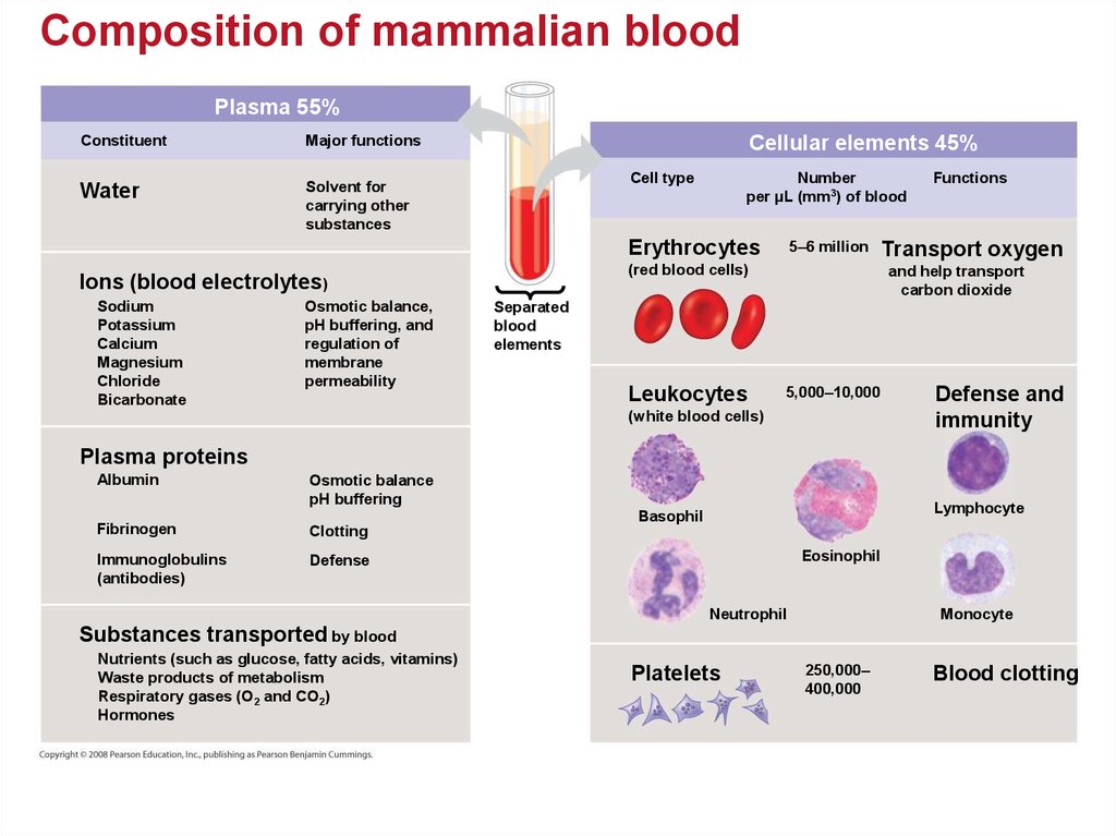
53. Blood Composition and Function
• Blood consists of several kinds of blood cellssuspended in a liquid matrix called plasma.
• The cellular elements: red blood cells, white
blood cells, and platelets occupy about 45% of
the volume of blood.
Copyright © 2008 Pearson Education, Inc., publishing as Pearson Benjamin Cummings
54.
Composition of mammalian bloodPlasma 55%
Constituent
Major functions
Water
Solvent for
carrying other
substances
Cellular elements 45%
Cell type
Number
per µL (mm3) of blood
Erythrocytes
(red blood cells)
Ions (blood electrolytes)
Sodium
Potassium
Calcium
Magnesium
Chloride
Bicarbonate
Osmotic balance,
pH buffering, and
regulation of
membrane
permeability
5–6 million
Functions
Transport oxygen
and help transport
carbon dioxide
Separated
blood
elements
Leukocytes
5,000–10,000
(white blood cells)
Defense and
immunity
Plasma proteins
Albumin
Osmotic balance
pH buffering
Lymphocyte
Basophil
Fibrinogen
Clotting
Immunoglobulins
(antibodies)
Defense
Eosinophil
Neutrophil
Monocyte
Substances transported by blood
Nutrients (such as glucose, fatty acids, vitamins)
Waste products of metabolism
Respiratory gases (O2 and CO2)
Hormones
Platelets
250,000–
400,000
Blood clotting
55. Plasma
• Blood plasma is about 90% water.• Among its solutes are inorganic salts in the
form of dissolved ions, sometimes called
electrolytes.
• Another important class of solutes is the
plasma proteins, which influence blood pH,
osmotic pressure, and viscosity. Various
plasma proteins function in lipid transport,
immunity, and blood clotting.
• Plasma transports nutrients, gases, and cell
waste.
Copyright © 2008 Pearson Education, Inc., publishing as Pearson Benjamin Cummings
56. Cellular Elements
• Suspended in blood plasma are two types ofcells:
– Red blood cells rbc = erythrocytes, transport
oxygen.
– White blood cells wbc = leukocytes, function
in defense.
• Platelets are fragments of cells that are
involved in blood clotting.
Copyright © 2008 Pearson Education, Inc., publishing as Pearson Benjamin Cummings
57.
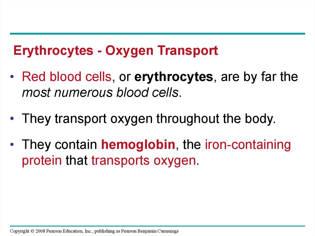
Erythrocytes - Oxygen Transport• Red blood cells, or erythrocytes, are by far the
most numerous blood cells.
• They transport oxygen throughout the body.
• They contain hemoglobin, the iron-containing
protein that transports oxygen.
Copyright © 2008 Pearson Education, Inc., publishing as Pearson Benjamin Cummings
58. Leukocytes - Defense
• There are five major types of white blood cells,or leukocytes: monocytes, neutrophils,
basophils, eosinophils, and lymphocytes.
• They function in defense by phagocytizing
bacteria and debris or by producing antibodies.
• They are found both in and outside of the
circulatory system.
Copyright © 2008 Pearson Education, Inc., publishing as Pearson Benjamin Cummings
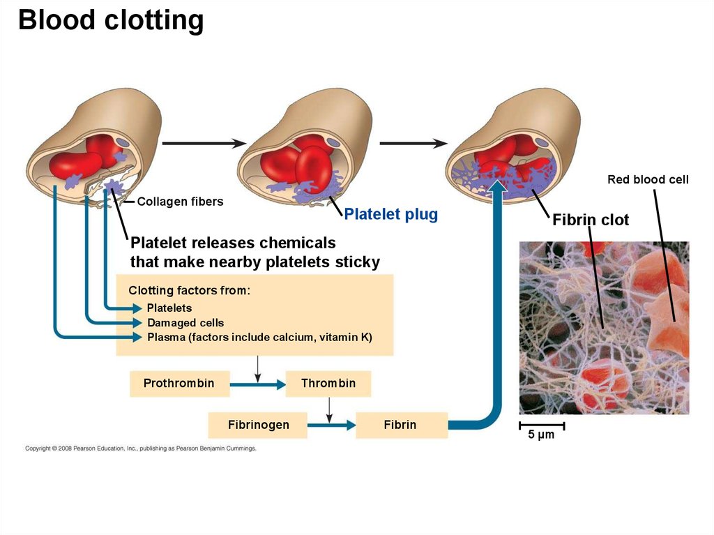
59. Platelets - Blood Clotting
• Platelets are fragments of cells and function inblood clotting.
• When the endothelium of a blood vessel is
damaged, the clotting mechanism begins.
• A cascade of complex reactions converts
fibrinogen to fibrin, forming a clot.
• A blood clot formed within a blood vessel is
called a thrombus and can block blood flow.
Copyright © 2008 Pearson Education, Inc., publishing as Pearson Benjamin Cummings
60.
Blood clottingRed blood cell
Collagen fibers
Platelet plug
Fibrin clot
Platelet releases chemicals
that make nearby platelets sticky
Clotting factors from:
Platelets
Damaged cells
Plasma (factors include calcium, vitamin K)
Prothrombin
Thrombin
Fibrinogen
Fibrin
5 µm
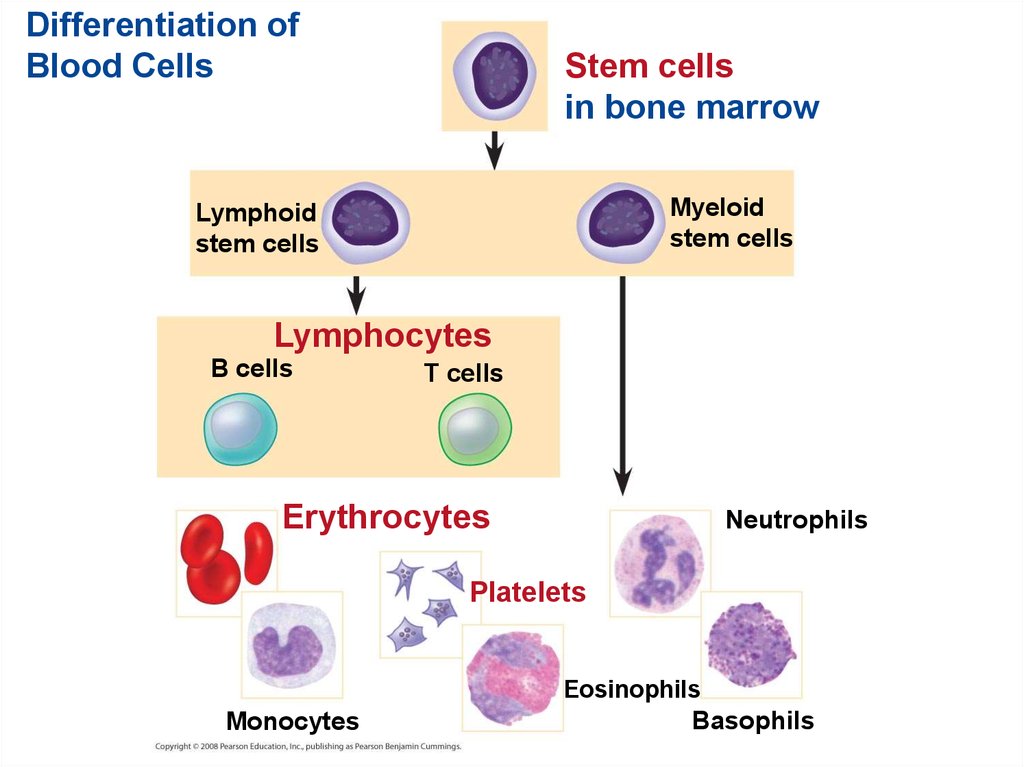
61. Stem Cells and the Replacement of Cellular Elements
• The cellular elements of blood wear out andare replaced constantly throughout a person’s
life.
• Erythrocytes, leukocytes, and platelets all
develop from a common source of stem cells
in the red marrow of bones.
• The hormone erythropoietin (EPO) stimulates
erythrocyte production when oxygen delivery is
low.
Copyright © 2008 Pearson Education, Inc., publishing as Pearson Benjamin Cummings
62.
Differentiation ofBlood Cells
Stem cells
in bone marrow
Myeloid
stem cells
Lymphoid
stem cells
Lymphocytes
B cells
T cells
Erythrocytes
Neutrophils
Platelets
Eosinophils
Monocytes
Basophils
63. Cardiovascular Disease = Disorders of the Heart and the Blood Vessels
• One type of cardiovascular disease, atherosclerosis,is caused by the buildup of plaque deposits within
arteries.
• A heart attack is the death of cardiac muscle tissue
resulting from blockage of one or more coronary
arteries.
• A stroke is the death of nervous tissue in the brain,
usually resulting from rupture or blockage of arteries
in the brain /head.
Copyright © 2008 Pearson Education, Inc., publishing as Pearson Benjamin Cummings
64.
AtherosclerosisConnective
tissue
Smooth
muscle
(a) Normal artery
Endothelium
50 µm (b) Partly
Plaque
clogged artery
250 µm
65. Treatment and Diagnosis of Cardiovascular Disease
• Cholesterol is a major contributor to atherosclerosis.• Low-density lipoproteins (LDLs) = “bad cholesterol,”
are associated with plaque formation.
• High-density lipoproteins (HDLs) = “good
cholesterol,” reduce the deposition of cholesterol.
• Hypertension = high blood pressure, promotes
atherosclerosis and increases the risk of heart attack
and stroke.
• Hypertension can be reduced by dietary changes,
exercise, and/or medication.
Copyright © 2008 Pearson Education, Inc., publishing as Pearson Benjamin Cummings
66. Gas exchange occurs across specialized respiratory surfaces
• Gas exchange supplies oxygen for cellular respirationand disposes of carbon dioxide. Gases diffuse down
pressure gradients in the lungs and other organs as a
result of differences in partial pressure.
• Partial pressure is the pressure exerted by a
particular gas in a mixture of gases. A gas diffuses
from a region of higher partial pressure to a region of
lower partial pressure: H --> L
• In the lungs and tissues, O2 and CO2 diffuse from
where their partial pressures are higher to where they
are lower.
Copyright © 2008 Pearson Education, Inc., publishing as Pearson Benjamin Cummings
67. Respiratory Media
• Animals can use air or water as a source of O2,or respiratory medium.
• In a given volume, there is less O2 available in
water than in air.
• Obtaining O2 from water requires greater
efficiency than air breathing.
Copyright © 2008 Pearson Education, Inc., publishing as Pearson Benjamin Cummings
68. Respiratory Surfaces
• Animals require large, moist respiratorysurfaces for exchange of gases between their
cells and the respiratory medium, either air or
water.
• Gas exchange across respiratory surfaces
takes place by diffusion.
• Respiratory surfaces vary by animal and can
include the outer surface, skin, gills, tracheae,
and lungs.
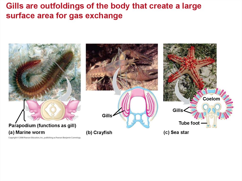
Copyright © 2008 Pearson Education, Inc., publishing as Pearson Benjamin Cummings
69.
Gills are outfoldings of the body that create a largesurface area for gas exchange
Coelom
Gills
Gills
Parapodium (functions as gill)
(a) Marine worm
Tube foot
(b) Crayfish
(c) Sea star
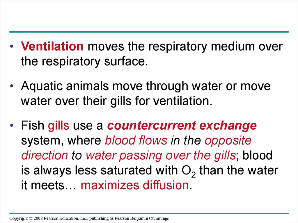
70.
• Ventilation moves the respiratory medium overthe respiratory surface.
• Aquatic animals move through water or move
water over their gills for ventilation.
• Fish gills use a countercurrent exchange
system, where blood flows in the opposite
direction to water passing over the gills; blood
is always less saturated with O2 than the water
it meets… maximizes diffusion.
Copyright © 2008 Pearson Education, Inc., publishing as Pearson Benjamin Cummings
71.
Structure and function of fish gillsFluid flow
through
gill filament
Oxygen-poor blood
Anatomy of gills
Oxygen-rich blood
Gill
Lamella
arch
Gill
arch
Water
flow
Gill filament
organization
Blood
vessels
Operculum
Water flow
between
lamellae
Blood flow through
capillaries in lamella
Countercurrent exchange
PO2 (mm Hg) in water
150 120 90 60 30
Gill filaments
Net diffusion
of O2from
water to
blood
140 110 80 50 20
PO2 (mm Hg) in blood
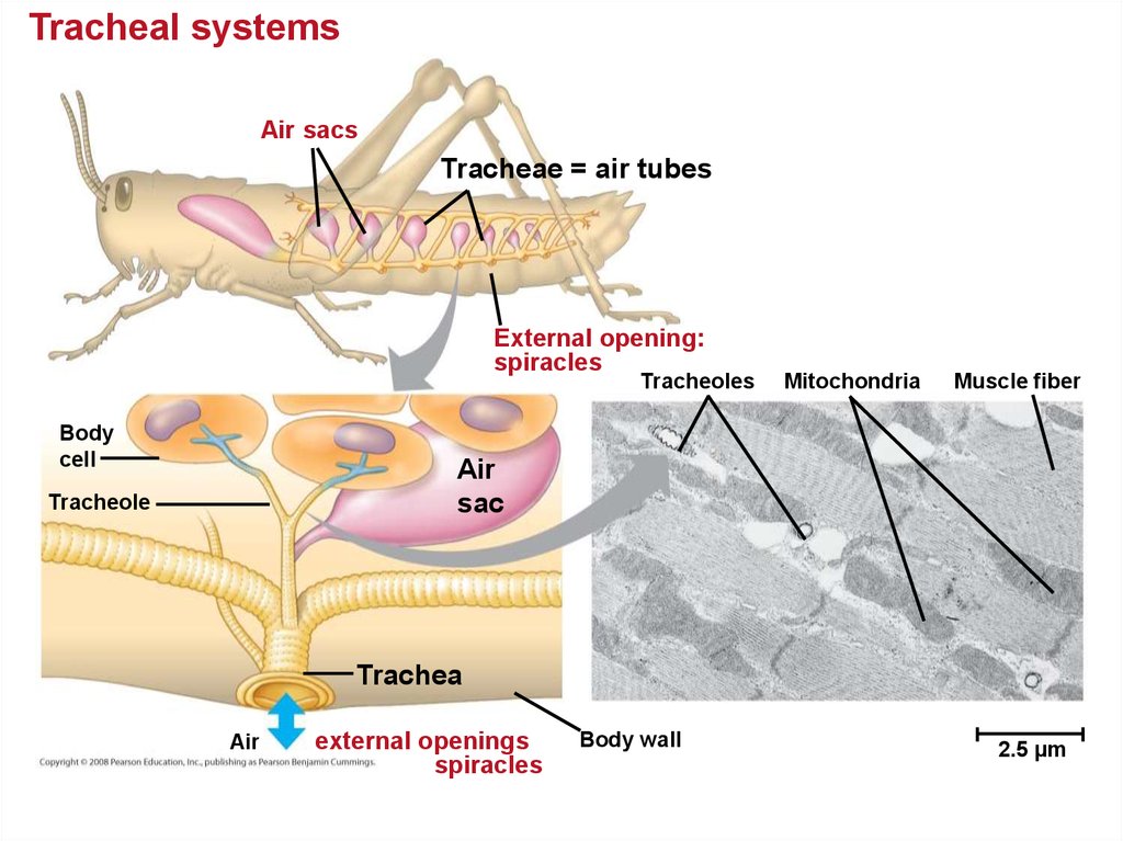
72. Tracheal Systems in Insects
• The tracheal system of insects consists of tinybranching tubes that penetrate the body.
• The tracheal tubes supply O2 directly to body
cells.
• The respiratory and circulatory systems are
separate.
• Larger insects must ventilate their tracheal
system to meet O2 demands.
Copyright © 2008 Pearson Education, Inc., publishing as Pearson Benjamin Cummings
73.
Tracheal systemsAir sacs
Tracheae = air tubes
External opening:
spiracles
Tracheoles
Body
cell
Mitochondria
Muscle fiber
Air
sac
Tracheole
Trachea
Air
external openings
spiracles
Body wall
2.5 µm
74. Lungs = Infoldings of the body surface
• The circulatory system (open or closed)transports gases between the lungs and the
rest of the body.
• The size and complexity of lungs correlate with
an animal’s metabolic rate.
Copyright © 2008 Pearson Education, Inc., publishing as Pearson Benjamin Cummings
75. Mammalian Respiratory Systems: A Closer Look
• A system of branching ducts / air tubesconveys air to the lungs.
• Air inhaled through the nostrils --> pharynx
--> larynx --> trachea --> bronchi -->
bronchioles --> alveoli = site of gas
exchange.
• Exhaled air passes over the vocal cords to
create sounds.
• Alveoli are wrapped by capillaries for GAS
EXCHANGE.
Copyright © 2008 Pearson Education, Inc., publishing as Pearson Benjamin Cummings
76.
Mammalian Respiratory SystemBranch of
pulmonary
vein
(oxygen-rich
blood)
Branch of
pulmonary
artery
(oxygen-poor
blood)
Terminal
bronchiole
Nasal
cavity
Pharynx
Larynx
(Esophagus)
Alveoli
Left
lung
Trachea
Right lung
Bronchus
Bronchiole
Diaphragm
Heart
SEM
50 µm
Colorized
SEM
50 µm
77. Breathing Ventilates the Lungs by Inhalation and Exhalation of Air
• Amphibians, such as a frog, ventilates its lungsby positive pressure breathing, which forces
air down the trachea.
• Mammals ventilate by negative pressure
breathing, which pulls air into the lungs by
varying volume / air pressure. Lung volume
increases as the rib muscles and diaphragm
contract.
• The tidal volume is the volume of air inhaled
with each breath. The maximum tidal volume is
the vital capacity. After exhalation, residual
volume of air remains in the lungs.
Copyright © 2008 Pearson Education, Inc., publishing as Pearson Benjamin Cummings
78.
Negative pressure breathing: H --> LRib cage
expands as
rib muscles
contract
Air
inhaled
Rib cage gets
smaller as
rib muscles
relax
Air
exhaled
Lung
Diaphragm
INHALATION
EXHALATION
Diaphragm contracts
(moves down)
Diaphragm relaxes
(moves up)
Volume increases
Pressure decreases
Air rushes in
Volume decreases
Pressure increases
Air rushes out
79. How a Bird Breathes
• Birds have eight or nine air sacs that functionas bellows that keep air flowing through the
lungs.
• Air passes through the lungs in one direction
only.
• Every exhalation completely renews the air in
the lungs.
Copyright © 2008 Pearson Education, Inc., publishing as Pearson Benjamin Cummings
80.
The Avian Respiratory SystemAir
Anterior
air sacs
Posterior
air sacs
Air
Trachea
Lungs
Lungs
Air tubes
(parabronchi)
in lung
INHALATION
Air sacs fill
EXHALATION
Air sacs empty;
Lungs Fill
1 mm
81. Control of Breathing in Humans
• In humans, the main breathing controlcenters are in two regions of the brain, the
medulla oblongata and the pons.
• The medulla regulates the rate and depth of
breathing in response to pH changes - CO2
levels in the cerebrospinal fluid.
• The medulla adjusts breathing rate and depth
to match metabolic demands.
• The pons regulates the tempo.
Copyright © 2008 Pearson Education, Inc., publishing as Pearson Benjamin Cummings
82.
• Sensors in the aorta and carotid arteriesmonitor O2 and CO2 concentrations in the
blood.
• These sensors exert secondary control over
breathing.
Copyright © 2008 Pearson Education, Inc., publishing as Pearson Benjamin Cummings
83.
Automaticcontrol of
breathing
Cerebrospinal
fluid
Pons
Breathing
control
centers
Medulla
oblongata
Carotid arteries
Aorta
Diaphragm
Rib muscles
84. Adaptations for gas exchange include pigments that bind and transport gases
• The metabolic demands of many organisms requirethat the blood transport large quantities of O2 and CO2
• Blood arriving in the lungs has a low partial pressure
of O2 and a high partial pressure of CO2 relative to air
in the alveoli.
• In the alveoli, O2 diffuses into the blood and CO2
diffuses into the air.
• In tissue capillaries, partial pressure gradients favor
diffusion of O2 into the interstitial fluids and CO2 into
the blood.
Copyright © 2008 Pearson Education, Inc., publishing as Pearson Benjamin Cummings
85.
Loading and unloading of respiratory gasesAlveolus
Alveolus
PCO2 = 40 mm Hg
PO2 = 100 mm Hg
PO2 = 40
PO2 = 100
PCO2 = 46
Circulatory
system
PO2 = 40
PCO2 = 40
Circulatory
system
PO2 = 100
PCO2 = 46
PO2 ≤ 40 mm Hg
PCO2 ≥ 46 mm Hg
Body tissue
(a)
Oxygen
PCO2 = 40
Body tissue
(b) Carbon
dioxide
86. Respiratory Pigments
• Respiratory pigments = proteins thattransport oxygen, greatly increase the amount
of oxygen that blood can carry.
• Arthropods and many molluscs have
hemocyanin with copper as the oxygen-binding
component.
• Most vertebrates and some invertebrates use
hemoglobin with iron = oxygen-binding
component contained within erythrocytes.
Copyright © 2008 Pearson Education, Inc., publishing as Pearson Benjamin Cummings
87. Hemoglobin
• A single hemoglobin molecule can carry fourmolecules of O2
• The hemoglobin dissociation curve shows
that a small change in the partial pressure of
oxygen can result in a large change in delivery
of O2
• CO2 produced during cellular respiration lowers
blood pH and decreases the affinity of
hemoglobin for O2
• This is called the Bohr shift.
Copyright © 2008 Pearson Education, Inc., publishing as Pearson Benjamin Cummings
88.
ChainsIron
Heme
Chains
Hemoglobin
89.
O2 saturation of hemoglobin (%)100
Dissociation
curves for
hemoglobin
at 37ºC
O2 unloaded
to tissues
at rest
80
O2 unloaded
to tissues
during exercise
60
40
20
0
0
20
Tissues during
exercise
40
60
80
Tissues
at rest
100
Lungs
PO2 (mm Hg)
(a) PO
2
and hemoglobin dissociation at pH 7.4
O2 saturation of hemoglobin (%)
100
pH 7.4
80
pH 7.2
Hemoglobin
retains less
O2 at lower pH
60
40
(higher CO2
concentration)
20
0
0
20
40
60
80
100
PO2 (mm Hg)
(b) pH
and hemoglobin dissociation
90. Carbon Dioxide Transport
• Hemoglobin also helps transport CO2 andassists in buffering.
• CO2 from respiring cells diffuses into the blood
and is transported either in blood plasma,
bound to hemoglobin, or as bicarbonate ions =
HCO3–.
Copyright © 2008 Pearson Education, Inc., publishing as Pearson Benjamin Cummings
91.
Body tissueCO transport
CO2 produced from tissues
2
Carbon dioxide
transport in the
blood
CO
Interstitial
fluid
2
Plasma
within capillary
CO2
Capillary
wall
CO2
H2 O
Red
H2CO3
Hb
blood
Carbonic acid
cell
HCO3– +
Bicarbonate
Hemoglobin
picks up
CO2 and H+
H+
HCO3–To lungs
CO2 transport
to lungs
HCO3–
HCO3– +
H+
H2CO3
H2 O
Hb
Hemoglobin
releases
CO2 and H+
CO2
CO2
CO2
CO2
Alveolar space in lung
92. Elite Animal Athletes
• Migratory and diving mammals have evolutionaryadaptations that allow them to perform extraordinary
feats.
• The extreme O2 consumption of the antelope-like
pronghorn underlies its ability to run at high speed
over long distances.
• Deep-diving air breathers stockpile O2 and deplete it
slowly.
• Weddell seals have a high blood to body volume ratio
and can store oxygen in their muscles in myoglobin
proteins.
Copyright © 2008 Pearson Education, Inc., publishing as Pearson Benjamin Cummings
93.
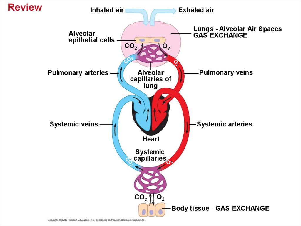
ReviewInhaled air
Alveolar
epithelial cells
Pulmonary arteries
Exhaled air
Lungs - Alveolar Air Spaces
GAS EXCHANGE
CO2
O2
Alveolar
capillaries of
lung
Systemic veins
Pulmonary veins
Systemic arteries
Heart
Systemic
capillaries
CO2
O2
Body tissue - GAS EXCHANGE
94. You should now be able to:
1. Compare and contrast open and closedcirculatory systems.
2. Compare and contrast the circulatory systems
of fish, amphibians, reptiles, and mammals or
birds.
3. Distinguish between pulmonary and systemic
circuits and explain the function of each.
4. Trace the path of a red blood cell through the
human heart, pulmonary circuit, and systemic
circuit.
Copyright © 2008 Pearson Education, Inc., publishing as Pearson Benjamin Cummings
95.
5. Define cardiac cycle and explain the role ofthe sinoatrial node.
6. Relate the structures of capillaries, arteries,
and veins to their function.
7. Define blood pressure and cardiac output and
describe two factors that influence each.
8. Explain how osmotic pressure and hydrostatic
pressure regulate the exchange of fluid and
solutes across the capillary walls.
Copyright © 2008 Pearson Education, Inc., publishing as Pearson Benjamin Cummings
96.
9. Describe the role played by the lymphaticsystem in relation to the circulatory system.
10. Describe the function of erythrocytes,
leukocytes, platelets, fibrin.
11. Distinguish between a heart attack and
stroke.
12. Discuss the advantages and disadvantages
of water and of air as respiratory media.
Copyright © 2008 Pearson Education, Inc., publishing as Pearson Benjamin Cummings
97.
13. For humans, describe the exchange of gasesin the lungs and in tissues.
14. Draw and explain the hemoglobin-oxygen
dissociation curve.
Copyright © 2008 Pearson Education, Inc., publishing as Pearson Benjamin Cummings