


 medicine
Similar presentations:


Кровоснабжение проводящей системы сердца
1. Кровоснабжение проводящей системы сердца
2.
A. Right & Left Coronary Arteries
B. Branches of Right Coronary Artery
1. Main
2. Posterior Descending Branch
3. Sinus Node branch
C. Branches of Left Coronary Artery
1. Main
2. Left Anterior Descending (LAD)
3. Circumflex
D. Blood Supply of Cardiac Conduction System
1. Sinus Node Artery Supplies Blood to Sinus Node
a. Origin of Sinus Node Artery
2. AV Nodal Artery Supplies Blood to the AV node
a. Origin of AV Nodal Artery
3. LAD supplies the right and left bundle branches
3.
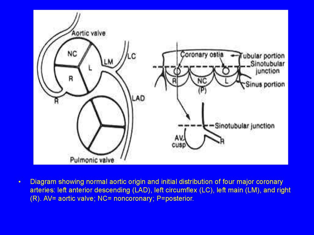
Diagram showing normal aortic origin and initial distribution of four major coronary
arteries: left anterior descending (LAD), left circumflex (LC), left main (LM), and right
(R). AV= aortic valve; NC= noncoronary; P=posterior.
4.
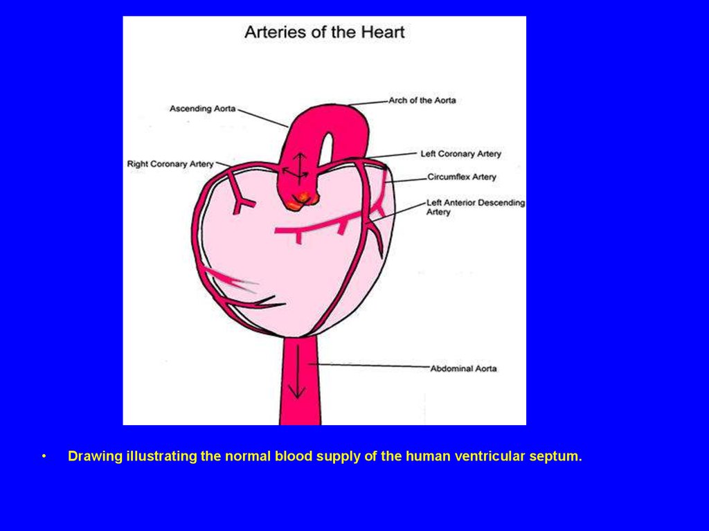
Drawing illustrating the normal blood supply of the human ventricular septum.
5.
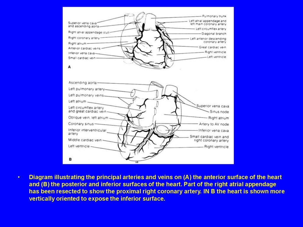
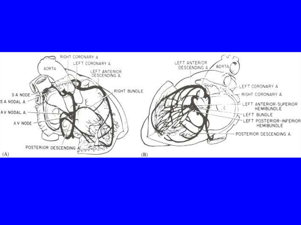
Diagram illustrating the principal arteries and veins on (A) the anterior surface of the heart
and (B) the posterior and inferior surfaces of the heart. Part of the right atrial appendage
has been resected to show the proximal right coronary artery. IN B the heart is shown more
vertically oriented to expose the inferior surface.
6.
Diagram showing myocardial perfusion patterns of major epicardial coronary
arteries as viewed from three tomographic cuts: four-chamber view, crosssectional view, and parasternal view, and parasternal long-axis. A= anterior;
ALPM= anterolateral papillary muscle; LC= left circumflex; LAD= left anterior
descending; LM= left main; LV= left ventricle; LVFW= left ventricular free wall;
P= posterior; PMPM= posteromedial papillary muscle; RV= right ventricle;
RVFW= right ventricular free wall; RVOFT= right ventricular outflow tract.
7.
Diagram showing arterial blood supply of the cardiac conduction system. The nodal artery
(NA) arises from the posterior descending artery (PD). The bulk of the arterial blood supply
to the right and left bundle branches comes from the left anterior descending artery (LAD).
8.
9.
10.
Schematic diagram of blood supply to cardiac conduction system. The first septal branch
of the left anterior descending (LAD) coronary artery supplies a critical portion of the
interventricular conduction system (red oval). AV, atrioventricular; marginal a., marginal
artery; PDA, posterior descending artery; RCA, right coronary artery; SA, sinoatrial.
Reproduced, with permission, from Harthorne JW, Pohost. GM, Electrical therapy of
cardiac dysrhythmias. In Levine, HJ (ed) Clinical Cardiovascular Physiology, New York,
Grune and Stratton: 1976. pp. 853–882