


")
")

")
")
")
")
")
")
")


")
")
 medicine
Similar presentations:


ECG - MI. Acute Coronary Syndromes Unstable Angina
1. ECG - MI
DR F AGYEKUMFOR
DR J AKAMAH
2.
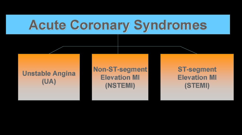
Acute Coronary SyndromesUnstable Angina
(UA)
Non-ST-segment
Elevation MI
(NSTEMI)
ST-segment
Elevation MI
(STEMI)
3. Acute Coronary Syndromes
Excessive demand or inadequate supply of oxygen andnutrients to the heart muscle
Associated with:
Plaque disruption
Thrombus formation
Vasoconstriction
4. Coronary Artery Occlusion
Patient’s clinical presentation and outcome depend on factorsincluding:
Amount of myocardium supplied by affected artery
Severity and duration of myocardial ischemia
Electrical instability of the ischemic myocardium
Degree and duration of coronary obstruction
Presence (and extent) or absence of collateral coronary circulation
5. Acute Coronary Syndromes
6. Ischemia, Injury, and Infarction
Main coronary arteries lie on theepicardial surface of the heart
This area is fed first before supplying
the inner layers with oxygenated
blood
7. Ischemia, Injury, and Infarction
Myocardial ischemiaImbalance between the metabolic needs of the myocardium (demand) and the flow
of oxygenated blood to it (supply)
Angina: The pain resulting from an imbalance between myocardial
oxygen supply and demand
1. Characteristic Quality and Duration: Retrosternal: Jaw, Left Arm, Neck
2. Provoked by Exertion or Emotional Stress
3. Relieved by Rest or Nitroglycerin
8. Ischemia, Injury, and Infarction
Myocardial ischemia delays repolarizationECG changes include temporary changes in the ST-segment and T
wave
When looking for evidence of infarction, most of the information is obtained
from analyzing a single, representative complex in each lead.
9. Ischemia, Injury, and Infarction
ST-segment depression is significant when the ST-segment is morethan ½ mm below the baseline at a point 0.04 sec to the right of the
J-point and is seen in two or more leads facing the same anatomic
area of the heart
10. Ischemia, Injury, and Infarction
Locate J-pointCompare ST-segment deviation to isoelectric line
11. Ischemia, Injury, and Infarction
Injured cells will die unless blood flow is quickly restoredMyocardial injury is viewed on the ECG as ST-segment elevation
in the leads facing the affected area
12. Ischemia, Injury, and Infarction
Injured cells will showST-segment elevation
in leads facing the
affected area
13. Ischemia, Injury, and Infarction
Suspect ventricularaneurysm if ST-segment
elevation persists for
more than a few months
after MI
14. Ischemia, Injury, and Infarction
Infarction occurs when blood flow to the heart muscle stops oris suddenly decreased long enough to cause cell death
Infarcted cells:
Cannot respond to an electrical stimulus
Do not provide any mechanical function
15. Myocardial Infarction—Diagnosis
Typical rise and gradual fall (troponin) or more rapid rise andfall (CK-MB) of biochemical markers of myocardial necrosis
with at least one of the following:
Ischemic symptoms
Development of pathologic Q waves on ECG
ECG changes (ST-segment elevation or depression)
Or coronary artery intervention
Pathologic findings of an acute MI
16. Infarction—ECG Changes
Non-ST-segment elevation MI (NSTEMI)ST-segment depression in leads facing the affected area
MI diagnosed if ECG changes are accompanied by elevations of
serum cardiac markers
17. Infarction—ECG Changes
Most patients with ST-segment elevation MI will develop Qwave MIAbnormal (pathologic) Q wave
>0.04 sec in duration and >1/3 the amplitude of the following R wave in that
lead
Indicates dead myocardial tissue, loss of electrical activity
18. Infarction—Indicative ECG Changes
19. Infarction—ECG Changes
ST-segment elevation“Smiley” face (upward concavity) is usually benign
Coved (“frowny face”) elevation is called an acute injury pattern
20. R-Wave Progression
Chest leads in anormal heart
As the electrode is
moved from right to
left:
R wave becomes taller
S wave becomes smaller
21. R-Wave Progression
V3 and V4 normallyrecord an
equiphasic (equally
positive and
negative) RS
complex
Transitional zone
22. Poor R-Wave Progression
A phrase used to describe Rwaves that decrease in size
from V1-V4
23. Layout of the 12-Lead ECG
Limb LeadsChest Leads
Standard Leads
Augmented Leads
V1-V3
V4-V6
Column I
Column II
Column III
Column IV
I: lateral
aVR: none
V1: septum
V4: anterior
II: inferior
aVL: lateral
V2: septum
V5: lateral
III: inferior
aVF: inferior
V3: anterior
V6: lateral
24. Indicative ECG Changes
Indicative changes are significant when they are seen in twoanatomically contiguous leads
Two leads are contiguous if:
They look at the same area of the heart
Or they are numerically consecutive chest leads
25. Indicative ECG Changes
26. Indicative ECG Changes
Which leads of a standard 12-lead ECG look at the inferiorwall of the left ventricle?
I: lateral
aVR: none
V1: septum
V4: anterior
II: inferior
aVL: lateral
V2: septum
V5: lateral
III: inferior
aVF: inferior
V3: anterior
V6: lateral
27. Which Leads Show ST-Segment Elevation?
Are they anatomically contiguous leads?28. ST-Segment Elevation is Present in II, III, aVF
They are anatomically contiguous; inferior MILateral
Septum
Inferior
Lateral
Inferior
Inferior
Septum
Anterior
Anterior
Lateral
Lateral
29. Reciprocal Changes
30. Localization of Infarction
31. Predicting the Site of Coronary Artery Occlusion
Leads II, III, and aVF = inferior wallSupplied by RCA in most of the population
Leads viewing areas supplied by the left coronary artery:
I, aVL, V5, V6 – lateral wall
V1-V2 – septum
V3-V4 – anterior wall
32. Assessing the Extent of Infarction
Evaluate how many leads are showing indicative changesChanges in only a few leads suggests a smaller infarction
In general, the more proximal the occlusion:
The larger the infarction
The greater the number of leads showing indicative changes
33. Specific Types of MIs
34. Anterior Wall MI (AWMI)
Leads V3 and V4 face anterior wall of left ventricleLeft main coronary artery supplies:
Left anterior descending artery (LAD)
Circumflex artery
Left main coronary artery occlusion
“Widow maker”
Often leads to cardiogenic shock and death without prompt
reperfusion
35. Anterior Wall MI (AWMI)
36. Evolution of Anteroseptal MI
Indicative changes in leads V2-4Left: At admission, hyperacute
phase is reflected by STsegment elevation
Middle: At 24 hours
Right: At 48 hours, pathologic Q
waves
37. Inferior Wall MI (IWMI)
38. Inferior Wall MI (IWMI)
39. Inferior Wall MI (IWMI)
40. Inferior Wall MI (IWMI)
41. Lateral Wall MI (LWMI)
Leads I, aVL, V5, and V6 view the lateral wall42. Lateral Wall MI (LWMI)
43. Lateral Wall MI (LWMI)
44. Septal MI
Leads V1 and V2 face the septal area of the leftventricle.
45. Septal Infarction Poor R-wave Progression
46. Posterior MI
47. Posterior MI
48. Posterior Chest Lead Placement
49. Posterior Infarction
Evolutionary changes in inferior andposterior MI
Left: Acute inferior and apical injury
Right: At 24 hours: Note tall R wave in
lead V1 not present in A, suggesting
posterior MI
Bottom: (V7-9) Posterior infarction
confirmed
50. Right Ventricular Infarction
51. Right Chest Leads
Right chest leads usedto view right ventricle
If time does not permit
obtaining all of the right
chest leads, V4R is lead
of choice
52. Right Ventricular Infarction (RVI)
Evolutionary changes in inferiorand right ventricular infarction
Left – At admission – acute phase
Middle – At 12 hours
Right – Right chest leads showing RVI
53. Right Ventricular Infarction (RVI)
Clinical triad of RVI:Hypotension
Jugular venous distention
Clear breath sounds
Only 10-15% of patients with RVI present with these signs and
symptoms