
 medicine
Similar presentations:


Ventricular tachycardias in the absence of structural heart disease
1.
VENTRICULAR TACHYCARDIAS IN THEABSENCE OF STRUCTURAL HEART DISEASE
Dr RAJESH K F
2.
10% of patients presenting with VT have no apparentstructural heart disease
VT in structurally normal hearts can be broadly considered
under
Non–life-threatening monomorphic VT
Life-threatening polymorphic VT
3.
NON–LIFE-THREATENING(TYPICALLY MONOMORPHIC)
Classified on basis of site of origin
Most common sites are ventricular outflow tracts and left
ventricular fascicles
4.
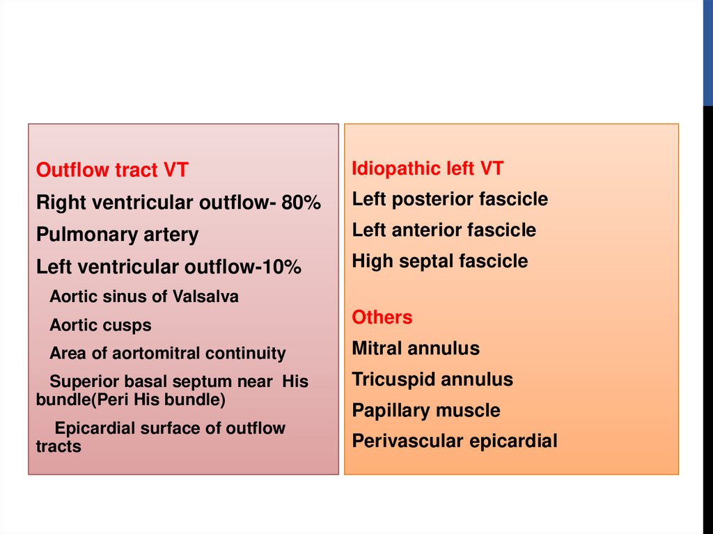
Outflow tract VTIdiopathic left VT
Right ventricular outflow- 80%
Left posterior fascicle
Pulmonary artery
Left anterior fascicle
Left ventricular outflow-10%
High septal fascicle
Aortic sinus of Valsalva
Aortic cusps
Others
Area of aortomitral continuity
Mitral annulus
Superior basal septum near His
bundle(Peri His bundle)
Epicardial surface of outflow
tracts
Tricuspid annulus
Papillary muscle
Perivascular epicardial
5.
OUTFLOW TRACT VTIdiopathic VT originate most commonly in outflow tract area
Nearly 80% of which originate from RVOT
Other outflow tract sites are rare
6.
PHENOTYPESPhenotypes are a continuum of the same focal cellular process
Premature ventricular complexes (PVCs)
Nonsustained,repetitive monomorphic VT(RMVT)
Paroxysmal, exercise-induced sustained VT
Considerable overlap observed among three phenotypes
Ablating one phenotype at a discrete site eliminates other two
Signature characteristic of sustained RVOT and LVOT is
tachycardia is termination by adenosine and verapamil
7.
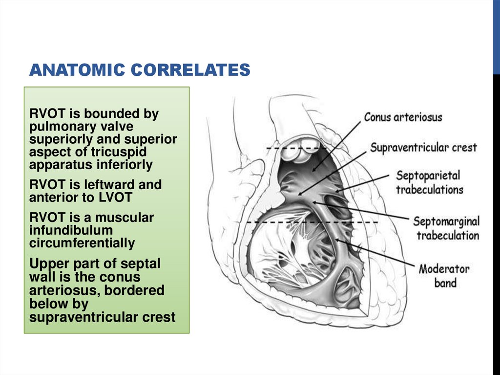
ANATOMIC CORRELATESRVOT is bounded by
pulmonary valve
superiorly and superior
aspect of tricuspid
apparatus inferiorly
RVOT is leftward and
anterior to LVOT
RVOT is a muscular
infundibulum
circumferentially
Upper part of septal
wall is the conus
arteriosus, bordered
below by
supraventricular crest
8.
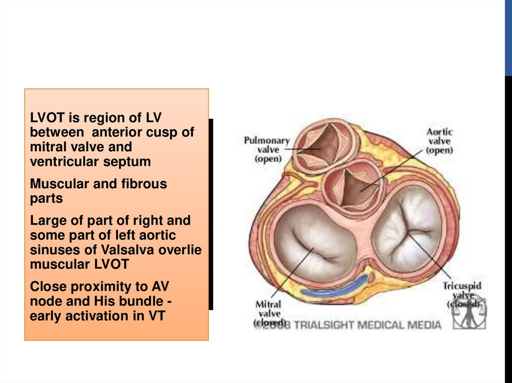
LVOT is region of LVbetween anterior cusp of
mitral valve and
ventricular septum
Muscular and fibrous
parts
Large of part of right and
some part of left aortic
sinuses of Valsalva overlie
muscular LVOT
Close proximity to AV
node and His bundle early activation in VT
9.
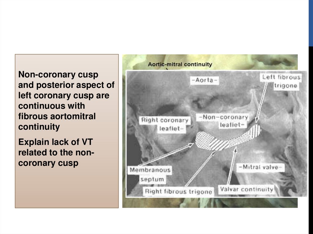
Non-coronary cuspand posterior aspect of
left coronary cusp are
continuous with
fibrous aortomitral
continuity
Explain lack of VT
related to the noncoronary cusp
10.
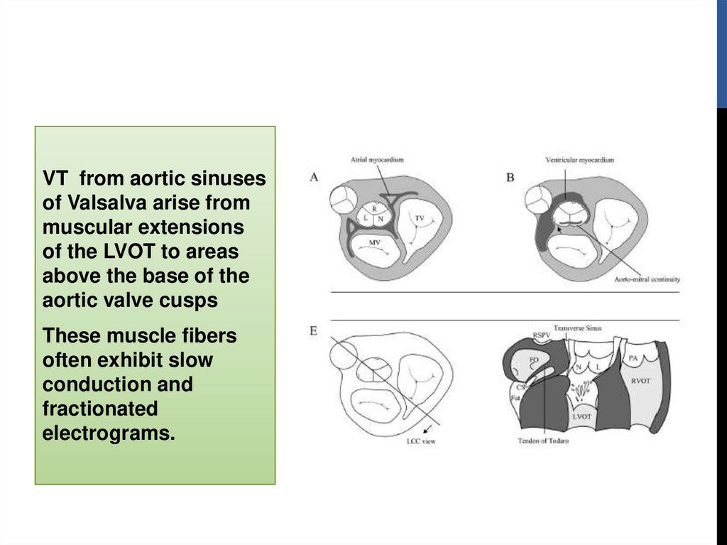
VT from aortic sinusesof Valsalva arise from
muscular extensions
of the LVOT to areas
above the base of the
aortic valve cusps
These muscle fibers
often exhibit slow
conduction and
fractionated
electrograms.
11.
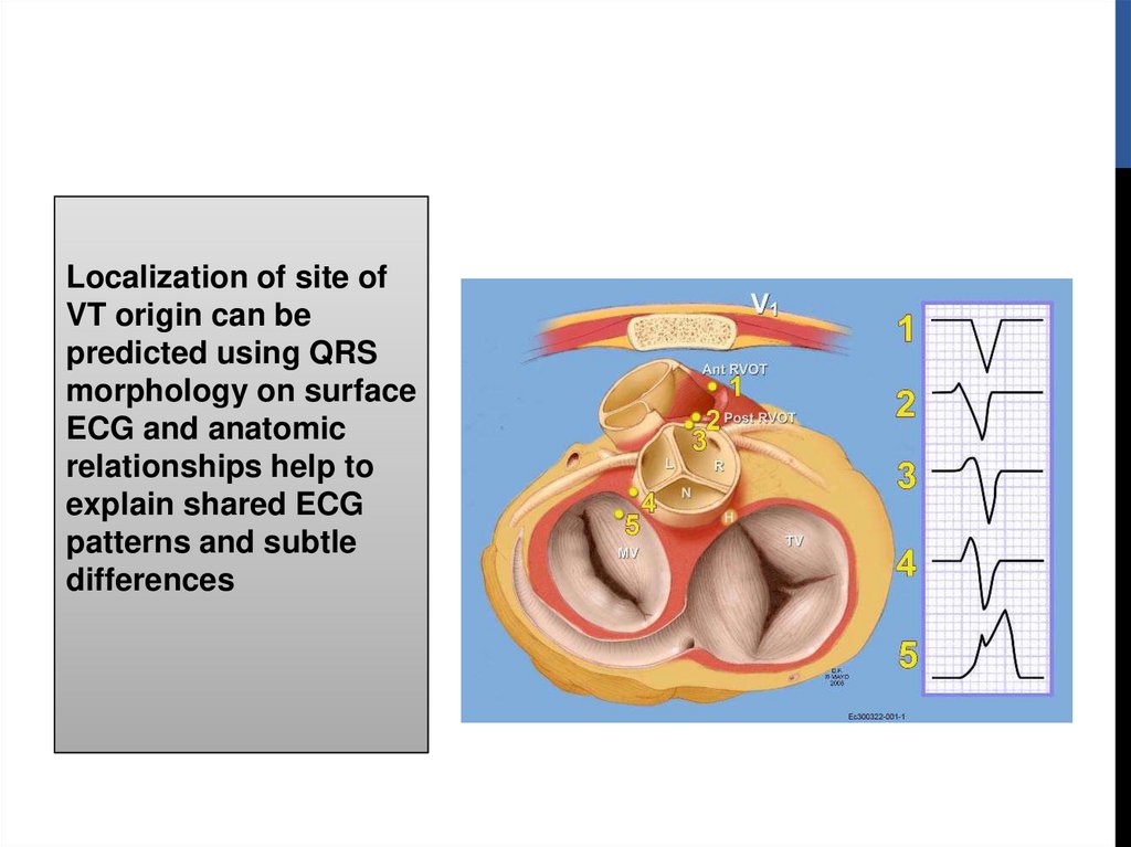
Localization of site ofVT origin can be
predicted using QRS
morphology on surface
ECG and anatomic
relationships help to
explain shared ECG
patterns and subtle
differences
12.
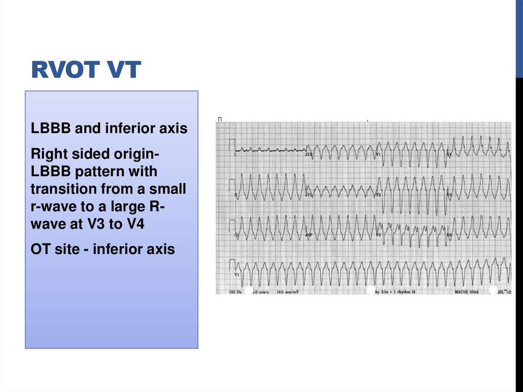
RVOT VTLBBB and inferior axis
Right sided originLBBB pattern with
transition from a small
r-wave to a large Rwave at V3 to V4
OT site - inferior axis
13.
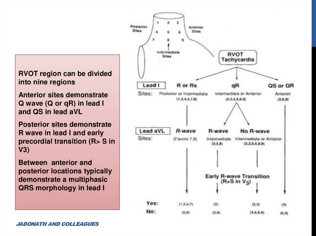
RVOT region can be dividedinto nine regions
Anterior sites demonstrate
Q wave (Q or qR) in lead I
and QS in lead aVL
Posterior sites demonstrate
R wave in lead I and early
precordial transition (R> S in
V3)
Between anterior and
posterior locations typically
demonstrate a multiphasic
QRS morphology in lead I
JADONATH AND COLLEAGUES
14.
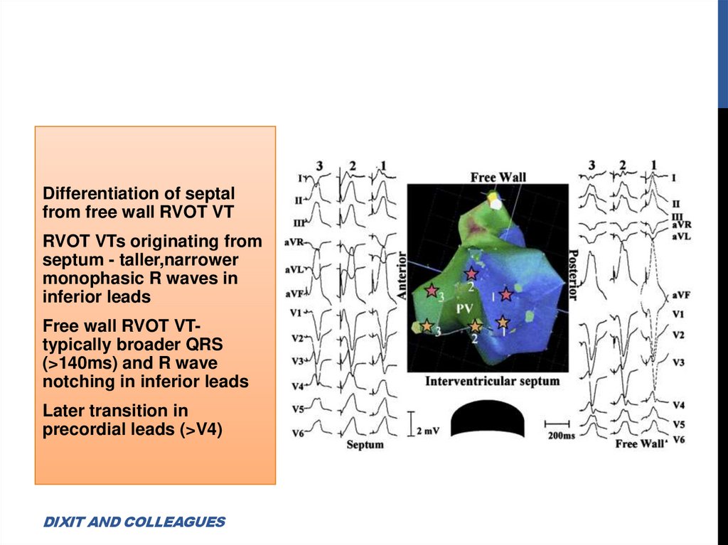
Differentiation of septalfrom free wall RVOT VT
RVOT VTs originating from
septum - taller,narrower
monophasic R waves in
inferior leads
Free wall RVOT VTtypically broader QRS
(>140ms) and R wave
notching in inferior leads
Later transition in
precordial leads (>V4)
DIXIT AND COLLEAGUES
15.
Anterior position of free wall relative to septum -Accounts fordeeper S wave in lead V2 than RVOT septum
Septal site associated with a Q/q wave in lead I, whereas a freewall site inscribes an R/r wave.
Caudal (> 2 cm from PV) Versus Cranial
VT arising >2 cm of the pulmonary valve near His bundle virtually
always has a negative QRS in lead aVL
16.
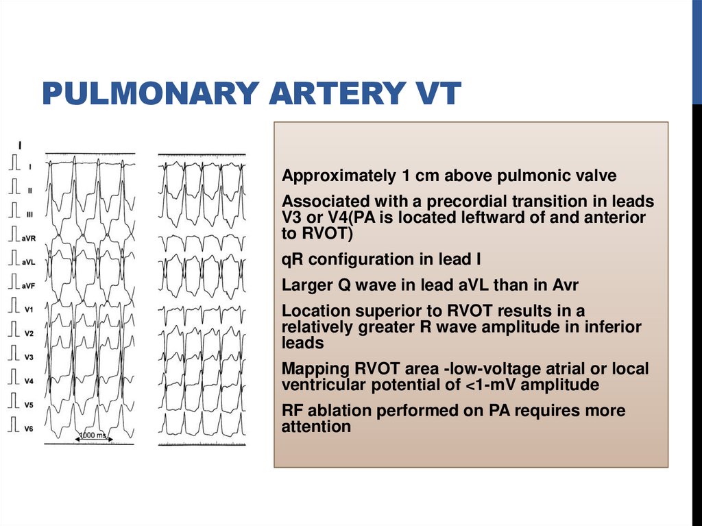
PULMONARY ARTERY VTApproximately 1 cm above pulmonic valve
Associated with a precordial transition in leads
V3 or V4(PA is located leftward of and anterior
to RVOT)
qR configuration in lead I
Larger Q wave in lead aVL than in Avr
Location superior to RVOT results in a
relatively greater R wave amplitude in inferior
leads
Mapping RVOT area -low-voltage atrial or local
ventricular potential of <1-mV amplitude
RF ablation performed on PA requires more
attention
17.
DIFFERENTIALDIAGNOSIS OF RVOT VT
Atriofascicular fibers (Mahaim fibers)
AVRT using Rt-sided accessory pathway
VT after repair of TOF
ARVD
18.
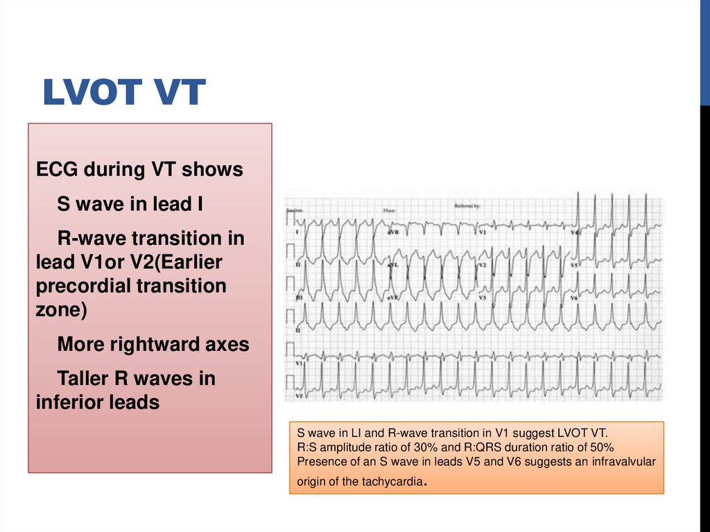
LVOT VTECG during VT shows
S wave in lead I
R-wave transition in
lead V1or V2(Earlier
precordial transition
zone)
More rightward axes
Taller R waves in
inferior leads
S wave in LI and R-wave transition in V1 suggest LVOT VT.
R:S amplitude ratio of 30% and R:QRS duration ratio of 50%
Presence of an S wave in leads V5 and V6 suggests an infravalvular
origin of the tachycardia.
19.
Shows one of the following depending on site of origina)Basal left interventricular or septal origin
LBBB morphology with an early precordial transition in lead V2 or
V3,S wave in lead V6 (due to activation of the left bundle Purkinje
system) and relatively narrow QRS complex
b)VT from region of left fibrous trigone (aortomitral valve
continuity)
RBBB morphology in V1 and broad monophasic R-waves across
precordium
20.
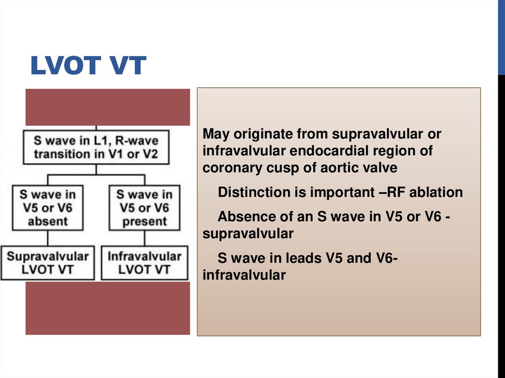
LVOT VTMay originate from supravalvular or
infravalvular endocardial region of
coronary cusp of aortic valve
Distinction is important –RF ablation
Absence of an S wave in V5 or V6 supravalvular
S wave in leads V5 and V6infravalvular
21.
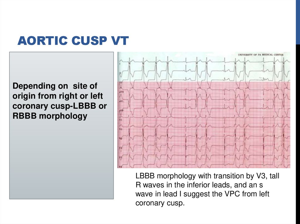
AORTIC CUSP VTDepending on site of
origin from right or left
coronary cusp-LBBB or
RBBB morphology
LBBB morphology with transition by V3, tall
R waves in the inferior leads, and an s
wave in lead I suggest the VPC from left
coronary cusp.
22.
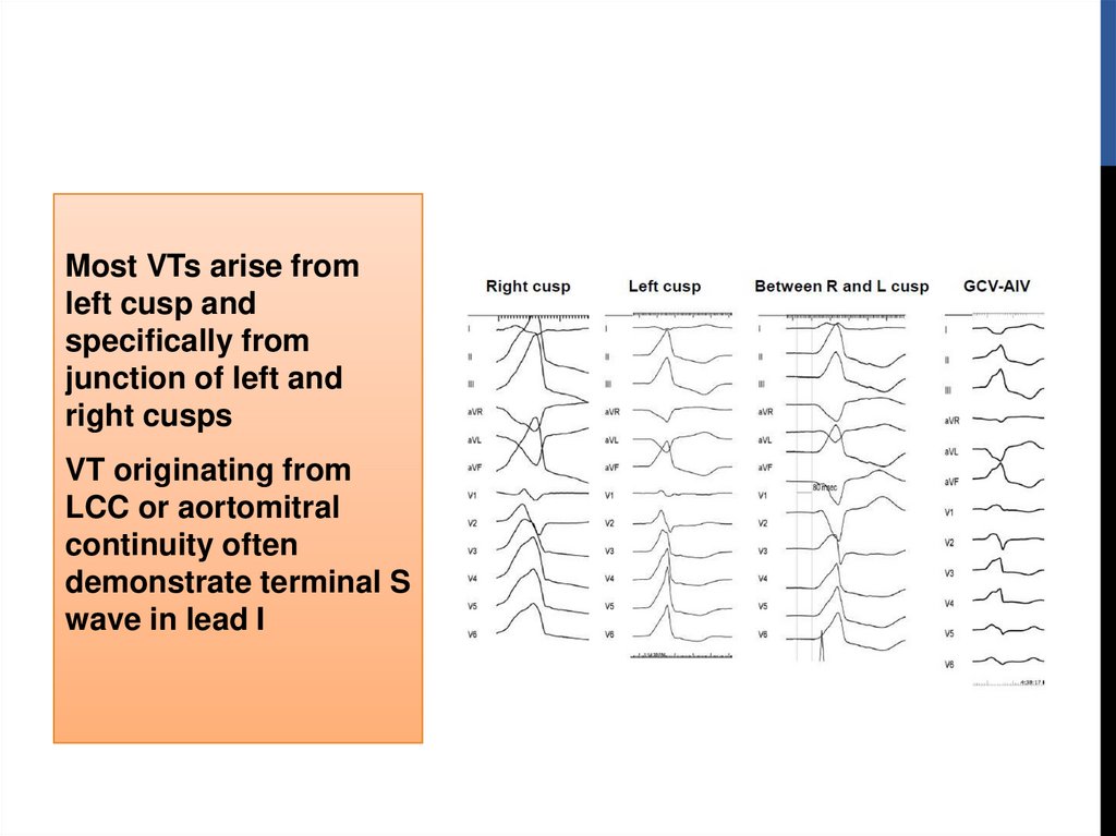
Most VTs arise fromleft cusp and
specifically from
junction of left and
right cusps
VT originating from
LCC or aortomitral
continuity often
demonstrate terminal S
wave in lead I
23.
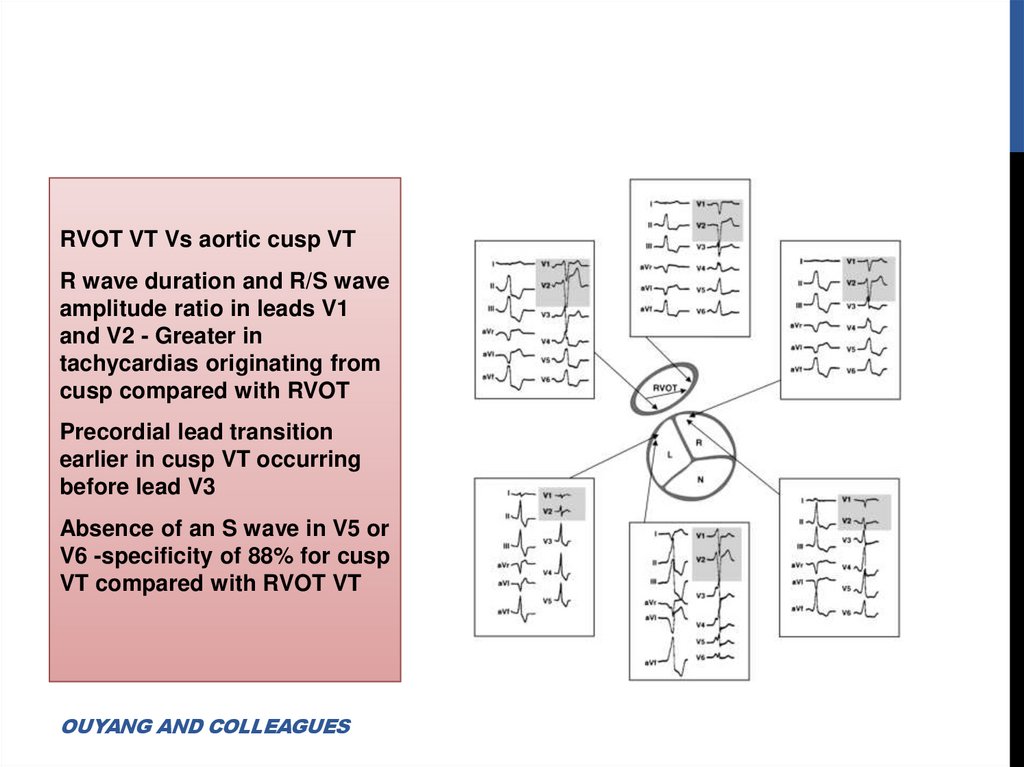
RVOT VT Vs aortic cusp VTR wave duration and R/S wave
amplitude ratio in leads V1
and V2 - Greater in
tachycardias originating from
cusp compared with RVOT
Precordial lead transition
earlier in cusp VT occurring
before lead V3
Absence of an S wave in V5 or
V6 -specificity of 88% for cusp
VT compared with RVOT VT
OUYANG AND COLLEAGUES
24.
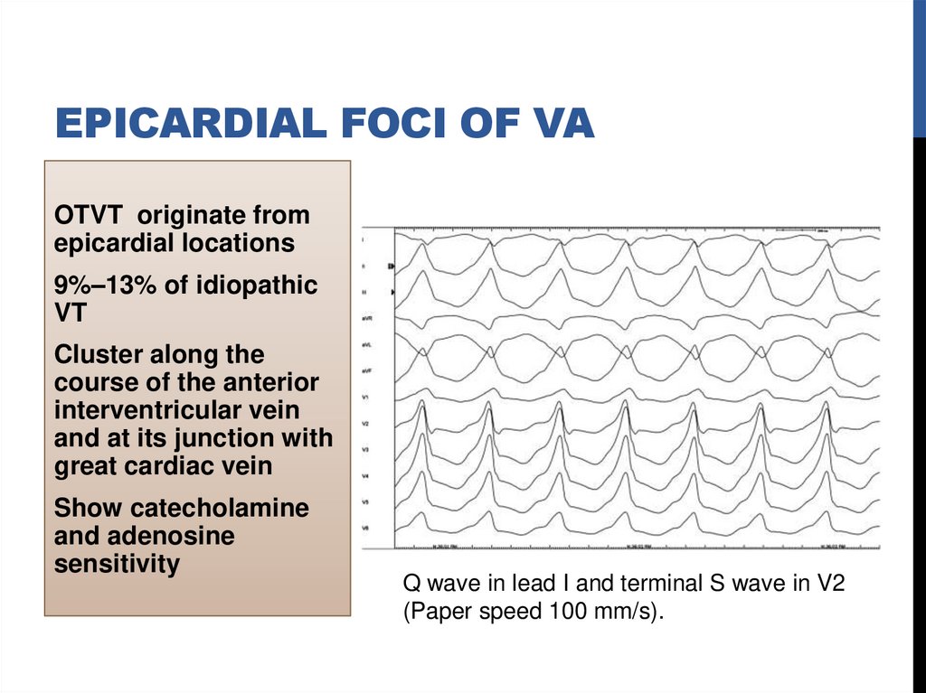
EPICARDIAL FOCI OF VAOTVT originate from
epicardial locations
9%–13% of idiopathic
VT
Cluster along the
course of the anterior
interventricular vein
and at its junction with
great cardiac vein
Show catecholamine
and adenosine
sensitivity
Q wave in lead I and terminal S wave in V2
(Paper speed 100 mm/s).
25.
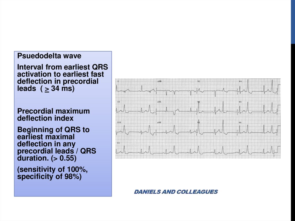
Psuedodelta waveInterval from earliest QRS
activation to earliest fast
deflection in precordial
leads ( > 34 ms)
Precordial maximum
deflection index
Beginning of QRS to
earliest maximal
deflection in any
precordial leads / QRS
duration. (> 0.55)
(sensitivity of 100%,
specificity of 98%)
26.
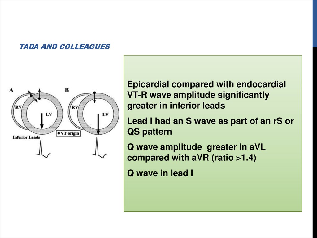
TADA AND COLLEAGUESEpicardial compared with endocardial
VT-R wave amplitude significantly
greater in inferior leads
Lead I had an S wave as part of an rS or
QS pattern
Q wave amplitude greater in aVL
compared with aVR (ratio >1.4)
Q wave in lead I
27.
MITRAL ANNULUS,TRICUSPID ANNULUS
PAPILLARY MUSCLE
PERIVASCULAR EPICARDIAL ECTOPY
28.
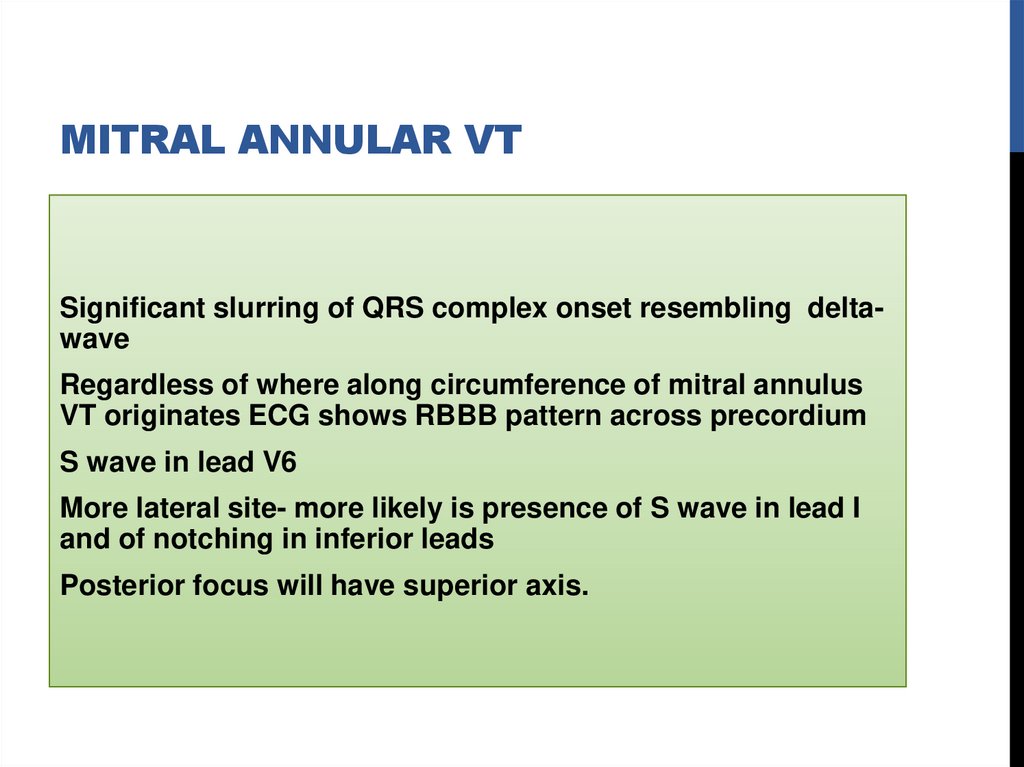
MITRAL ANNULAR VTSignificant slurring of QRS complex onset resembling deltawave
Regardless of where along circumference of mitral annulus
VT originates ECG shows RBBB pattern across precordium
S wave in lead V6
More lateral site- more likely is presence of S wave in lead I
and of notching in inferior leads
Posterior focus will have superior axis.
29.
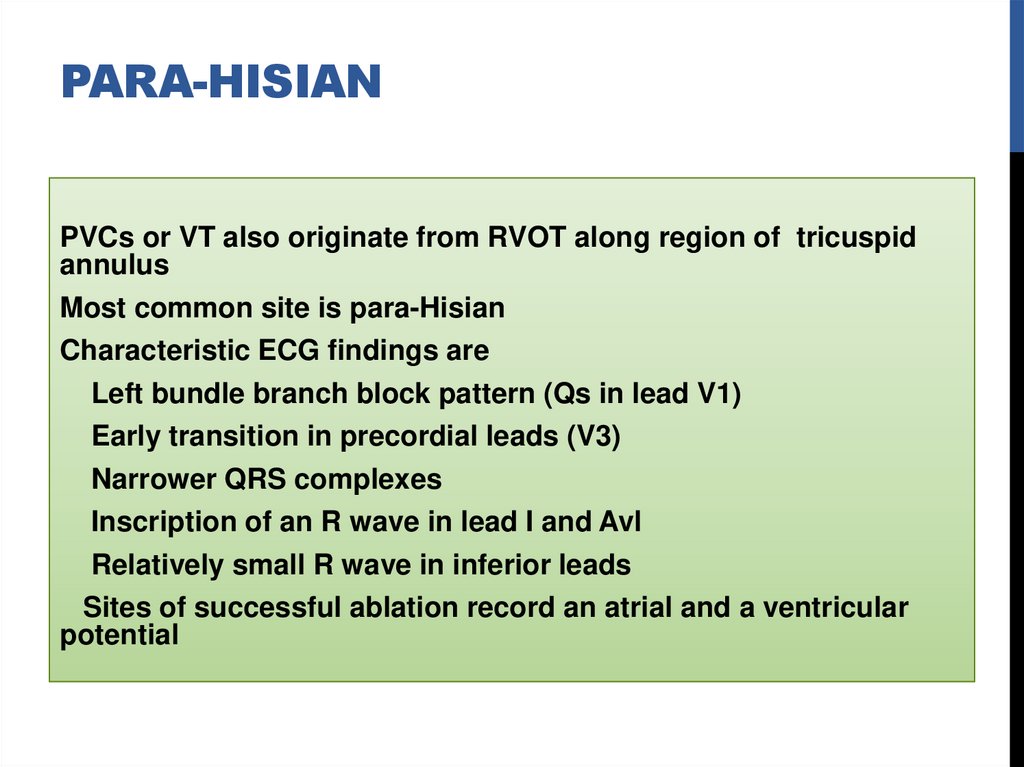
PARA-HISIANPVCs or VT also originate from RVOT along region of tricuspid
annulus
Most common site is para-Hisian
Characteristic ECG findings are
Left bundle branch block pattern (Qs in lead V1)
Early transition in precordial leads (V3)
Narrower QRS complexes
Inscription of an R wave in lead I and Avl
Relatively small R wave in inferior leads
Sites of successful ablation record an atrial and a ventricular
potential
30.
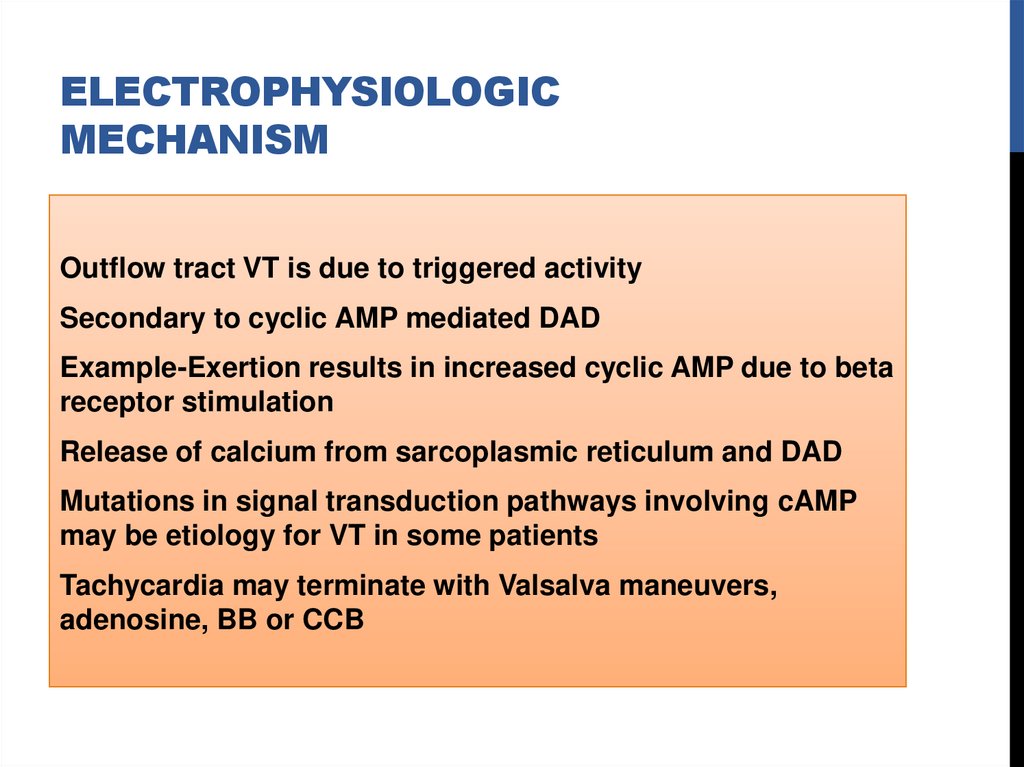
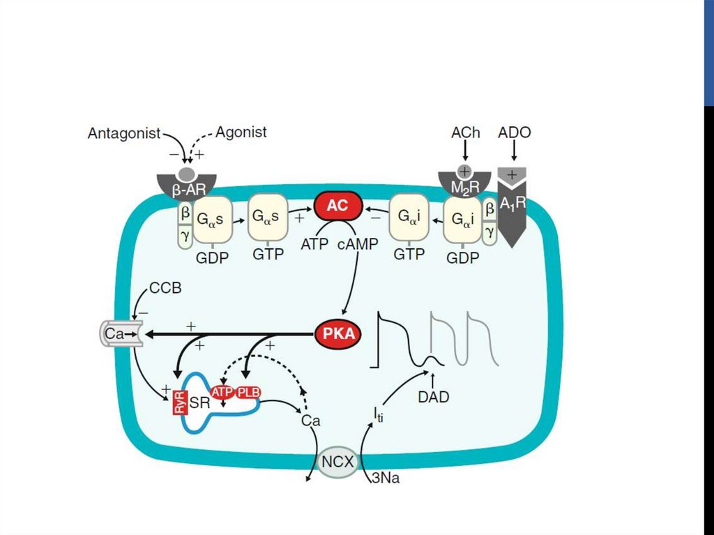
ELECTROPHYSIOLOGICMECHANISM
Outflow tract VT is due to triggered activity
Secondary to cyclic AMP mediated DAD
Example-Exertion results in increased cyclic AMP due to beta
receptor stimulation
Release of calcium from sarcoplasmic reticulum and DAD
Mutations in signal transduction pathways involving cAMP
may be etiology for VT in some patients
Tachycardia may terminate with Valsalva maneuvers,
adenosine, BB or CCB
31.
32.
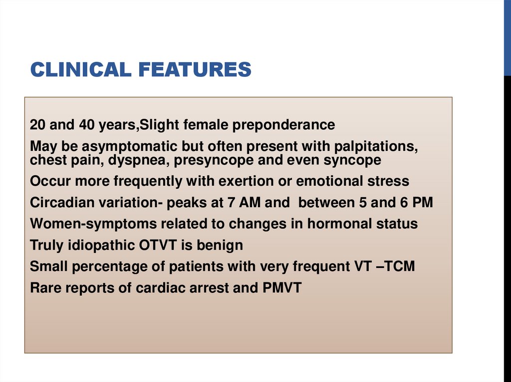
CLINICAL FEATURES20 and 40 years,Slight female preponderance
May be asymptomatic but often present with palpitations,
chest pain, dyspnea, presyncope and even syncope
Occur more frequently with exertion or emotional stress
Circadian variation- peaks at 7 AM and between 5 and 6 PM
Women-symptoms related to changes in hormonal status
Truly idiopathic OTVT is benign
Small percentage of patients with very frequent VT –TCM
Rare reports of cardiac arrest and PMVT
33.
TREATMENTMay respond acutely to carotid sinus massage, Valsalva
maneuvers or intravenous adenosine or verapamil
Long-term oral therapy with either BB or CCB
Nonresponders (33%)- class I or III antiarrhythmic agents
34.
RFAWhen medical therapy is ineffective or not tolerated
High success rate (>80%)
Ablation of epicardial or aortic sinuses of Valsalva sites is
highly effective
Technically challenging and carries higher risks -proximity to
coronary arteries
35.
Tachycardia localization12-lead ECG
Intracardiac activation
Pace mapping
36.
BIPOLAR ACTIVATIONMAPPING
OTVTs are mediated by triggered activity
Electrogram at site of origin typically precedes onset of QRS
by approximately 20 msec
Exception -cusp VT, prepotentials (~50 msec) may be seen
during VPCs that correspond to late potentials during sinus
rhythm
37.
PACE MAPPINGUseful because typically site of origin is focal and because
underlying tissue is normal
Performed with a low output
Result in a small discrete area of depolarization
Mapping performed at site of origin of clinical arrhythmia,
ECG should mimic clinical arrhythmia perfectly (12/12,
including notches)
38.
ELECTROANATOMIC RECREATION OF 3DANATOMY
Helpful for catheter mapping and localization of site of origin
Incessant VT- 3D anatomy should ideally be created during
tachycardia which should be able to localize earliest site to a
small region (<5 mm) with centrifugal activation
Typically pace mapping from this region should achieve a
perfect match
39.
Predictors for successful ablationSingle VT morphology
Accurate pace maps
Absence of a deltalike wave at beginning of QRS during
tachycardia
Ability to use pace mapping and activation mapping
40.
Some tachycardias arise from epicardium, necessitateablation from great cardiac vein or epicardium itself using
pericardial puncture technique
Coronary angiography is performed before ablation on
epicardium or in aortic sinus
41.
Complications during outflow tract VT ablation are rareRBBB (1%)
Cardiac perforation
Damage to the coronary artery (LAD) - cusp region ablation
Overall recurrence rate is approximately 10%
42.
IDIOPATHIC LEFT VTThree varieties
left posterior fascicular VT -RBBB and LAD (90%)
left anterior fascicular VT -RBBB and RAD
high septal fascicular VT -relatively narrow QRS and normal axis
43.
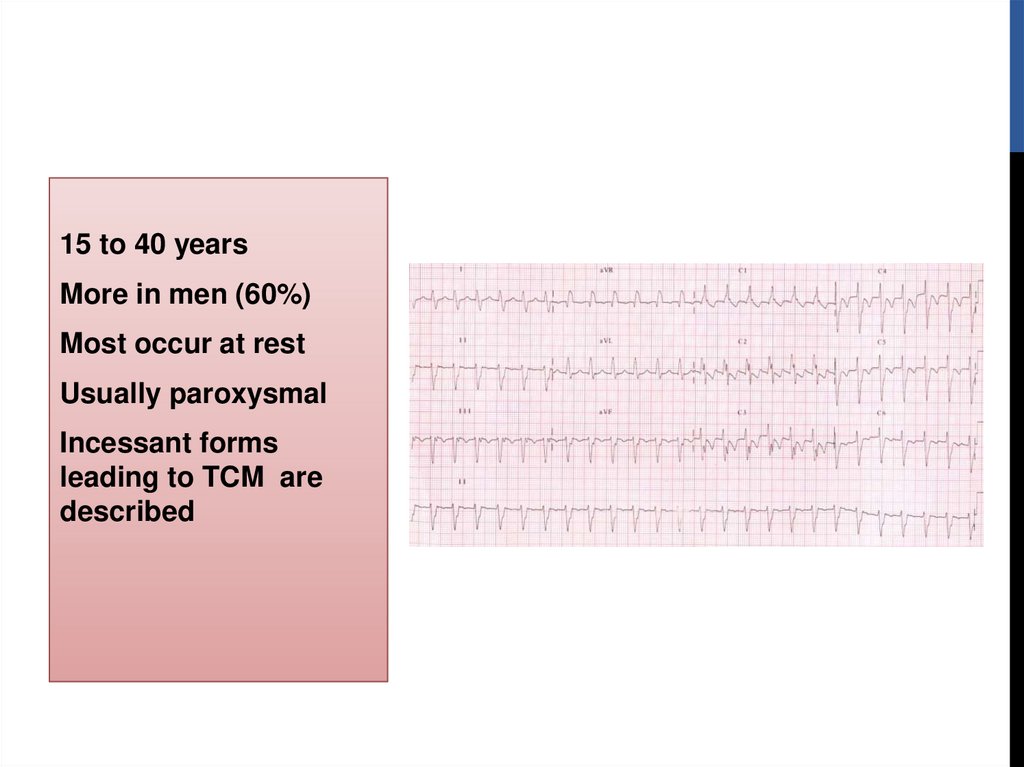
15 to 40 yearsMore in men (60%)
Most occur at rest
Usually paroxysmal
Incessant forms
leading to TCM are
described
44.
ELECTROPHYSIOLOGICMECHANISM
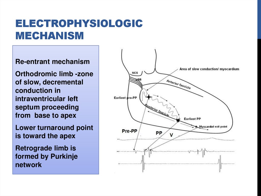
Re-entrant mechanism
Orthodromic limb -zone
of slow, decremental
conduction in
intraventricular left
septum proceeding
from base to apex
Lower turnaround point
is toward the apex
Retrograde limb is
formed by Purkinje
network
45.
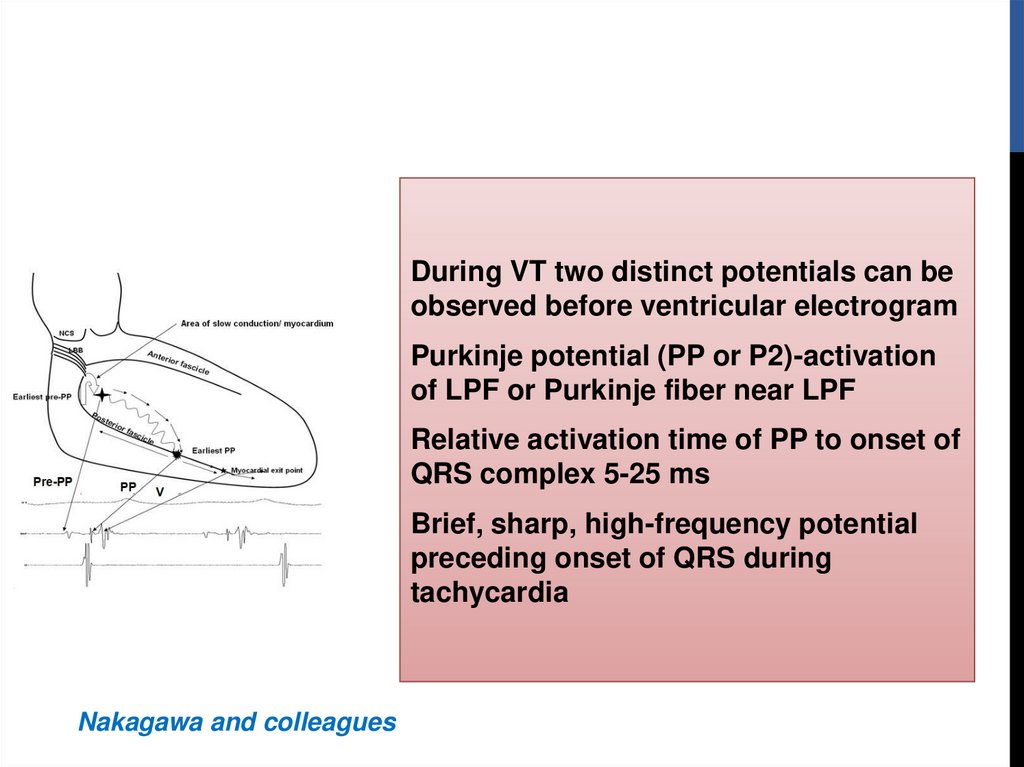
During VT two distinct potentials can beobserved before ventricular electrogram
Purkinje potential (PP or P2)-activation
of LPF or Purkinje fiber near LPF
Relative activation time of PP to onset of
QRS complex 5-25 ms
Brief, sharp, high-frequency potential
preceding onset of QRS during
tachycardia
Nakagawa and colleagues
46.
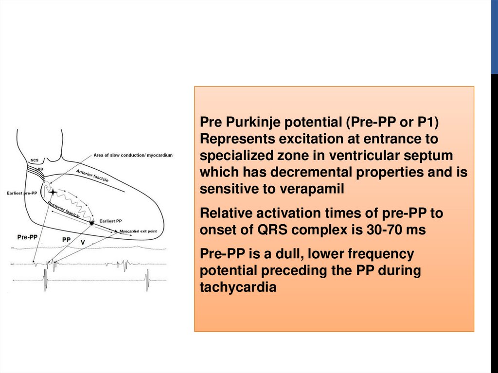
Pre Purkinje potential (Pre-PP or P1)Represents excitation at entrance to
specialized zone in ventricular septum
which has decremental properties and is
sensitive to verapamil
Relative activation times of pre-PP to
onset of QRS complex is 30-70 ms
Pre-PP is a dull, lower frequency
potential preceding the PP during
tachycardia
47.
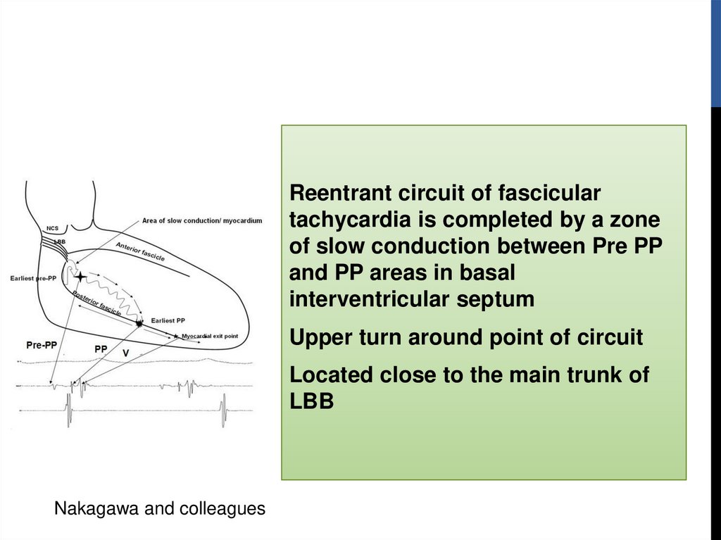
Reentrant circuit of fasciculartachycardia is completed by a zone
of slow conduction between Pre PP
and PP areas in basal
interventricular septum
Upper turn around point of circuit
Located close to the main trunk of
LBB
Nakagawa and colleagues
48.
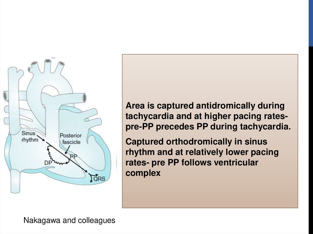
Area is captured antidromically duringtachycardia and at higher pacing ratespre-PP precedes PP during tachycardia.
Captured orthodromically in sinus
rhythm and at relatively lower pacing
rates- pre PP follows ventricular
complex
Nakagawa and colleagues
49.
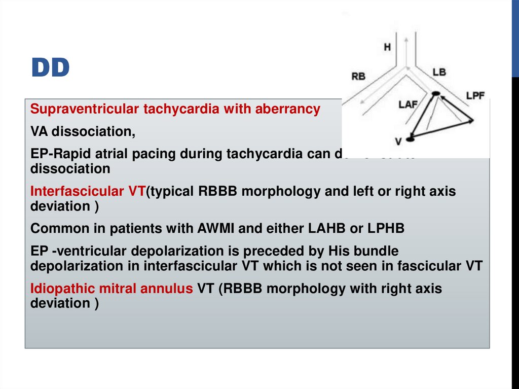
DDSupraventricular tachycardia with aberrancy
VA dissociation,
EP-Rapid atrial pacing during tachycardia can demonstrate AV
dissociation
Interfascicular VT(typical RBBB morphology and left or right axis
deviation )
Common in patients with AWMI and either LAHB or LPHB
EP -ventricular depolarization is preceded by His bundle
depolarization in interfascicular VT which is not seen in fascicular VT
Idiopathic mitral annulus VT (RBBB morphology with right axis
deviation )
50.
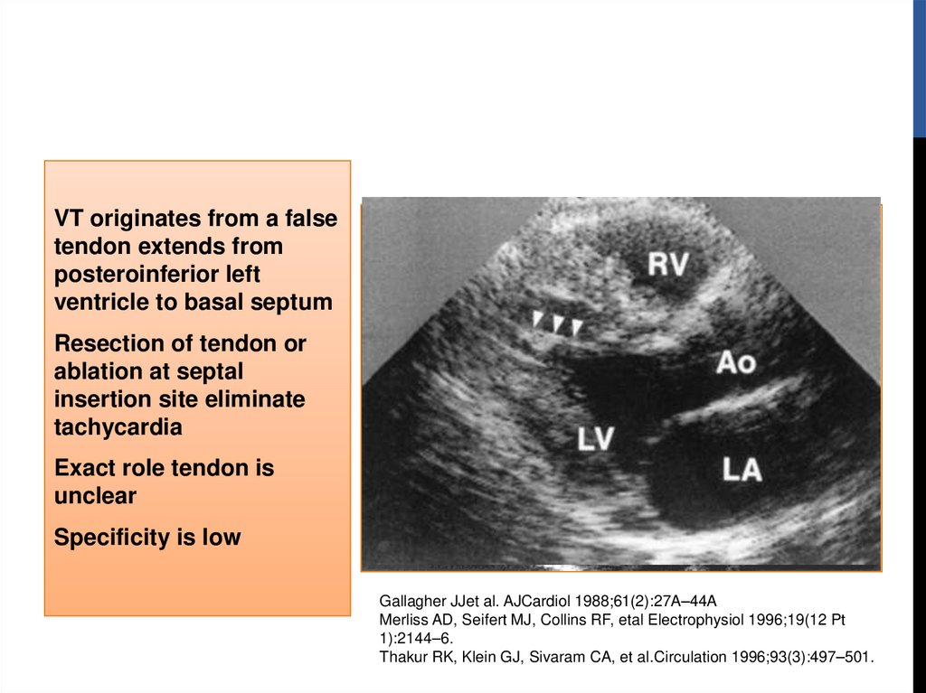
VT originates from a falsetendon extends from
posteroinferior left
ventricle to basal septum
Resection of tendon or
ablation at septal
insertion site eliminate
tachycardia
Exact role tendon is
unclear
Specificity is low
Gallagher JJet al. AJCardiol 1988;61(2):27A–44A
Merliss AD, Seifert MJ, Collins RF, etal Electrophysiol 1996;19(12 Pt
1):2144–6.
Thakur RK, Klein GJ, Sivaram CA, et al.Circulation 1996;93(3):497–501.
51.
ECGBaseline 12-lead ECG is normal in most patients
Exit near the area of the left posterior fascicle
RBBB + left superior frontal plane axis
Relatively narrow QRS duration (<140 msec)
RS interval <80 msec
Exit near the area of the left anterior fascicle
RBBB+ right frontal plane axis
52.
Long-term prognosis is very goodPatients who have incessant tachycardia may develop
tachycardia related cardiomyopathy
Intravenous verapamil is effective in acutely terminating VT
Mild to moderate symptoms oral –verapamil
BB and class I and III antiarrhythmic agents useful in some
Medical therapy is often ineffective in patients who have
severe symptoms
53.
RADIOFREQUENCYABLATION
Associated with significant symptoms or who are intolerant
or resistant to medical therapy
Strategies employed to identify the ideal site for ablation
Pace mapping
Endocardial activation mapping
Identifying diastolic Purkinje potentials (MC approach)
Identifying late diastolic potentials
When VT is noninducible-ablation during sinus rhythm
using electroanatomic mapping may be considered
54.
LIFE-THREATENING(TYPICALLY POLYMORPHIC VT)
Rare
Generally occurs with genetic ion channel disorders
Associated with SCD
Abnormalities exist at molecular level
55.
LIFE-THREATENING(TYPICALLY POLYMORPHIC VT)
Long QT Syndrome
Brugada Syndrome
CPVT
Short QT Syndrome
56.
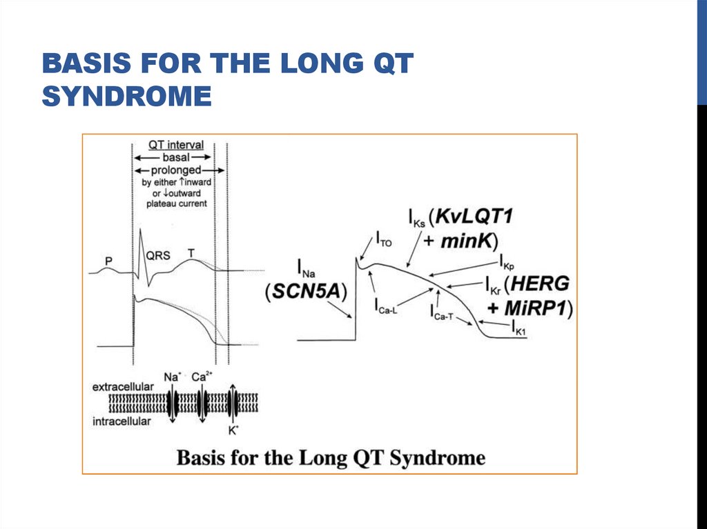
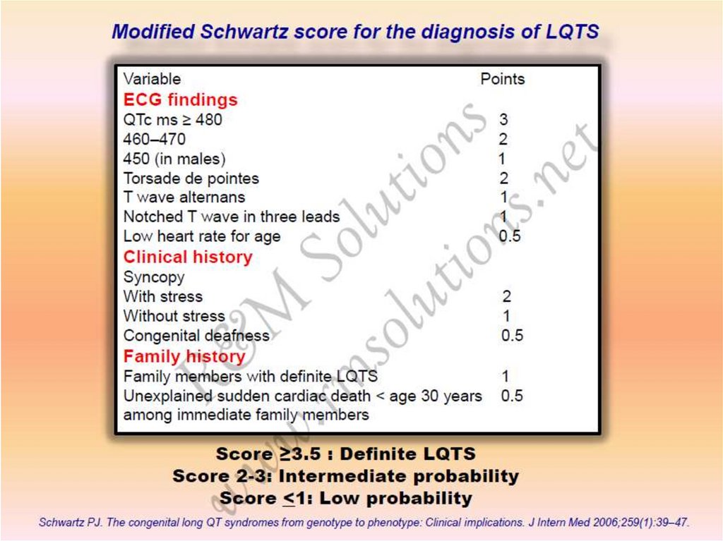
LONG QT SYNDROMECorrected QT interval 440 ms in men and 460 ms in women
with or without morphological abnormalities of the T waves
Decrease in outward potassium currents or increase in
inward sodium currents
Prolongs repolarization phase of cardiac action potential
Result in prolongation of QT interval
Predisposition to EAD and torsade de pointes VT
Twelve different genes described
LQT1, LQT2, and LQT3 account for 90%
57.
BASIS FOR THE LONG QTSYNDROME
58.
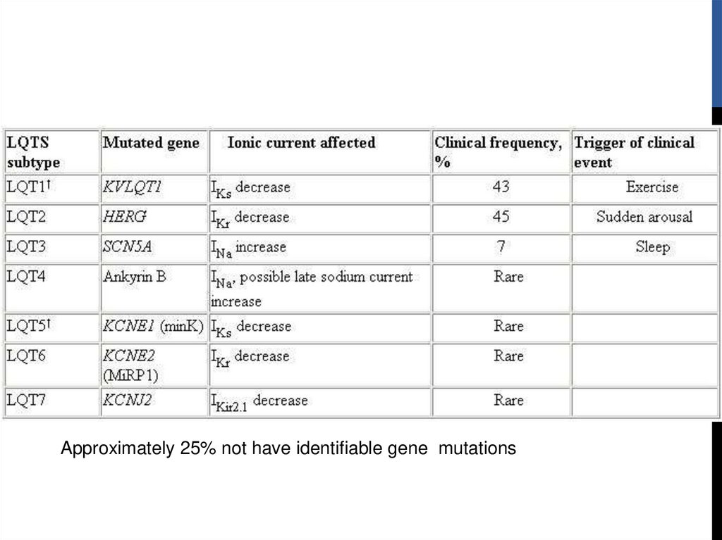
Approximately 25% not have identifiable gene mutations59.
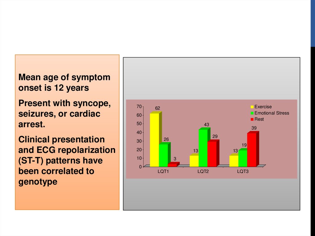
Mean age of symptomonset is 12 years
Present with syncope,
seizures, or cardiac
arrest.
70
Exercise
Emotional Stress
Rest
62
60
50
43
40
Clinical presentation
and ECG repolarization
(ST-T) patterns have
been correlated to
genotype
30
39
29
26
19
20
13
10
13
3
0
LQT1
LQT2
LQT3
60.
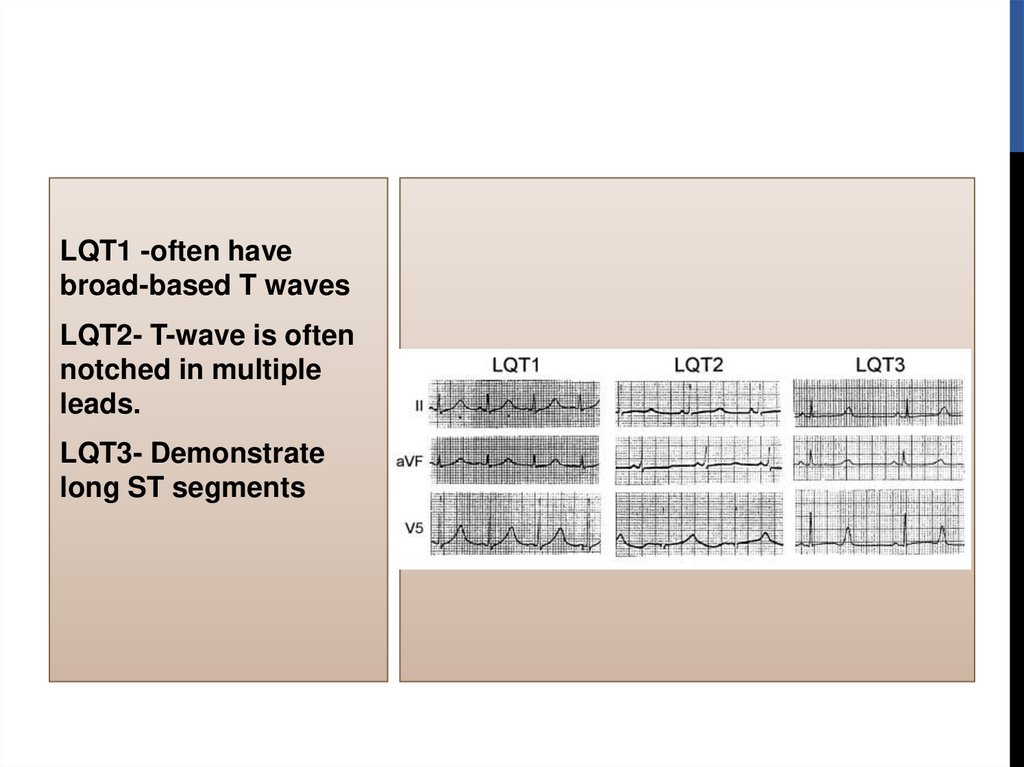
LQT1 -often havebroad-based T waves
LQT2- T-wave is often
notched in multiple
leads.
LQT3- Demonstrate
long ST segments
61.
62.
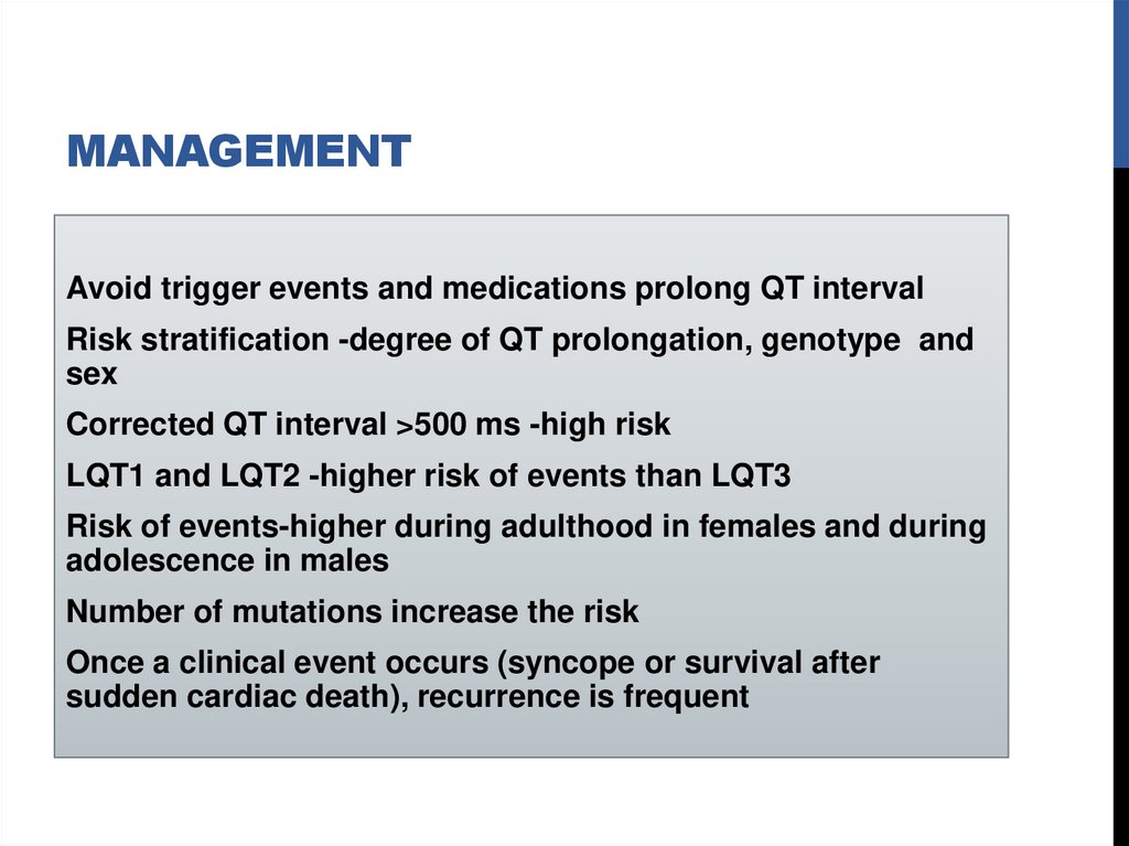
MANAGEMENTAvoid trigger events and medications prolong QT interval
Risk stratification -degree of QT prolongation, genotype and
sex
Corrected QT interval >500 ms -high risk
LQT1 and LQT2 -higher risk of events than LQT3
Risk of events-higher during adulthood in females and during
adolescence in males
Number of mutations increase the risk
Once a clinical event occurs (syncope or survival after
sudden cardiac death), recurrence is frequent
63.
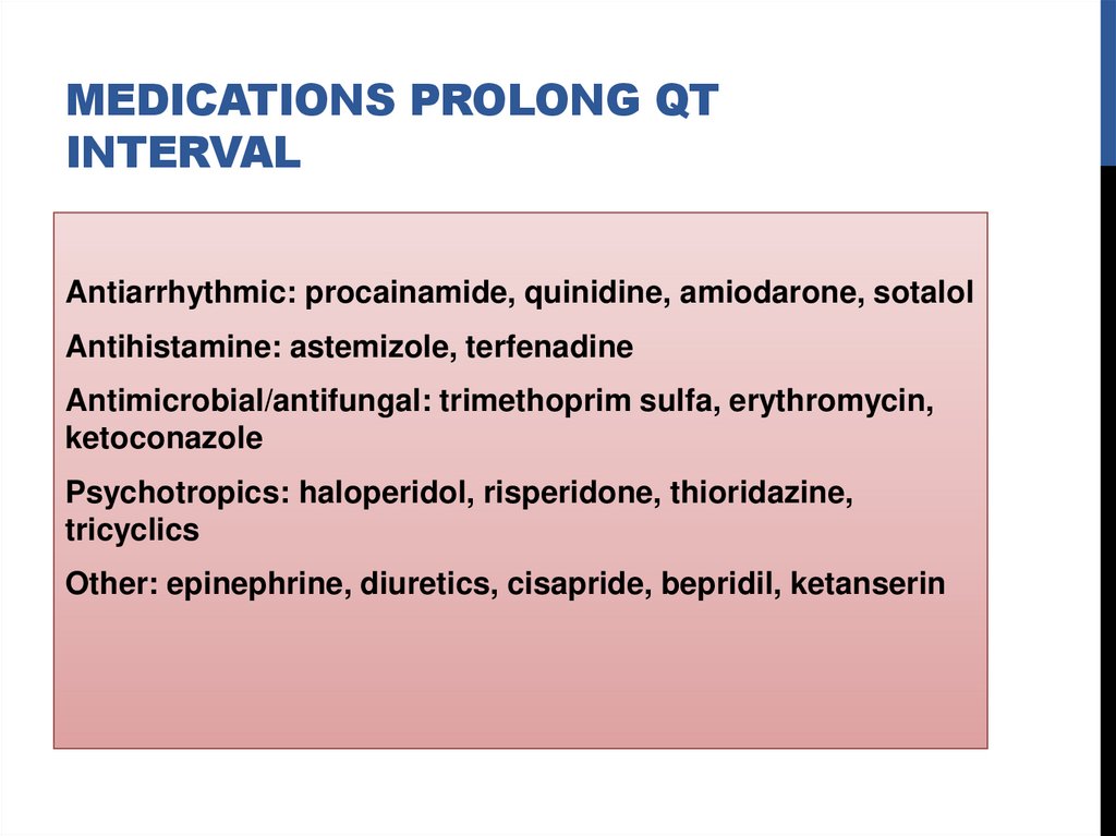
MEDICATIONS PROLONG QTINTERVAL
Antiarrhythmic: procainamide, quinidine, amiodarone, sotalol
Antihistamine: astemizole, terfenadine
Antimicrobial/antifungal: trimethoprim sulfa, erythromycin,
ketoconazole
Psychotropics: haloperidol, risperidone, thioridazine,
tricyclics
Other: epinephrine, diuretics, cisapride, bepridil, ketanserin
64.
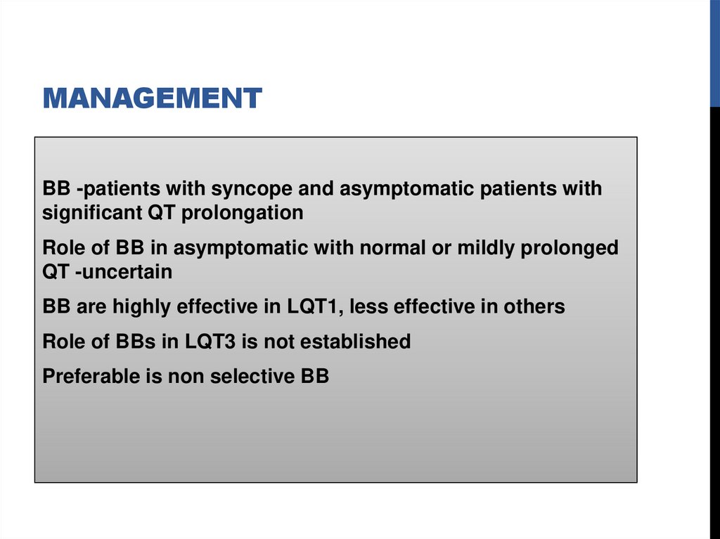
MANAGEMENTBB -patients with syncope and asymptomatic patients with
significant QT prolongation
Role of BB in asymptomatic with normal or mildly prolonged
QT -uncertain
BB are highly effective in LQT1, less effective in others
Role of BBs in LQT3 is not established
Preferable is non selective BB
65.
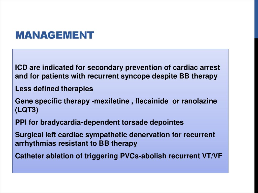
MANAGEMENTICD are indicated for secondary prevention of cardiac arrest
and for patients with recurrent syncope despite BB therapy
Less defined therapies
Gene specific therapy -mexiletine , flecainide or ranolazine
(LQT3)
PPI for bradycardia-dependent torsade depointes
Surgical left cardiac sympathetic denervation for recurrent
arrhythmias resistant to BB therapy
Catheter ablation of triggering PVCs-abolish recurrent VT/VF
66.
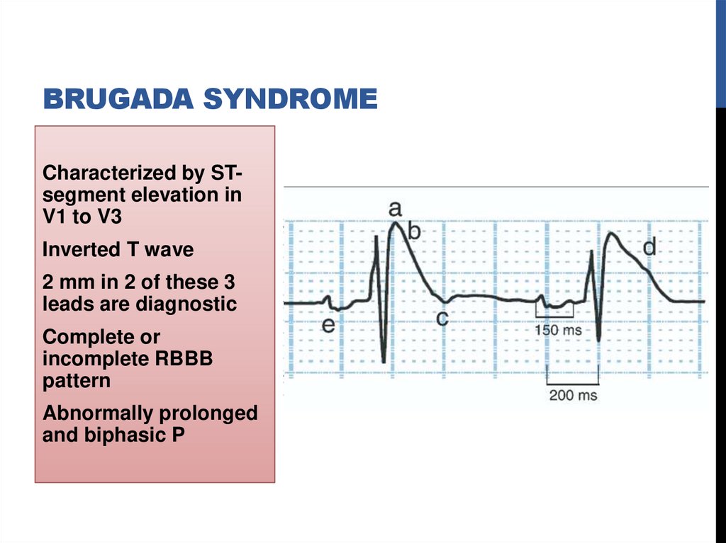
BRUGADA SYNDROMECharacterized by STsegment elevation in
V1 to V3
Inverted T wave
2 mm in 2 of these 3
leads are diagnostic
Complete or
incomplete RBBB
pattern
Abnormally prolonged
and biphasic P
67.
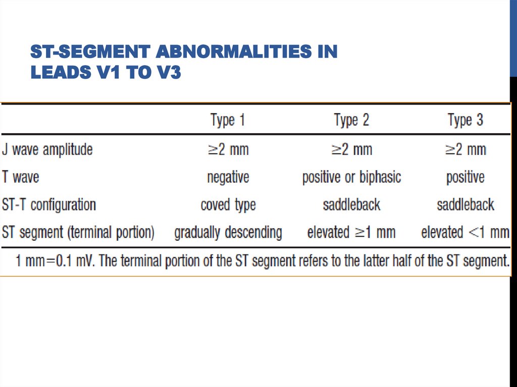
ST-SEGMENT ABNORMALITIES INLEADS V1 TO V3
68.
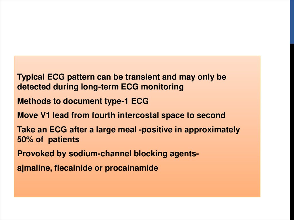
Typical ECG pattern can be transient and may only bedetected during long-term ECG monitoring
Methods to document type-1 ECG
Move V1 lead from fourth intercostal space to second
Take an ECG after a large meal -positive in approximately
50% of patients
Provoked by sodium-channel blocking agentsajmaline, flecainide or procainamide
69.
CLINICAL PRESENTATION0.12% to 0.14% in general population
Syncope or cardiac arrest
Predominantly in men in third and fourth decade
SCD in young men,typically occurs at night
Prone to atrial fibrillation and sinus node dysfunction
Precipitated by a febrile state, vagotonic agents, a-adrenergic
agonists,BBs, TCAs, hypokalaemia,alcohol and cocaine
toxicity
70.
Risk of SCD with Brugada syndrome is substantialRisk of recurrent events during 4 years of follow-up
62% for those with cardiac arrest
19% for those with syncope.
Asymptomatic group -8% event rate during 2 years
Brugada P, Brugada R, Brugada J. Sudden death in patients and relatives with the
syndrome of right bundle branch block, ST segment elevation in the precordial leads
V(1) to V(3) and sudden death. Eur Heart J 2000;21:321-6.
71.
TREATMENTDrugs inhibit Ito (such as quinidine) and increase calcium
current (such as isoproterenol) are effective
Lowdose quinidine may be used to treat frequent VAs in
patients who already have an ICD
Quinidine and isoproterenol may be useful in VT storms
Catheter ablation of triggering PVCs and ablation of RVOT
epicardial musculature successful in abolishing recurrent
VT/VF in a small number of patients
Dimethyl lithospermate B (Danshen’s extract)
cilostazol
72.
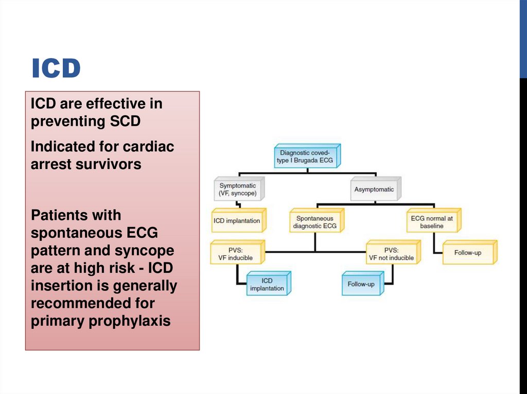
ICDICD are effective in
preventing SCD
Indicated for cardiac
arrest survivors
Patients with
spontaneous ECG
pattern and syncope
are at high risk - ICD
insertion is generally
recommended for
primary prophylaxis
73.
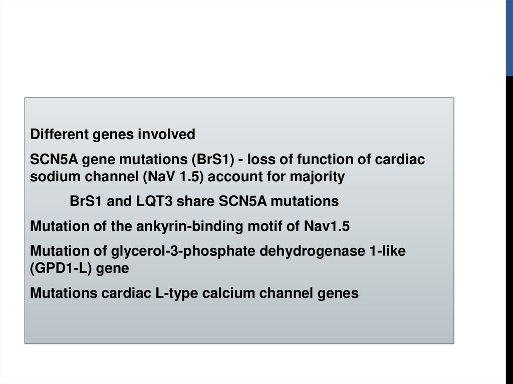
Different genes involvedSCN5A gene mutations (BrS1) - loss of function of cardiac
sodium channel (NaV 1.5) account for majority
BrS1 and LQT3 share SCN5A mutations
Mutation of the ankyrin-binding motif of Nav1.5
Mutation of glycerol-3-phosphate dehydrogenase 1-like
(GPD1-L) gene
Mutations cardiac L-type calcium channel genes
74.
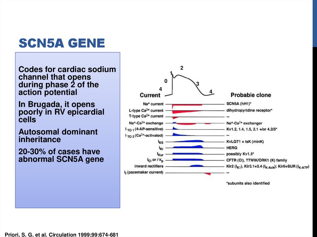
SCN5A GENECodes for cardiac sodium
channel that opens
during phase 2 of the
action potential
In Brugada, it opens
poorly in RV epicardial
cells
Autosomal dominant
inheritance
20-30% of cases have
abnormal SCN5A gene
Priori, S. G. et al. Circulation 1999;99:674-681
2
1
0
4
3
4
75.
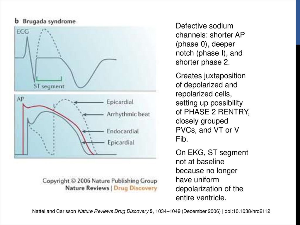
Defective sodiumchannels: shorter AP
(phase 0), deeper
notch (phase I), and
shorter phase 2.
Creates juxtaposition
of depolarized and
repolarized cells,
setting up possibility
of PHASE 2 RENTRY,
closely grouped
PVCs, and VT or V
Fib.
On EKG, ST segment
not at baseline
because no longer
have uniform
depolarization of the
entire ventricle.
Nattel and Carlsson Nature Reviews Drug Discovery 5, 1034–1049 (December 2006) | doi:10.1038/nrd2112
76.
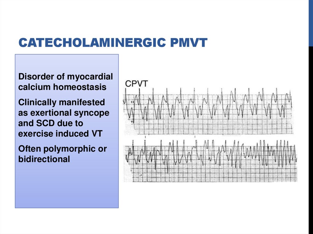
CATECHOLAMINERGIC PMVTDisorder of myocardial
calcium homeostasis
Clinically manifested
as exertional syncope
and SCD due to
exercise induced VT
Often polymorphic or
bidirectional
77.
Autosomal dominant (50% )-mutation of cardiac ryanodinereceptor (RyR2 gene)
Autosomal recessive (3% to 5% )-mutations of calsequestrin
2 gene (CASQ2)
78.
Ryanodine receptor spans membrane of sarcoplasmicreticulum
Releases calcium triggered by calcium entry into cell through
L-type calcium channels
Calsequestrin-protein sequestrates calcium ions within
sarcoplasmic reticulum
RyR2 and CASQ2 mutations cause intracellular calcium
overload and DAD -basis of arrhythmogenesis
79.
Resting ECG is unremarkable, prominent U waves may beseen
Typical VT patterns are reproducible with exercise or
catecholamine infusion
VAs typically appear during sinus tachycardia rates of 120
beats/min to 130 beats/min, with progressive frequency of
PVCs followed by bursts of polymorphic or bidirectional VT
Mean age for presentation with syncope is 4 years
EP study is not helpful in risk stratification
80.
Medical management-BB46% may have recurrent events while receiving therapy
CCB -limited effectiveness
Flecainide (blocks RyR2 receptor) also used
81.
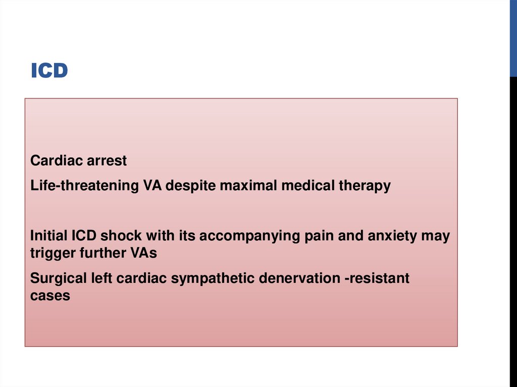
ICDCardiac arrest
Life-threatening VA despite maximal medical therapy
Initial ICD shock with its accompanying pain and anxiety may
trigger further VAs
Surgical left cardiac sympathetic denervation -resistant
cases
82.
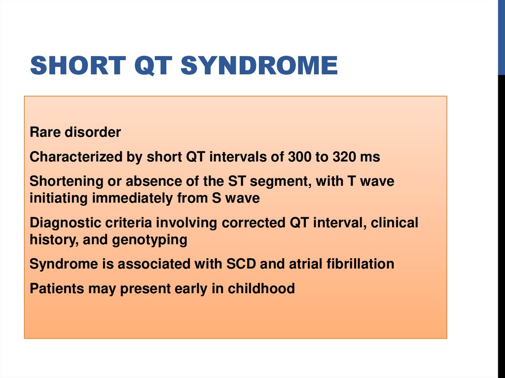
SHORT QT SYNDROMERare disorder
Characterized by short QT intervals of 300 to 320 ms
Shortening or absence of the ST segment, with T wave
initiating immediately from S wave
Diagnostic criteria involving corrected QT interval, clinical
history, and genotyping
Syndrome is associated with SCD and atrial fibrillation
Patients may present early in childhood
83.
84.
ICD implantation for secondary and primary preventionPreliminary observations suggest quinidine might be useful
85.
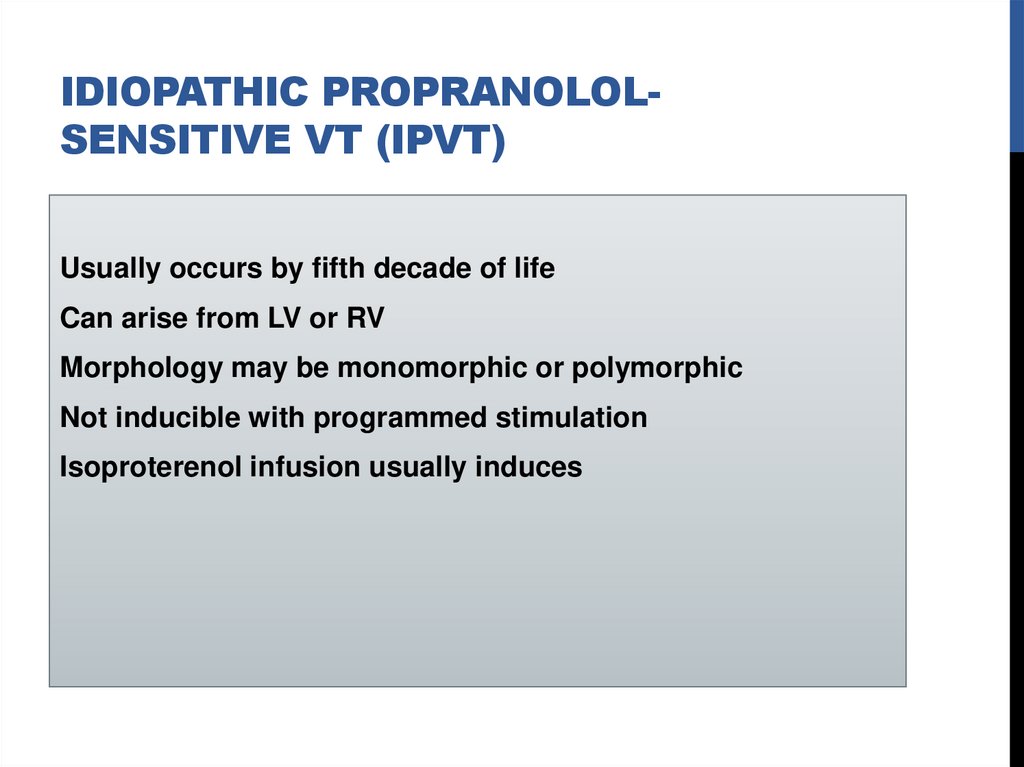
IDIOPATHIC PROPRANOLOLSENSITIVE VT (IPVT)Usually occurs by fifth decade of life
Can arise from LV or RV
Morphology may be monomorphic or polymorphic
Not inducible with programmed stimulation
Isoproterenol infusion usually induces
86.
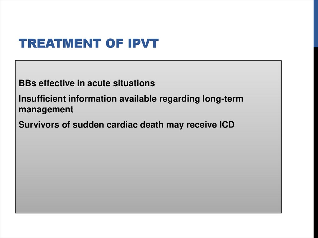
TREATMENT OF IPVTBBs effective in acute situations
Insufficient information available regarding long-term
management
Survivors of sudden cardiac death may receive ICD
87.
88.
89.
REFERENCESZIPES 5th EDITION
BRAUNWALD 9TH EDITION
HURST 13TH EDITION
VENTRICULAR ARRHYTHMIAS IN NORMAL HEARTS,SHUAIB LATIF, MD
Cardiol Clin 26 (2008) 367–380
VENTRICULAR TACHYCARDIA IN STRUCTURALLY NORMAL HEARTS:
RECOGNITION AND MANAGEMENT,P NATHANI
Supplement of JAPI • April 2007 • vol. 55
VENTRICULAR TACHYCARDIA IN THE ABSENCE OF STRUCTURAL
HEART DISEASE KOMANDOOR SRIVATHSAN, MD,
IPEJ (ISSN 0972-6292), 5(2): 106-121 (2005)
VENTRICULAR ARRHYTHMIAS IN THE ABSENCE OF STRUCTURAL
HEART DISEASE ERIC N. PRYSTOWSKY, MD,
Vol. 59, No. 20, 2012 ISSN 0735-1097 JACC
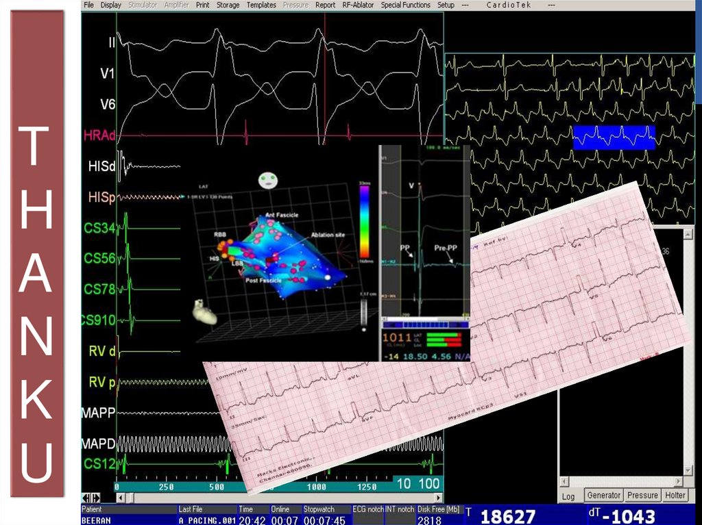
90.
TH
A
N
K
U