")

 substance – a gel colloid system from water, salts and organic substances: glycoproteins,")


 biology
Similar presentations:


Соединительныя ткани
1. Соединительные ткани - Connective tissues (consist of cells and intercellular substance)
Собственно соединительные тканиActually connective tissues
Волокнистая
Collagenic
Скелетные ткани
Skeletal tissues
Со специальными свойствами
Хрящевая
With special properties
Cartilage
• Ретикулярная - reticular
• Жировая - adipose
•Слизистая - mucous
Рыхлая неоформленная
Loose collagen
Плотная - Dense collagen
• оформленная - regular type
• неоформленная - irregular type
Костная
Bone
• Ретикулофиброзная
reticulofibrosus
• Пластинчатая
lamellaris
• Гиалиновый - hyaline
• Эластический - elastic
• Волокнистый - fibrous
2. Функции соединительных тканей
1. Опорная (капсулы органов,сухожилии, фасции, скелет)
2. Трофическая (обмен веществ
между кровью и клетками)
3. Защитная (механич. защита,
прочность органов, фагоцитоз
макрофагами, участие в
воспалении и иммунном ответе)
Functions of
connective tissues
1. Basic - make capsules of organs,
tendons, fascia, skeleton.
2. Trophic - metabolism between
blood and cells.
3. Protective - mechanical protection,
durability of organs, phagosytosis by
macrophages, participates in an
inflammation and immunity.
4. Кроветворная (микроокружение для клеток гемопоэза)
4. Hemopoietic - a microenvironment
5. Пластическая (адаптирует к
5. Plastic - adapts organs at change of
изменяющимся условиям за счёт
изменения обмена веществ)
for hemopoiesis cells.
conditions due to change of
a metabolism, participates in
regeneration.
3.
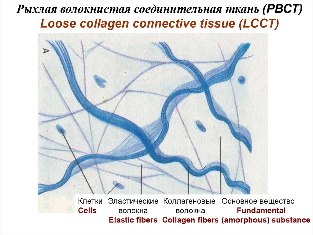
Рыхлая волокнистая соединительная ткань (РВСТ)Loose collagen connective tissue (LCCT)
Клетки Эластические Коллагеновые Основное вещество
Cells
волокна
волокна
Fundamental
Elastic fibers Collagen fibers (amorphous) substance
4. The fundamental (amorphous) substance – a gel colloid system from water, salts and organic substances: glycoproteins,
glycosaminoglycans (GAG) andproteoglycans.
Glycoproteins are the proteins connected with oligosaccharines,
connect cells with fibres, happen soluble and insoluble structural,
species-specific.
Proteoglycans are the proteins connected with GAG.
Glycosaminoglycans (GAG) are sour high-polymer
combinations, synthesized by fibroblasts. Distinguish 5 groups of
GAG.
5.
Основное веществоFundamental (amorphous)
substance
Вода - water
Неорганические вещества - salts
Органические вещества - organic substances :
• Гликозаминогликаны (ГАГ)
glycosaminoglycans (GAG)
Гликопротеиды (белки + олигосахариды)
glycoproteins (proteins + oligosaccharines)
Протеогликаны (белки + ГАГ)
proteoglycans (proteins + GAG)
6.
Гликозаминогликаны (ГАГ)Сульфатированные, гидрофобные:
1 группа – хондроитинсульфаты А,В,С
2 группа – дерматансульфаты
3 группа – кератансульфаты
4 группа – гепарансульфаты и гепарин
Не сульфатирована, гидрофильна:
5 группа – гиалуроновая кислота
Glycosaminoglycans (GAG)
4 groups are sulfatated, connected with
proteins, are a part of proteoglycans:
1) chondroitinsulfats A,B,C,
2) dermatansulfats,
3) keratansulfats,
4) heparansulfats and heparin.
5-th group is not sulfatated:
- hyaluronic acid
It has the greatest molecular weight,
can be free and connected with
proteins.
GAG define permeability of a tissue for water and solutions.
7.
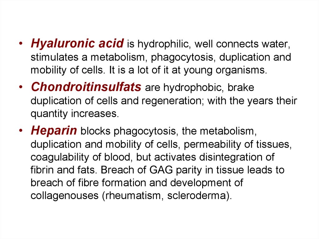
• Hyaluronic acid is hydrophilic, well connects water,stimulates a metabolism, phagocytosis, duplication and
mobility of cells. It is a lot of it at young organisms.
• Chondroitinsulfats are hydrophobic, brake
duplication of cells and regeneration; with the years their
quantity increases.
• Heparin blocks phagocytosis, the metabolism,
duplication and mobility of cells, permeability of tissues,
coagulability of blood, but activates disintegration of
fibrin and fats. Breach of GAG parity in tissue leads to
breach of fibre formation and development of
collagenouses (rheumatism, scleroderma).
8.
Коллагеновые волокна (из белка коллагена)Collagen fibers (from protein collagen)
Collagenic fibers are
• very strong,
• oxiphilic,
• tape-like,
• not anastomose,
• lay freely,
• slightly twisting.
9.
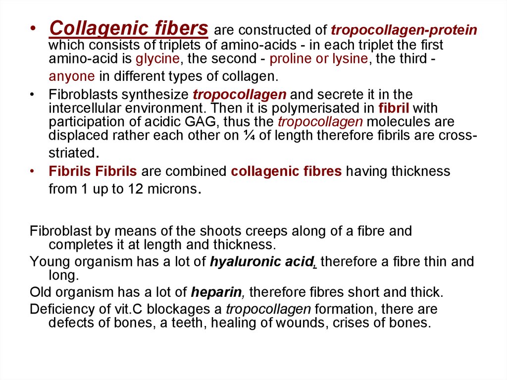
• Collagenic fibers are constructed of tropocollagen-proteinwhich consists of triplets of amino-acids - in each triplet the first
amino-acid is glycine, the second - proline or lysine, the third anyone in different types of collagen.
• Fibroblasts synthesize tropocollagen and secrete it in the
intercellular environment. Then it is polymerisated in fibril with
participation of acidic GAG, thus the tropocollagen molecules are
displaced rather each other on ¼ of length therefore fibrils are crossstriated.
• Fibrils Fibrils are combined collagenic fibres having thickness
from 1 up to 12 microns.
Fibroblast by means of the shoots creeps along of a fibre and
completes it at length and thickness.
Young organism has a lot of hyaluronic acid, therefore a fibre thin and
long.
Old organism has a lot of heparin, therefore fibres short and thick.
Deficiency of vit.C blockages a tropocollagen formation, there are
defects of bones, a teeth, healing of wounds, crises of bones.
10.
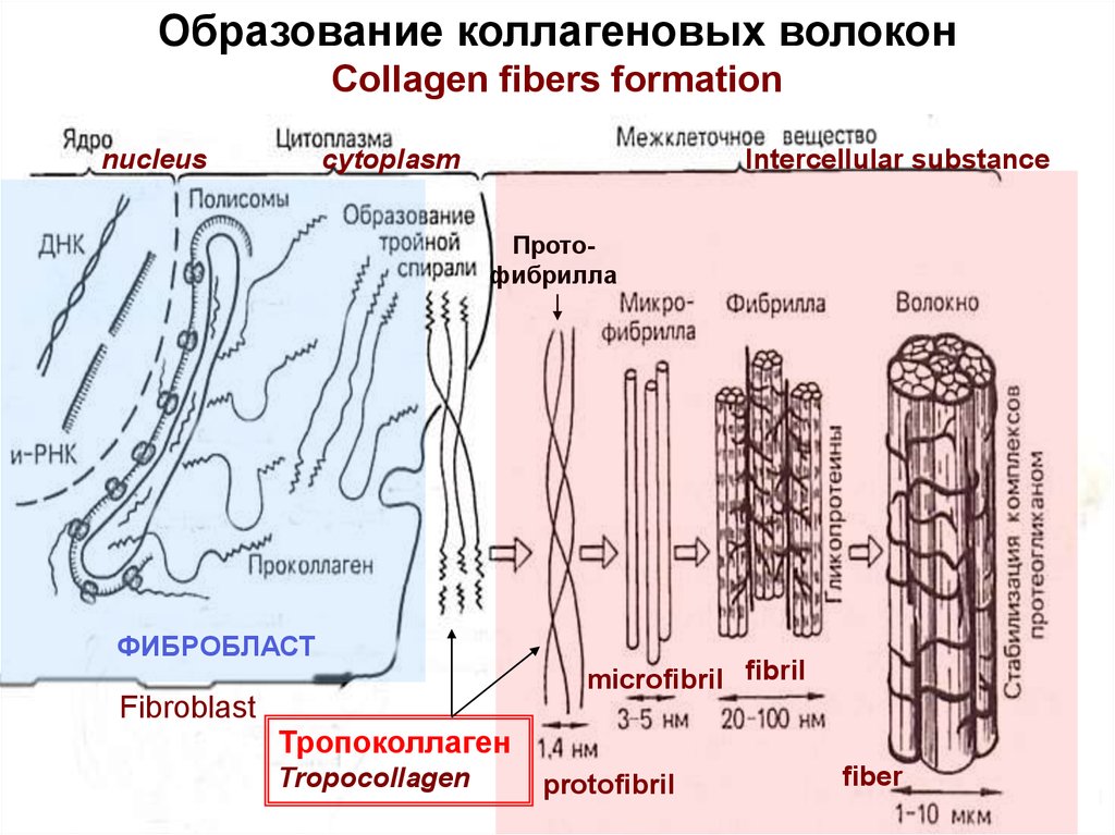
Образование коллагеновых волоконCollagen fibers formation
nucleus
cytoplasm
Intercellular substance
Протофибрилла
ФИБРОБЛАСТ
microfibril fibril
Fibroblast
Тропоколлаген
Tropocollagen
protofibril
fiber
11.
Фибрилла (тропоколлаген)Fibril (tropocollagen)
Молекула тропоколлагена
tropocollagen molecule
Зазоры заполнены
гликозаминогликанами
(Intervals fill in GAG)
64 нм
12. Типы коллагена Types of collagen
Толстые 1 тип – в соединительной ткани, в костях, зубах Thickволокна 1 type - in a connective tissue, bones and a teeth fibers
2 тип – в хрящах, в стекловидном теле глаза
2 type - in cartilages and vitreous body of an eye
Тонкие
волокна
3 тип – в ретикулярной и рыхлой соед. ткани
3 type - in reticular and loose collagen tissue
4 тип – в базальных мембранах эпителия
4 type - in basal membrane of epithelium
5 тип – в базальных мембранах эндотелия
5 type - in basal membrane of endothelium
Thin
fibers
13.
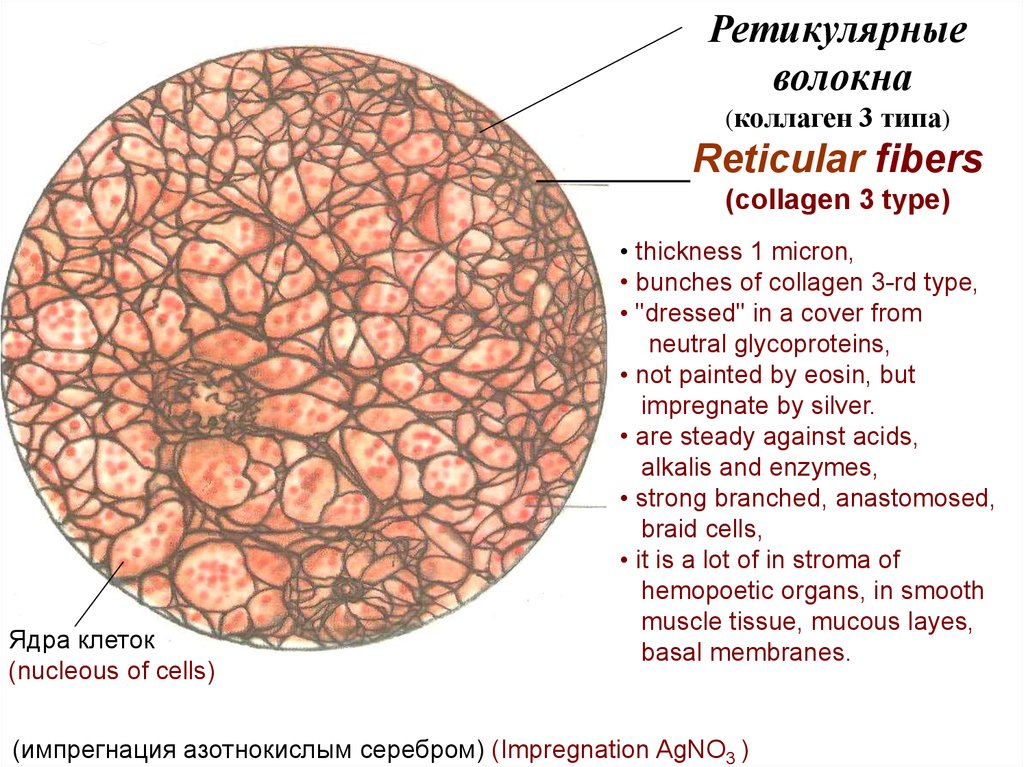
Ретикулярныеволокна
(коллаген 3 типа)
Reticular fibers
(collagen 3 type)
Ядра клеток
(nucleous of cells)
• thickness 1 micron,
• bunches of collagen 3-rd type,
• "dressed" in a cover from
neutral glycoproteins,
• not painted by eosin, but
impregnate by silver.
• are steady against acids,
alkalis and enzymes,
• strong branched, anastomosed,
braid cells,
• it is a lot of in stroma of
hemopoetic organs, in smooth
muscle tissue, mucous layes,
basal membranes.
(импрегнация азотнокислым серебром) (Impregnation AgNO3 )
14.
Эластические волокна (из белка эластина)Elastic fibers (protein elastin) • are more thin and light
Анастомозы
волокон
Anastomosis
of fibers
then collagenic fibers,
• less strong,
• are well stretched,
• rectilinear,
• branching and anastomosing.
• Are painted by orsein,
• collapse only by elastase,
• elasticity is provided by means
of derivative amino acids –
desmosin and isodesmosin.
15.
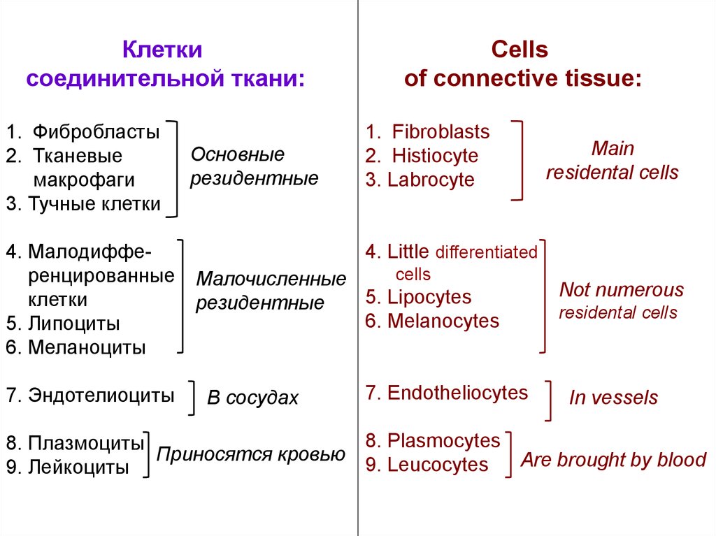
Клеткисоединительной ткани:
1. Фибробласты
2. Тканевые
макрофаги
3. Тучные клетки
4. Малодифференцированные
клетки
5. Липоциты
6. Меланоциты
7. Эндотелиоциты
Основные
резидентные
Cells
of connective tissue:
1. Fibroblasts
2. Histiocyte
3. Labrocyte
Main
residental cells
4. Little differentiated
cells
Малочисленные
5. Lipocytes
резидентные
6. Melanocytes
В сосудах
7. Endotheliocytes
Not numerous
residental cells
In vessels
8. Plasmocytes
8. Плазмоциты
Приносятся кровью
Are brought by blood
9. Leucocytes
9. Лейкоциты
16.
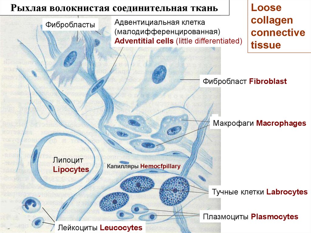
Рыхлая волокнистая соединительная тканьФибробласты
Адвентициальная клетка
(малодифференцированная)
Adventitial cells (little differentiated)
Loose
collagen
connective
tissue
Фибробласт Fibroblast
Макрофаги Macrophages
Липоцит
Lipocytes
Капилляры Hemocfpillary
Тучные клетки Labrocytes
Плазмоциты Plasmocytes
Лейкоциты Leucocytes
17.
Lymphocytes, eosinophis, basophilsprevalence at the immune answer,
neutrophils - at a acute inflammation.
Plasmocytes happens much at a chronic
inflammation, at an allergy.
Plasmocytes have:
• a dense nucleus with
arrangement of dense chromatin
in the form of spokes in a wheel,
• basophilic cytoplasm,
• well developed a rough
endoplasmic reticulum,
• a light court yard about a nucleus
(a site where KG is located).
Плазмоциты
Plasmocytes
Светлый дворик
light court yard
18.
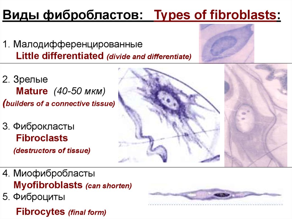
Виды фибробластов: Types of fibroblasts:1. Малодифференцированные
Little differentiated (divide and differentiate)
2. Зрелые
Mature (40-50 мкм)
(builders of a connective tissue)
3. Фиброкласты
Fibroclasts
(destructors of tissue)
4. Миофибробласты
Myofibroblasts (can shorten)
5. Фиброциты
Fibrocytes (final form)
19.
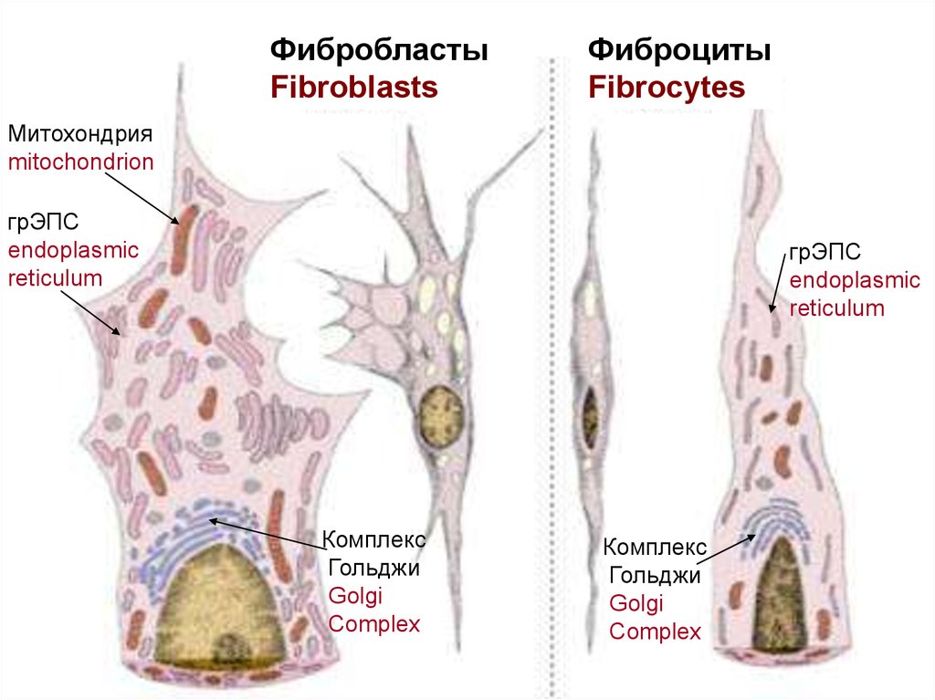
ФибробластыFibroblasts
Фиброциты
Fibrocytes
Митохондрия
mitochondrion
грЭПС
endoplasmic
reticulum
грЭПС
endoplasmic
reticulum
Комплекс
Гольджи
Golgi
Complex
Комплекс
Гольджи
Golgi
Complex
20.
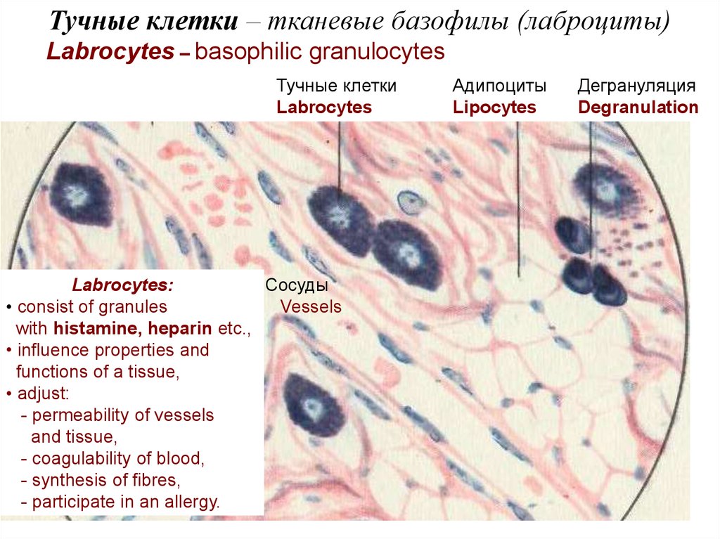
Тучные клетки – тканевые базофилы (лаброциты)Labrocytes – basophilic granulocytes
Тучные клетки
Labrocytes
Labrocytes:
Сосуды
• consist of granules
Vessels
with histamine, heparin etc.,
• influence properties and
functions of a tissue,
• adjust:
- permeability of vessels
and tissue,
- coagulability of blood,
- synthesis of fibres,
- participate in an allergy.
Адипоциты
Lipocytes
Дегрануляция
Degranulation
21.
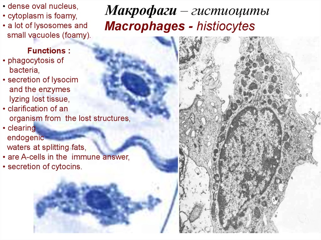
• dense oval nucleus,• cytoplasm is foamy,
• a lot of lysosomes and
small vacuoles (foamy).
Макрофаги – гистиоциты
Macrophages - histiocytes
Functions :
• phagocytosis of
bacteria,
• secretion of lysocim
and the enzymes
lyzing lost tissue,
• clarification of an
organism from the lost structures,
• clearing
endogenic
waters at splitting fats,
• are A-cells in the immune answer,
• secretion of cytocins.
22.
Бактерии (bacteria)Псевдоподии макрофага
Pseudopodia of macrophage
23. Мононуклеарная фагоцитарная система
Гистиоциты
Альвеолярные макрофаги лёгких
Звёздчатые клетки Купфера печени
Береговые клетки лимфоузлов
Макрофаги ретикулярной ткани
Перитониальные макрофаги
Гигантские клетки инородных тел
Остеокласты
Микроглия нервной ткани
Клетки мезангиума почек
Клетки Лангерганса в эпидермисе
Mononuclear
phagocytic system
Histiocytes
Alveolar lung macrophages
Stellate Kupffer’s cells of a liver
Reticular cells of lymphatic
node sinus
Macrophages of reticular tissue
Peritoneal macrophages
Gigantic cells of alien bodies
Osteoclasts
Microglia of nerve tissue
Renal mesangium cells
Langergans’s cells of epidermis