


")


")


 medicine
Similar presentations:


")


Leishmaniasis. Department of Infectious Diseases Leishmaniasis
1. Leishmaniasis
Department of Infectious DiseasesLeishmaniasis
Professor Kutmanova A.Z.
2.
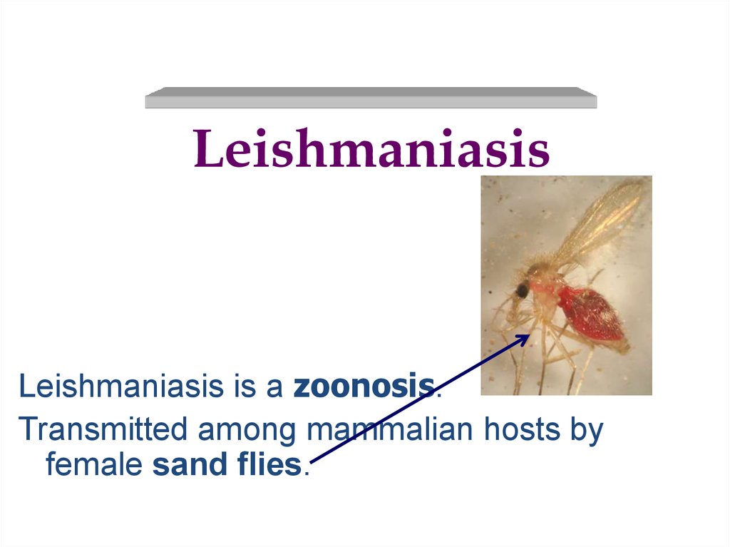
LeishmaniasisLeishmaniasis is a zoonosis.
Transmitted among mammalian hosts by
female sand flies.
3. Leishmaniasis
Species Pathogenic in HumansLeishmania donovani (complex) (VL)
Leishmania tropica (CL)
Leishmania major (CL)
Leishmania aethiopica (CL)
Leishmania mexicana (Complex) (CL)
Leishmania brazilliensis (complex) (MCL)
4. Three important Species
Leishmania donovani (VL )VISCERAL LEISHMANIASIS : involving endothelial tissue liver,
spleen, and bone marrow.
Leishmania tropica (CL)
OLD WORLD CUTANEOUS LEISHMANIASIS
: involving epithelial
cells the skin at the site of a sand fly bite.
Leishmania brazilliensis (MCL)
NEW WORLD MUCO CUTANEOUS LEISHMANIASIS
: involving
mucous membranes of the mouth and nose after
spread from a nearby cutaneous lesion.
5.
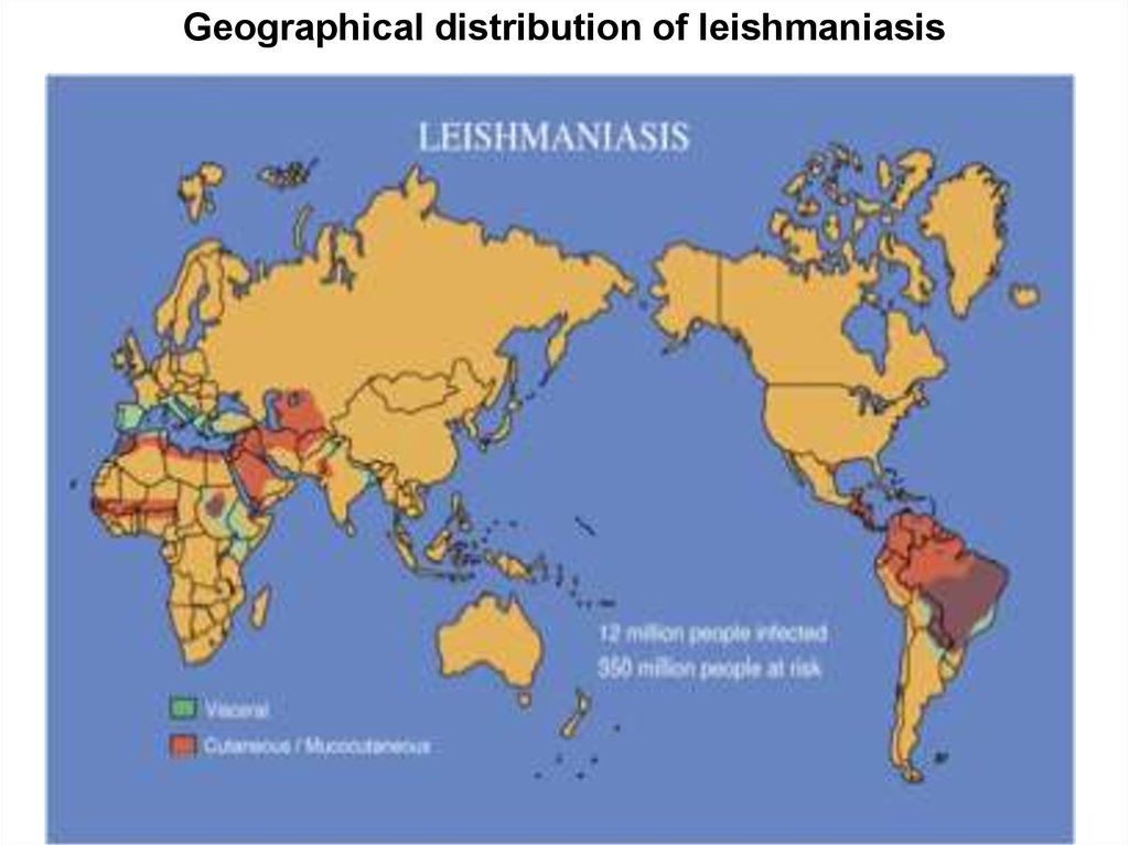
Geographical distribution of leishmaniasis6.
7.
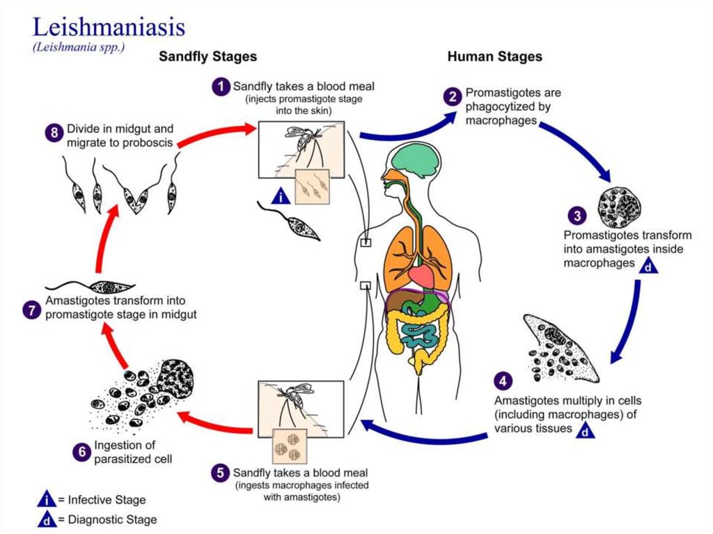
8. Life Cycle of leishmaniasis
PromastigoteAmasitgote
Transformation
9.
Promastigote stagePromastigote stage inside the Sandfly
flagella
Sand fly : Vectors Intermediate
host, transmitted disease
10.
Promastigotes inrosettes in a culture of
an orient sore on N.N.N.
medium (Giemsa stain).
11.
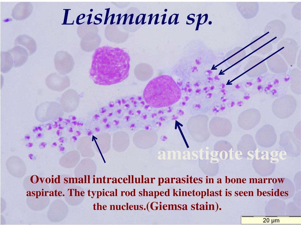
Leishmania sp.amastigote stage
Ovoid small intracellular parasites in a bone marrow
aspirate. The typical rod shaped kinetoplast is seen besides
the nucleus.(Giemsa stain).
12. Life cycle
13.
14.
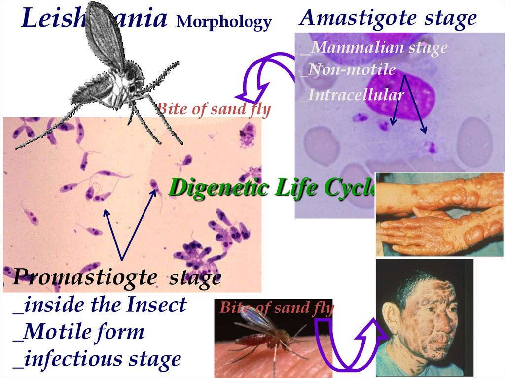
Leishmania Morphology Amastigote stage_Mammalian stage
_Non-motile
Bite of sand fly
_Intracellular
Digenetic Life Cycle
Promastiogte stage
_inside the Insect
_Motile form
_infectious stage
Bite of sand fly
15. Transmission of Leishmaniasis
_ by sand flies._ artificial transmission of leishmania
via the sharing of contaminated
syringes and needles, from one
intravenous drug user to another.
Rarely, Leishmaniasis is spread from
a pregnant woman to her baby
(Materno-fetal transplacental
transmission).
Blood transfusion or contaminated
needles also can spread
Leishmaniasis.
16. Cutaneous Leishmaniasis
Cutaneous forms of thedisease normally produce
skin ulcers on the exposed
parts of the body such as
the face, arms and legs. The
disease can produce a large
number of lesions
17. A cutaneous leishmaniasis lesion on the arm.
Some people have swollenlymph glands near the sores.
For example, the glands
under the arm can swell if
the sores are on the arm or
hand.
The skin sores will heal by
themselves, but this can take
months or years. The sores can
leave ugly scars.
18. Cutaneous Leishmaniasis
19. Baghdad-boil, 2004
The Baghdad boilBaghdad-boil, 2004
Several hundred US soldiers
in Iraq.
20. Leishmania tropica
Leishmania• Causes ulceration of the skin
called Cutaneous Leshmaniasis
tropica• Dry or urban C.L.
• Dry sore that may persist for
several months before healing,
then person is immune
• Some people “vaccinate” their
children against Leshmaniasis.
• Rarely can cause infections of
the viscera
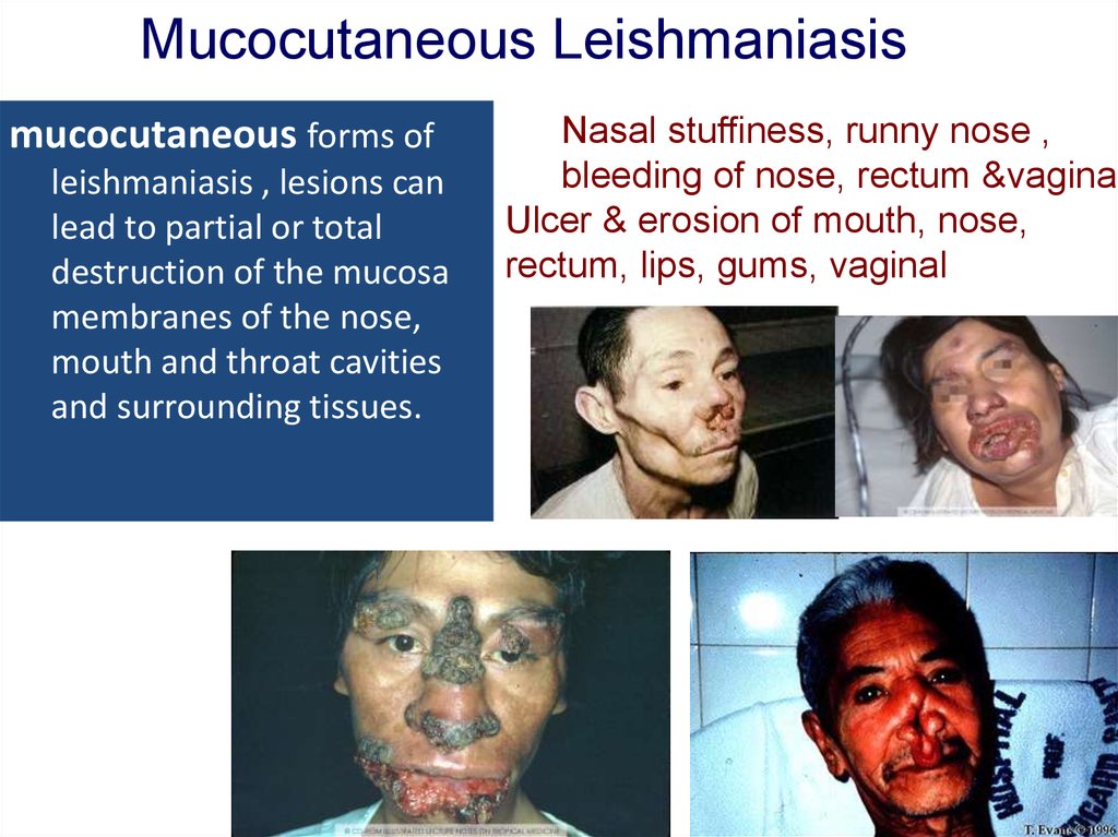
21. Mucocutaneous Leishmaniasis
Mucocutaneous leishmaniasis (Espundia)Leishmania braziliensis &
L . maxicana
22.
Mucocutaneous Leishmaniasismucocutaneous forms of
leishmaniasis , lesions can
lead to partial or total
destruction of the mucosa
membranes of the nose,
mouth and throat cavities
and surrounding tissues.
Nasal stuffiness, runny nose ,
bleeding of nose, rectum &vagina.
Ulcer & erosion of mouth, nose,
rectum, lips, gums, vaginal
23. Visceral Leishmaniasis
Visceral disease (Kala-azar)24. Visceral disease (Kala-azar)
Most severe form of disease, the disease typically starts with irregularbouts of fever, chills, and general anemia
Since leishmaniasis is primarily a disease of the reticulo-endothelial system,
replacement of infected cells produces hyperplasia and consequent enlargement of
the visceral organs associated with the system (e.g., spleen and liver) .
Hepatosplenomegaly
25. Post Kala Azar Dermal Leishmanoid
Normally develops <2 years after recoveryRestricted to skin, rare but varies
geographically
• Some people recover spontaneously
• Some people who were treated later develop
Post-Kala- azar dermal leishmanoid
26. Hepatosplenomegaly
Post Kala AzarDermal
Leishmanoid
27.
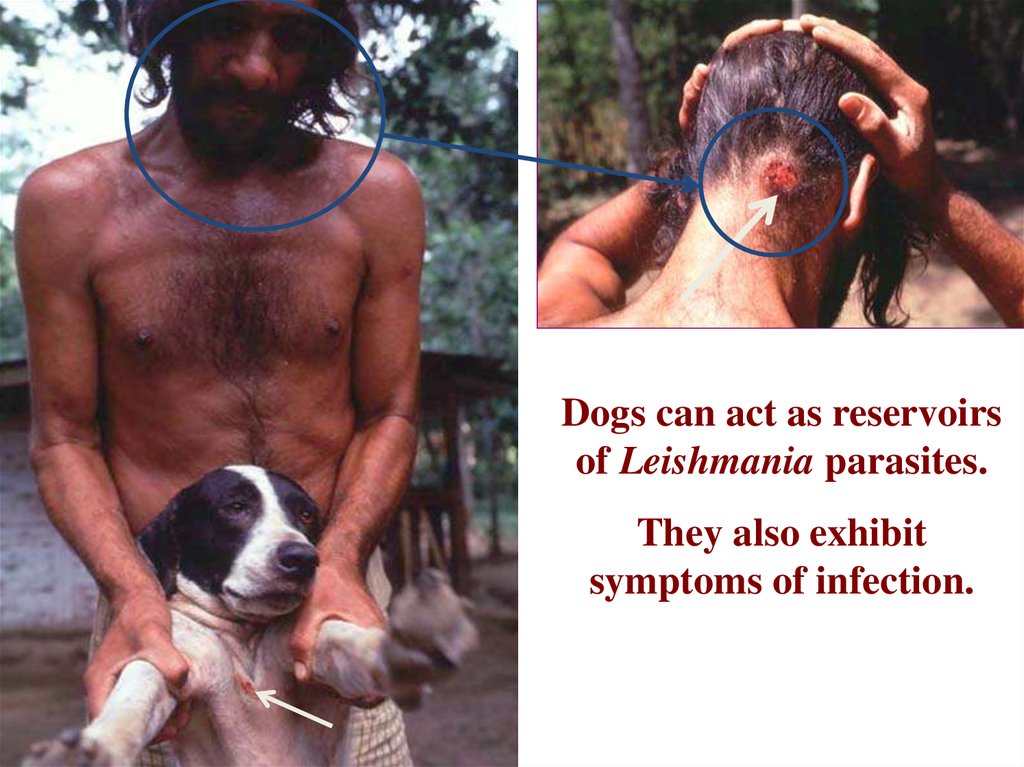
Dogs can act as reservoirsof Leishmania parasites.
They also exhibit
symptoms of infection.
28. Diagnosis
Diagnosing Leishmaniasis can bedifficult Sometimes the Lab tests are
negative even if a person has
Leishmaniasis.
29. Diagnosis
1. Clinical Diagnosis: signs & symptomsPatient history (travel, vectors)
2. Laboratory Diagnosis :
30.
Laboratory Diagnosis of leishmaniasis :Cutaneous leishmaniasis :
– Tissue sample (scraping, aspirate or punch biopsy) for
smear and culture
Visceral leishmaniasis :
– Bone marrow biopsy or splenic aspirate for smear and
culture.(N.N.N) V.L.(anemia , leukopenia ,
glubuline/albumine is high (Hypergammaglobulinia)
– Serology ( ELISA ) ( IFAT ).
– PCR
– Skin test
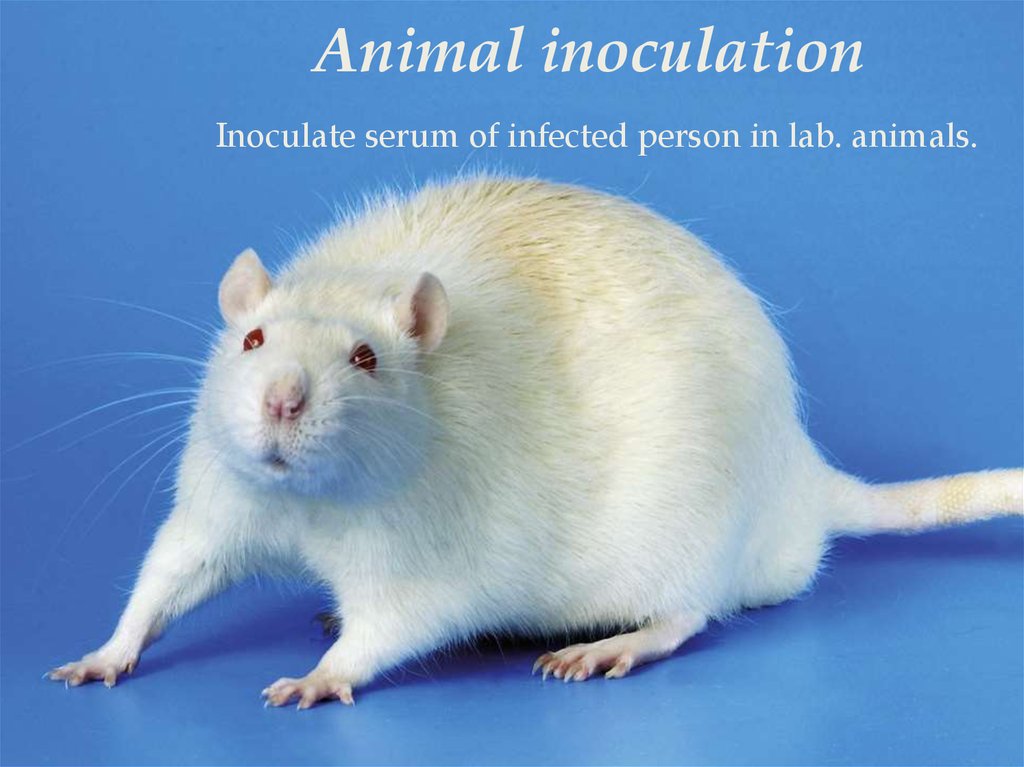
– Inoculate serum of infected person in lab. animals.
31.
Animal inoculationInoculate serum of infected person in lab. animals.
32. Cutaneous and mucocutaneous treatment
• Antimony components : Meglumine antimoniate(Glucantime) and Sodium stibogluconate (Pentostam) are
drugs of choice.
– 20 mg/kg/d IV or IM for 20d
- Pentamidine, Paromomycin are alternative drugs for CL
- Amphotricine B for antimony resistant MCL
• Fluconazole may decrease healing time
33. Visceral leishmaniasis treatment
Pentostam or Glucantime 20 mg /kg/d IV or IM for 28d
Amphotricin B: 0.5-1 mg/kg IV daily 15-20d
Liposomal Amphotricin B (Ambisome): 3 mg/kg/d IV on days 1-5, day
14 and day 21
– Low toxicity and high stability, better delivery
• Alternative: Pentamidine (4mg/kg three times weekly, between 5-25 weeks
), Parmomycine
34. Visceral leishmaniasis treatment (con.)
• Miltefosine (Impavido) (2.5 mg/kg /d p.o. for 28 d)– It was developed for cancer therapy at first
– The only oral drug
– safer and more tolerable drug (less toxicity for bone
marrow and haematopoietic progenitor cells)
– teratogenic
35.
36. Leishmaniasis control
• Vector control• Decrease of susceptibility:
– insecticides
Childhood age, malnutrition and
– insecticide impregnated bed
Immunosuppression are
susceptibility factors for VL.
nets (IIB)
– eliminating of childhood
• Case finding treatment
malnutrition
• Aniaml reservoir control
– try to produce an efficient
– Treatment or killing of
vaccine
seropositive dogs
– Rodent killing