


 medicine
Similar presentations:


Medical protozology
1. MEDICAL PROTOZOLOGY
2.
Protozoa (singular, protozoan), from the Greek‘protos’ and ‘zoon’ meaning “first animal”, are members
of eukaryotic protists.
They may be distinguished from other eukaryotic
protists by their ability to move at some stage of their life
cycle and by their lack of cell wall.
3.
Occurrence of protozoaProtozoa are found in all moist habitats. They are
common in sea, in soil and in fresh water.
These organisms occur generally as a single cell.
Colonies of protozoa might also occur in which
individual cells are joined by cytoplasmic threads and
form aggregates of independent cells.
However, distinct types of protozoa, include a
resistant cyst (non-motile) stage to survive adverse
environmental conditions, such as desiccation, low
nutrient supply, and even anaerobiosis.
4.
Morphology of protozoaProtozoa are predominantly microscopic, ranging in size from 2
to more than 100μm.
Morphologically, they are within a mass of protoplasm,
consisting of a true membrane – bound nucleus and cytoplasm.
The nucleus contains clumped or dispersed chromatin and
central nucleolus or karyosome, which are useful structures to
distinguish protozoan species from one another based on the shape,
size and distribution of these structures.
Reproduction and regeneration of protozoa
As a general rule, protozoa multiply by asexual reproduction.
This is not to say that sexual processes are absent in the protozoa.
Some parasitic forms may have an asexual phase in one host and a
sexual phase in another host.
5.
Importance of protozoaProtozoa serve as an important link in the food chain
and ecological balance of many communities in wetland
& aquatic environments.
They are also important in biological sewage
treatment, which involves both anaerobic digestion
and/or aeration.
In addition, protozoa are important laboratory
organisms in research areas, by which their asexual
reproduction enables clones to be established with the
same genetic make-up.
These are useful in the study of cell cycles and
nucleic acid biosynthesis during cell division.
6.
TransmissionIn most parasitic protozoa, the developmental stages are
often transmitted from one host to another within a cyst.
The reproduction process is also related to the formation of
the cyst. Asexual reproduction of some ciliates and
flagellates is associated with cyst formation, and sexual
reproduction of Sporozoa invariably results in a cyst.
Pathogenic protozoa can spread from one infected person to
another by:
• Faecal – oral transmission of contaminated foods and
water.
• Insect bit inoculums or rubbing infected insect faeces
on the site of bite.
• Sexual intercourse
7.
PathogenesisProtozoan organisms are virtually always acquired from an
exogenous source, and as such, they have evolved numerous ways
to enter the body of the human host. Factors that are important for
pathogenecity include:
• Attachment to the host tissue followed by replication to
establish colonization.
• Toxic products released by parasitic protozoa.
• Shifting of antigenic expression to evade the immune response
and inactivate host defenses.
8.
Classification of ProtozoaProtozoa of medical importance are classified based
on their morphology and locomotive system as described
below:
Amoebas - Entamoeba histolytica (Amoebiasis)
Flagellates - Giarda lamblia (Giardiasis or
lambliosis), Trichomonas vaginalis (Trichomoniasis),
Trypanosoma spp (Tripanosomiasis), Leishmania spp.
(Leishmaniasis)
Ciliates - Balantidium coli (Balantidiasis)
Sporozoa (Coccidian) - Toxoplasma gondii
(Toxoplasmosis), Plasmodium spp. (Malaria).
9. 1. Parasitic amoeba
10.
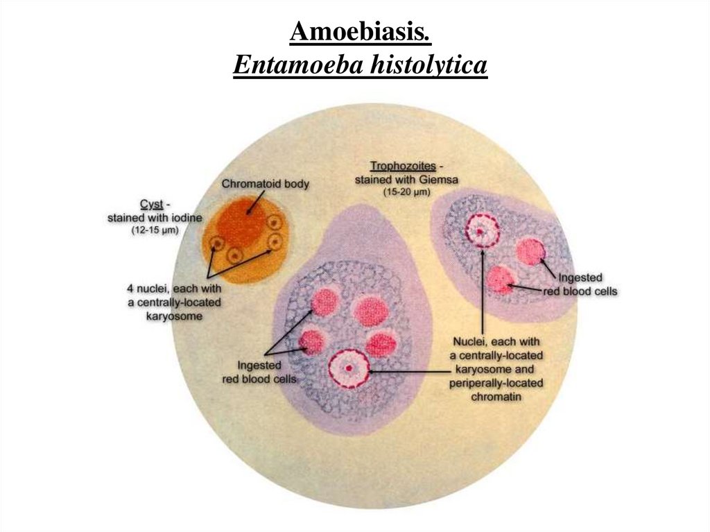
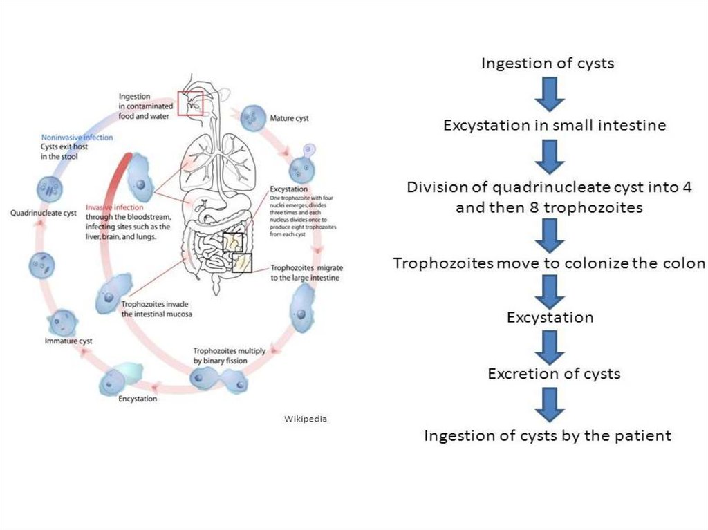
Amoebiasis.Entamoeba histolytica
11.
12.
Symptoms:• Abdominal pain,
• Mild diarrhea, bloody diarrhea,
• Perforation and tissue death. This last
complication may cause peritonitis.
• People affected may develop anemia due to loss
of blood, weakness
13.
Invasion of the intestinal lining causes amoebic bloodydiarrhea or amoebic colitis. If the parasite reaches the bloodstream
it can spread through the body, most frequently ending up in the
liver where it causes amoebic liver abscesses.
Disease occurs when amoeba comes in contact with the cells
lining the intestine. It then secretes the same substances it uses to
digest bacteria, which include enzymes that destroy cell
membranes and proteins. This process can lead to penetration and
digestion of human tissues, resulting first in flack-shaped ulcers in
the intestine.
Entamoeba histolitica ingests the destroyed cells by
phagocytosis and is often seen with red blood cells inside when
viewed in stool samples.
14.
Transmission:• It is usually transmitted by fecal-oral route, but it can
also be transmitted indirectly through contact with dirty
hands or objects as well as by anal-oral contact.
• Infection is spread through ingestion of the cyst form
of the parasite. This form found in feces. Trophozoites
may also be present in stool. These are rarely the source
of new infections.
• Contaminated food (vegetables and fruits) and water.
• Soil cultivation.
15.
Diagnosis:• Microscopy of feces
• Serological tests. Serology becomes positive about 2
weeks after invasion.
Treatment:
Entamoeba histolitica infections occur in both the
intestine and in tissue of the intestine and/or liver. As a
result, two different classes of drugs are needs to treat
the infection, one for each location. Such anti-amoebic
drugs are known as amoebicides.
16.
Prevention:• Wash hands with soap and hot running water
• Clean bathrooms and toilets often
• Avoid sharing towels or face washers
• Clean vegetables and fruits
• Boil water or treat with iodine tablets
• Avoid eating street foods especially in public places
where others are sharing sauces in one container.
• Cysts are usually resistant to chlorination; therefore,
sedimentation and filtration of water supplies are necessary
to reduce the incidence of infection.
17. 2. Parasitic ciliates
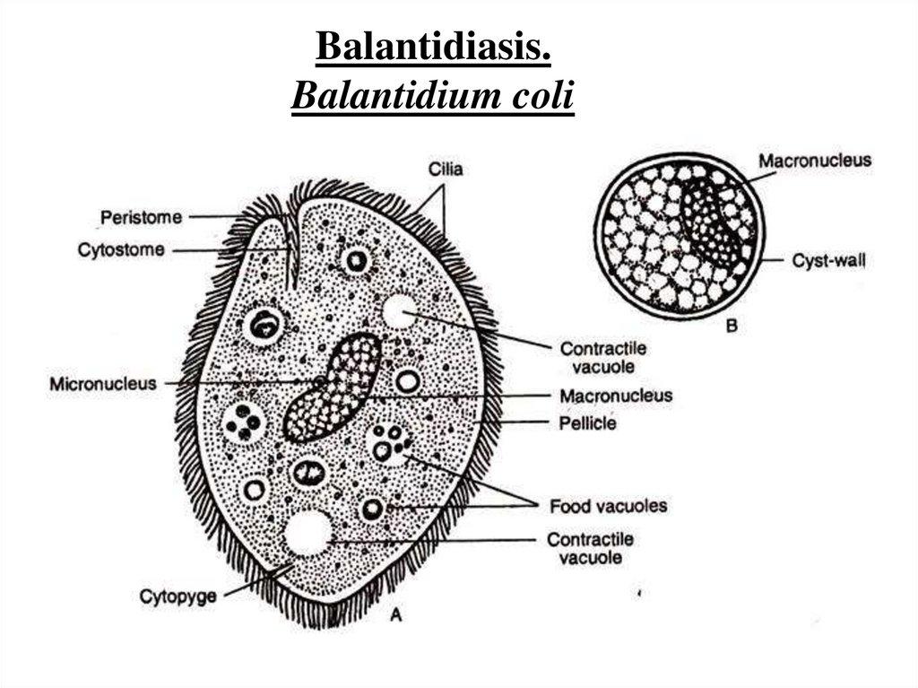
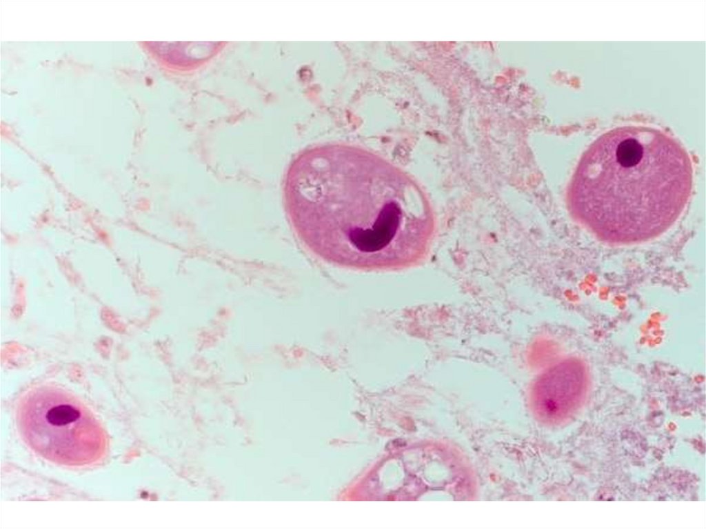
18.
Balantidiasis.Balantidium coli
19.
20.
21.
Symptoms:Can be local due to involvement of the intestinal
mucosa, systemic in nature and include either diarrhea or
constipation
• Frequent loose muco-purulent, then bloody stools
• Tenesmus
• Pain in the colon, formation of ulcers
• Loss of appetite
• Nausea
• General weakness, sometimes increasing temperature
• Peritonitis
22.
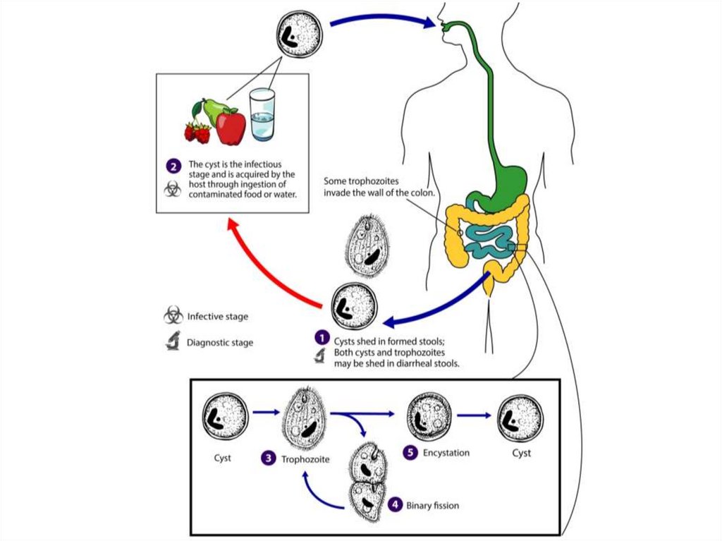
Transmission:The main source of infection is the pig. But man,
releasing ciliates can infect others. Cysts under favorable
conditions persist in the feces for several weeks and
unable to get into the human digestive tract with
contaminated food, water, vegetables and also via
contaminated hands. Flies can also carry the cysts.
Diagnosis:
Microscopic examination of stool
Treatment:
Metronidazole
23.
Prevention:• Is the same as in other intestinal infections: measures
that prevent pollution of the environment by feces of
humans and pigs. In pig farms the need for good
maintenance of pigs, cleaning pig, composting of feces.
Special attention should be given to the timely
identification and treatment of carriers and patients with
this infection.
• Purification of drinking water
• Proper handling of food
• Careful disposal of humans feces
• Monitoring the contacts of balantidiasis patients
24.
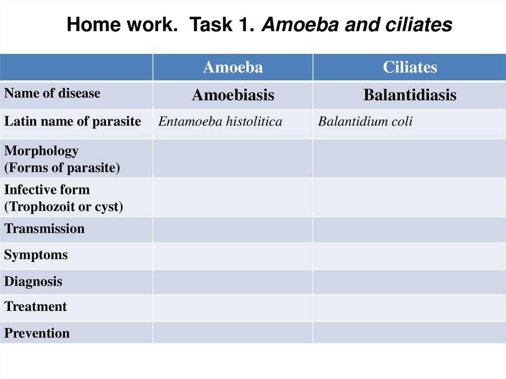
Home work. Task 1. Amoeba and ciliatesName of disease
Latin name of parasite
Morphology
(Forms of parasite)
Infective form
(Trophozoit or cyst)
Transmission
Symptoms
Diagnosis
Treatment
Prevention
Amoeba
Ciliates
Amoebiasis
Balantidiasis
Entamoeba histolitica
Balantidium coli
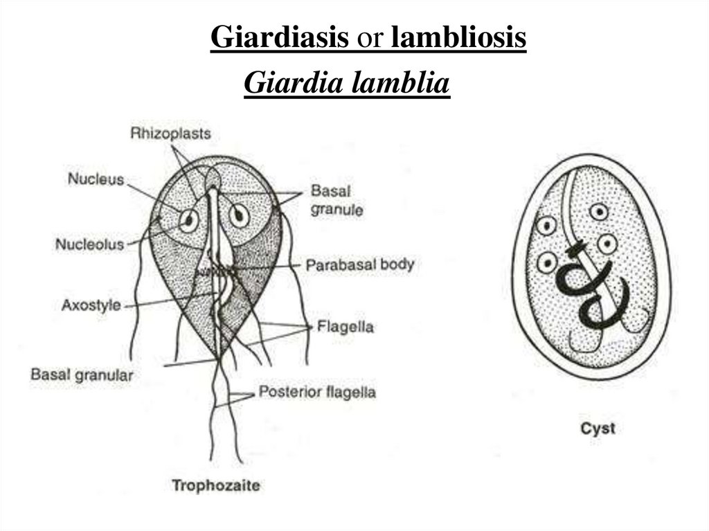
25. 3. Parasitic Flagellates: 3.1. Intestinal and vaginal Flagellates
26.
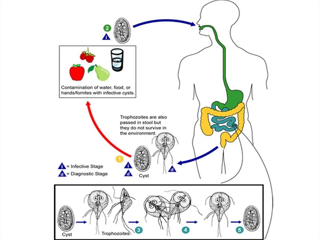
Giardiasis or lambliosisGiardia lamblia
27.
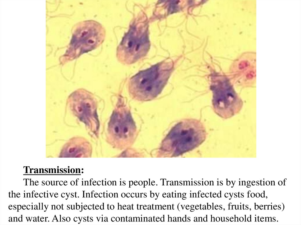
Transmission:The source of infection is people. Transmission is by ingestion of
the infective cyst. Infection occurs by eating infected cysts food,
especially not subjected to heat treatment (vegetables, fruits, berries)
and water. Also cysts via contaminated hands and household items.
28.
29.
Symptoms:The main habitat of giardia in human body are duodenum and
initial part of colon. Parasites attached to the villi of the mucous
membrane of the small intestine and, apparently, eat food parietal
digestion.
Mechanical irritation of interoreceptors Giardia the small
intestine can lead to reflex disturbance of its function, which
apparently can be cause of a violation of the physiology of
digestion.
Diarrhea, smelling gas, headaches, weight loss, fatigue, loss of
appetite, fell sick in general, abdominal pain, particularly
cramping, bloating, nausea with/or without vomiting, malaise.
Fever is unusual.
30.
Diagnosis:• Antigene testing of the stool for the presence of giardial
proteins
• Examination of stool under microscope for cyst or trophzoites
• Examination of fluid from the duodenum
• Biopsy of the small intestine
Treatment:
Antiparasitic drugs – metronidazole, tinidazole, paromomycin
Prevention:
• Washing hands
• Don’t swallow water from rivers and lakes
• Drink clean water, don’t drink tap water
• Avoid eating uncooked food. Clean vegetables, fruits and
berries before eating.
31.
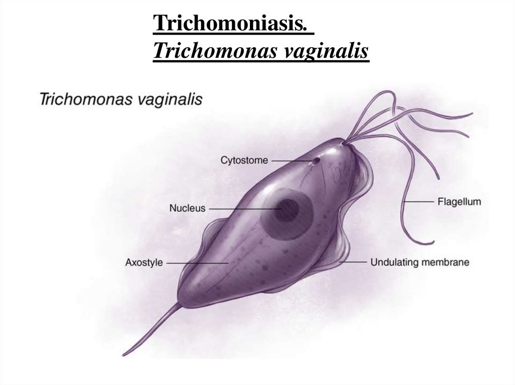
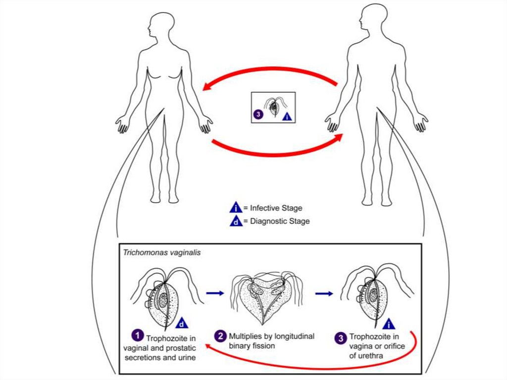
Trichomoniasis.Trichomonas vaginalis
32.
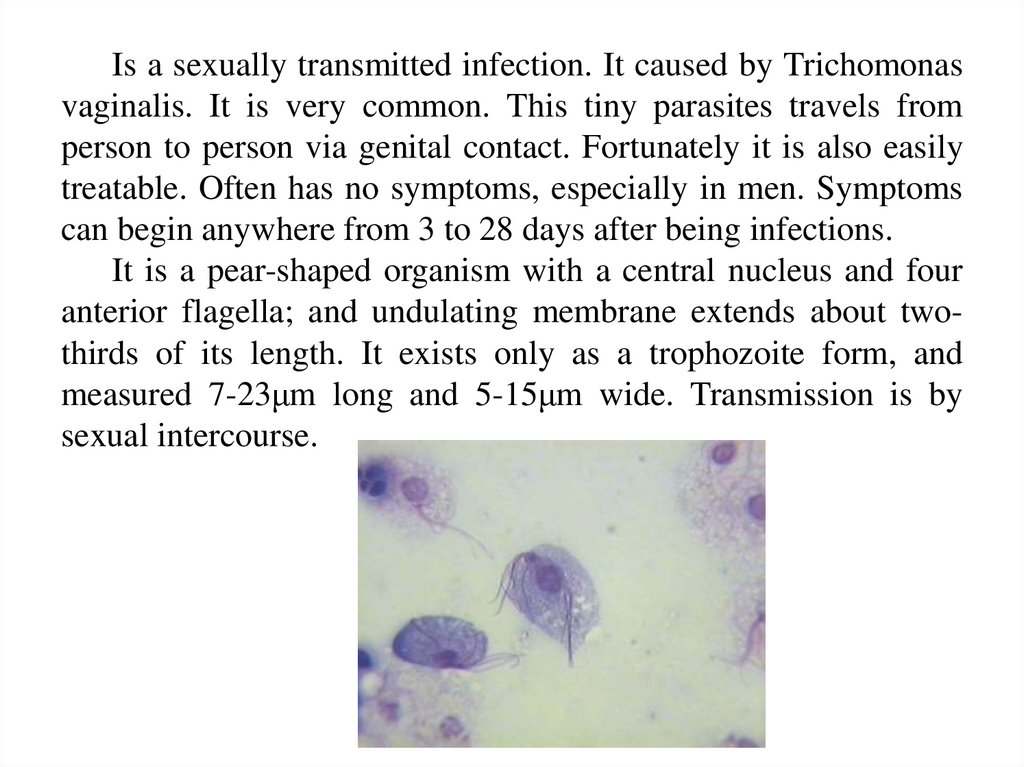
Is a sexually transmitted infection. It caused by Trichomonasvaginalis. It is very common. This tiny parasites travels from
person to person via genital contact. Fortunately it is also easily
treatable. Often has no symptoms, especially in men. Symptoms
can begin anywhere from 3 to 28 days after being infections.
It is a pear-shaped organism with a central nucleus and four
anterior flagella; and undulating membrane extends about twothirds of its length. It exists only as a trophozoite form, and
measured 7-23μm long and 5-15μm wide. Transmission is by
sexual intercourse.
33.
34.
Symptoms:• Vaginal discharge, which may be white, gray, yellow or
green and usually has unpleasant smell.
• Genital redness or swelling
• Pain during urination or sexual intercourse.
• An urge to urinate frequently
• Discharge from the urethra
• Burning during urination
• An urge to urinate frequently
35.
Diagnosis:Physical test and laboratory test: cell culture, DNA
examing samples of vaginal fluids.
Treatment:
Metronidazole is the drug of choice. If resistant
cases occur, re-treatment with higher doses is required.
Prevention:
- Both male and female sex partners must be treated
to avoid reinfection
- Good personal hygiene, avoidance of shared toilet
articles and clothing.
- Safe sexual practice.
36.
Home work. Task 2.Amoeba, Ciliates, intestinal and vaginal flagellates
Name of disease
Amoebiasis
Balantidiasis
Giardiasis
Trichomoniasis
Latin name of
parasite
Entamoeba
histolitica
Balantidium
coli
Giardia
lamblia
Tricomonis
vaginalis
Morphology
(Forms of parasite)
Infective form
(Trophozoit or cyst)
Transmission
Symptoms
Diagnosis
Treatment
Prevention
37.
3. Parasitic Flagellates:3.2. Hemoflagellates
38.
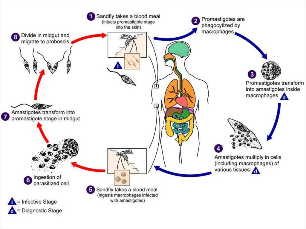
Leishmaniasis.
Is a disease caused by protozoan parasites of the genus
Leishmania and spread by the bite of certain types of sandflies.
Definitive host: human
Intermediate host: sandfly
Symptoms:
The disease can present in three main ways: cutaneous,
mucocutaneous, or visceral.
1. The cutaneous form presents with skin ulcers,
2. while the mucocutaneous form presents with ulcers of the skin,
mouth, and nose.
3. Visceral form starts with skin ulcers and then later presents with
fever, low red blood cells, and enlarged liver or spleen.
39.
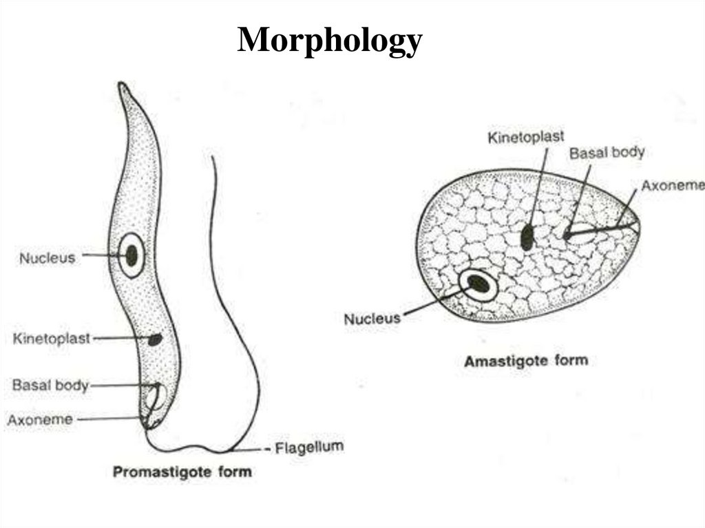
Morphology40.
41.
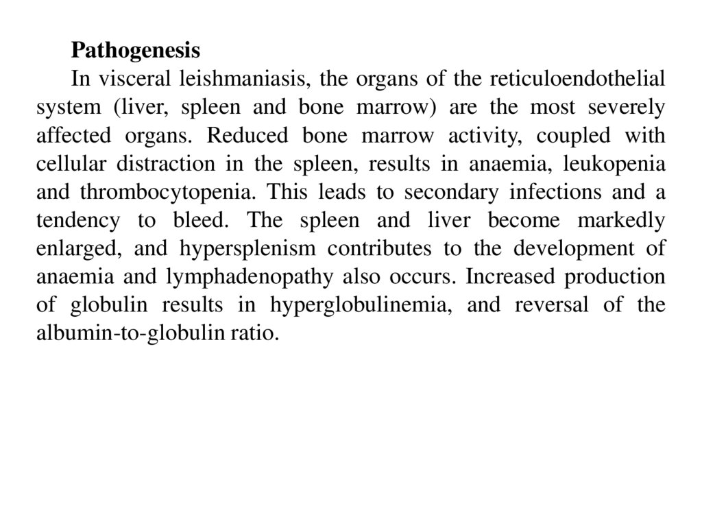
PathogenesisIn visceral leishmaniasis, the organs of the reticuloendothelial
system (liver, spleen and bone marrow) are the most severely
affected organs. Reduced bone marrow activity, coupled with
cellular distraction in the spleen, results in anaemia, leukopenia
and thrombocytopenia. This leads to secondary infections and a
tendency to bleed. The spleen and liver become markedly
enlarged, and hypersplenism contributes to the development of
anaemia and lymphadenopathy also occurs. Increased production
of globulin results in hyperglobulinemia, and reversal of the
albumin-to-globulin ratio.
42.
Clinical featuresSymptoms begin with intermittent fever, weakness, and
diarrhea; chills and sweating that may resemble malaria symptoms
are also common early in the infection. As organisms proliferate
and invade cells of the liver and spleen, marked enlargement of the
organs, weight loss, anemia, and emaciation occurs. With
persistence of the disease, deeply pigmented, granulomatous lesion
of skin, referred to as post-kala-azar dermal leishmaniasis, occurs.
Untreated visceral leishmaniasis is nearly always fatal as a result of
secondary infection.
43.
44.
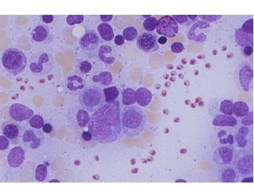
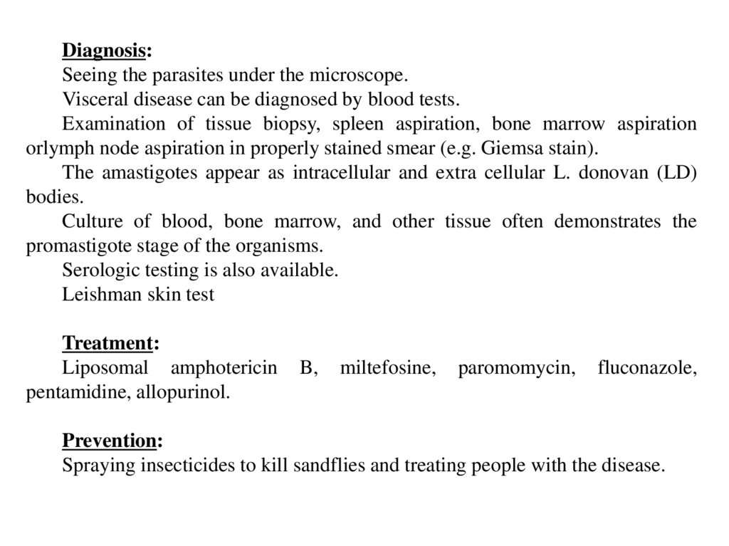
Diagnosis:Seeing the parasites under the microscope.
Visceral disease can be diagnosed by blood tests.
Examination of tissue biopsy, spleen aspiration, bone marrow aspiration
orlymph node aspiration in properly stained smear (e.g. Giemsa stain).
The amastigotes appear as intracellular and extra cellular L. donovan (LD)
bodies.
Culture of blood, bone marrow, and other tissue often demonstrates the
promastigote stage of the organisms.
Serologic testing is also available.
Leishman skin test
Treatment:
Liposomal amphotericin
pentamidine, allopurinol.
B,
miltefosine,
paromomycin,
fluconazole,
Prevention:
Spraying insecticides to kill sandflies and treating people with the disease.
45.
TrypanasomiasisThis disease caused by Tripanosoma species. In humans this
include African and South-American types.
Trypanosoma brucei complex – African trypanosomiasis
(sleeping sickness)
Trypanosoma cruzi – American trypanosomiasis (Chagas’
disease)
These species may have amastigote, promastigote, epimastigote,
and trypomastigote stages in their life cycle. In human trypanosomes
of the African form, however, the amastigote and promastigote
stages of development are absent.
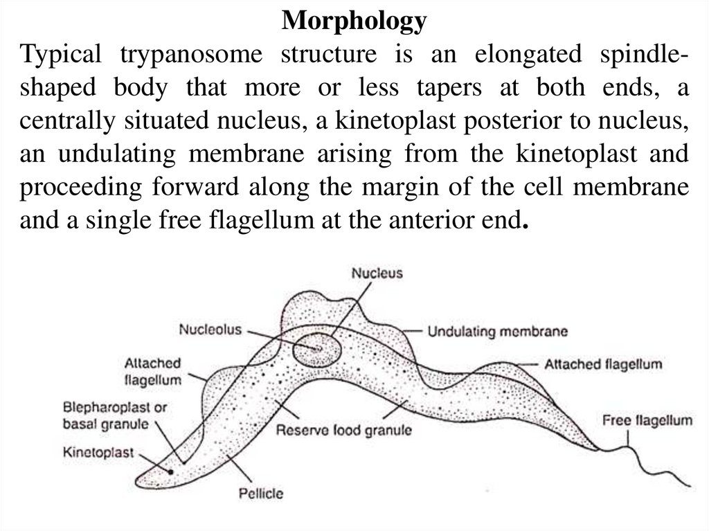
46.
MorphologyTypical trypanosome structure is an elongated spindleshaped body that more or less tapers at both ends, a
centrally situated nucleus, a kinetoplast posterior to nucleus,
an undulating membrane arising from the kinetoplast and
proceeding forward along the margin of the cell membrane
and a single free flagellum at the anterior end.
47.
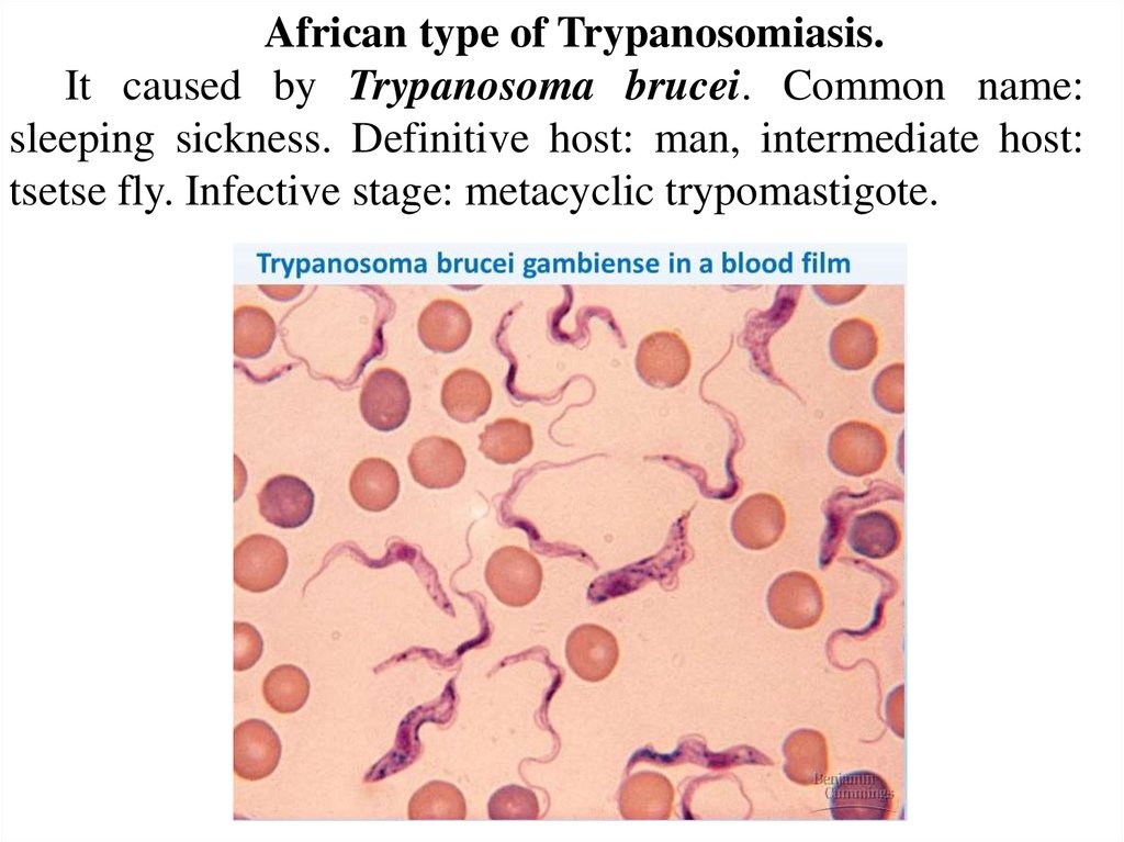
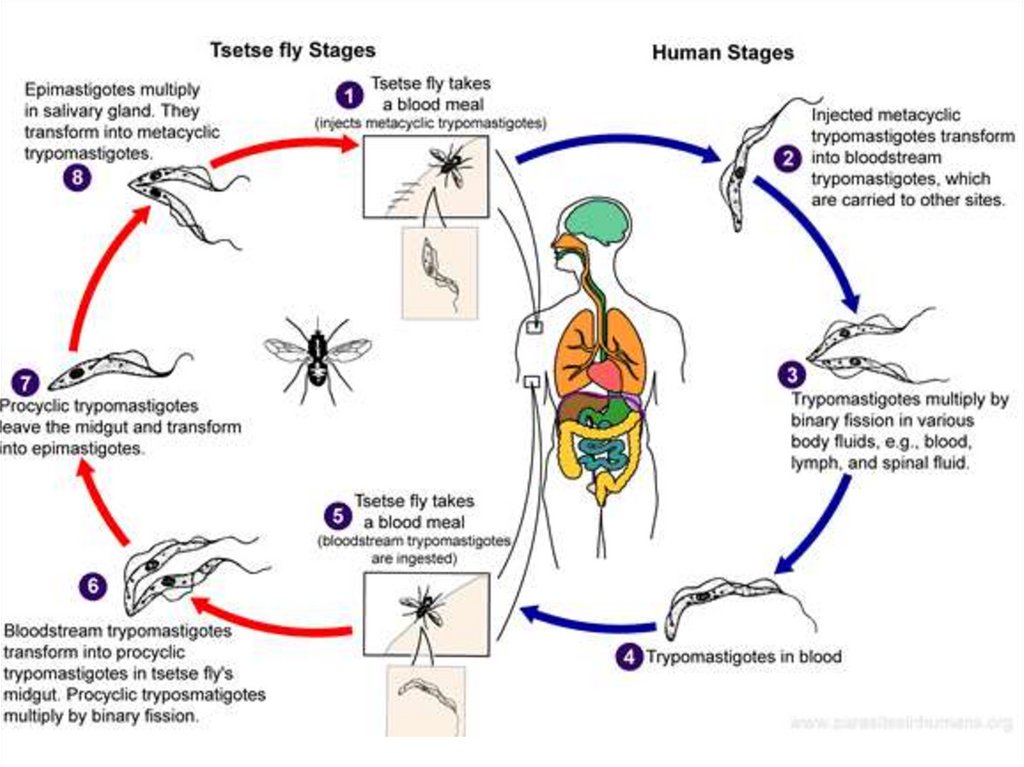
African type of Trypanosomiasis.It caused by Trypanosoma brucei. Common name:
sleeping sickness. Definitive host: man, intermediate host:
tsetse fly. Infective stage: metacyclic trypomastigote.
48.
SymptomsTransmission by tsetse fly.
The tsetse fly bite erupts into a red chancre sore and within a
few weeks, the person can experience fever, swollen lymph glands,
blood in the urine, aching muscles and joints, headaches and
irritability. In first phase, the patient has only intermittent bouts of
fever with lymphadenopathy together with other non-specific signs
and symptoms.
The second and third stage is marked by involvement of the
central nervous system with extensive neurological effects like
changes in personality, alteration of the biological clock, confusion,
slurred speech, seizures and difficulty in walking and talking. Also
general toxic symptoms, anemia, bone pain. These problems can
develop over many years and if not treated, the person dies.
49.
50.
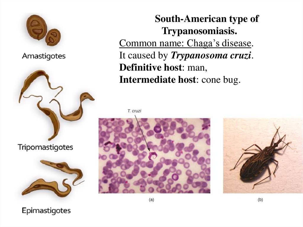
South-American type ofTrypanosomiasis.
Common name: Chaga’s disease.
It caused by Trypanosoma cruzi.
Definitive host: man,
Intermediate host: cone bug.
51.
52.
Symptoms of an acute form:
Blood and reticulo-endothelial cells predominantly involved
Fever
Oedema (lymph blockage)
Lymphadenopathy
Enlargement of liver and spleen
Sometimes encephalitis
Symptoms of chronic form:
• General toxic symptoms and focal signs depending on localization
(toxic depression of bone marrow, anemia, hepatomegaly,
splenomegaly, lymphadenopathy)
• Predominantly cardiac and CNS manifestations (myocarditis,
tachycardia, heart block, encephalitis, general or focal CNS signs and
symptoms)
• May be asymptomatic.
53.
Diagnosis:Often missed in the first phase of the disease due to non-specific nature of
symptoms. Examine thin or thick stained preparations for trypomastigotes. Wet
preparations should also be examined to look for motile organisms that leave the
blood stream and become difficult to find. Biopsy of lymph nodes, liver, spleen,
or bone marrow may demonstrate organisms in amastigote stage. Xenodiagnosis which consists of allowing an uninfected, laboratory-raised reduviid bug to feed
on the patient and, after several weeks, examining the intestinal contents of the
bug for the organism.
Treatment:
Pentamidine and suramin are used for treatment in the first stage.
Melarsoprol, nifurtimox and eflornithine are used in the second stage.
Prevention:
Spraying insecticides, repellents, using mosquito nets.
Treating infected person and exclusion of donors by screening blood.
Development of vaccine.
54.
Home work. Task 3. HemoflagellatesName of disease
Latin name of
parasite
Morphology
(Forms of
parasite)
Infective form
Transmission
Symptoms
Diagnosis
Treatment
Prevention
Leishmaniasis
Trypanosomiasis
Leishmania donovani Trypanosoma brucei
Trypanosoma cruzi
55.
4. Sporozoa56.
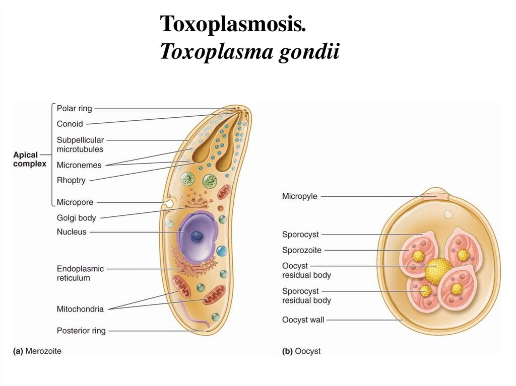
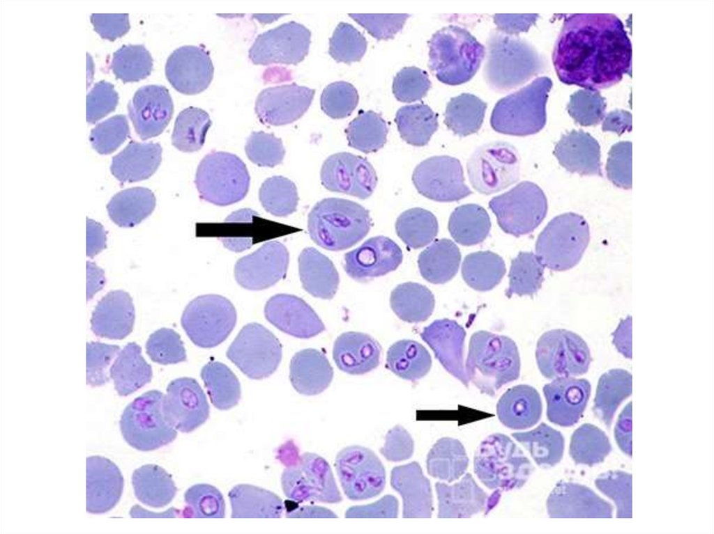
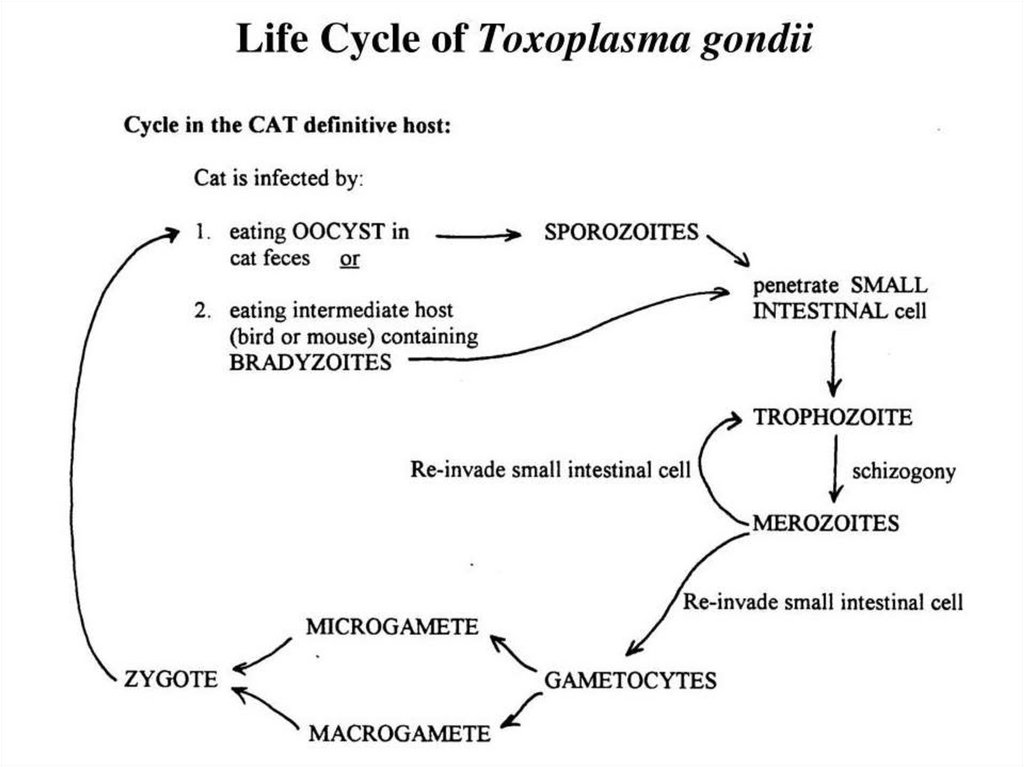
Toxoplasmosis.Toxoplasma gondii
57.
58.
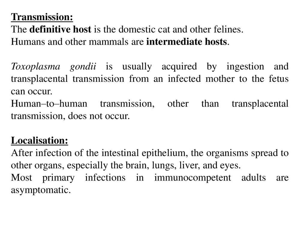
Transmission:The definitive host is the domestic cat and other felines.
Humans and other mammals are intermediate hosts.
Toxoplasma gondii is usually acquired by ingestion and
transplacental transmission from an infected mother to the fetus
can occur.
Human–to–human transmission, other than transplacental
transmission, does not occur.
Localisation:
After infection of the intestinal epithelium, the organisms spread to
other organs, especially the brain, lungs, liver, and eyes.
Most primary infections in immunocompetent adults are
asymptomatic.
59.
60.
Symptoms:Infection has 3 stages.
• Acute toxoplasmosis. It is often asymptomatic. However,
symptoms may manifest and are often influenza-like: swollen
lymph nodes, headaches, fever, fatigue or muscle aches and pains
that last for a months or more. People with weakened immune
system are likely to experience headache, confusion, poor
coordination, seizures, lung problems that may resemble
tuberculosis or pneumonia. Acute encephalopathy, chorioretinitis,
lymphadenopathy, myocarditis, hepatosplenomegaly. It is harmful
for pregnant woman and cause fetus death.
• Latent toxoplasmosis. This stage associated with numerous
disease burdens, neural alterations, and subtle gender-dependent
behavioral changes in immunocompetent humans.
• Cutaneous toxoplasmosis. Roseola and erythema multiforme
eruptions, prurigo-like nodules, urticarial, maculopapular lesions.
Newborns may have punctate macules, ecchymoses, or “blueberry
muffin” lesions.
61.
Diagnosis:Biological, serological, histological or molecular
methods.
May be detected in blood, amniotic fluid or cerebrospinal
fluid by PCR. Used tests to measure IgG antibody and the
modified direct agglutination test. In contrast to IgG IgM
antibodies can be used to detect acute form.
Treatment:
Acute form – clindamycin, spiramycin, latent form –
clindamycin, atovaguone, spiramycin, pyrimethamine,
sufradiazine.
Prevention:
• Personal hygiene
• Be careful with cats, control cat’s health
• Control populations of rodents (may be used for
transmission)
62.
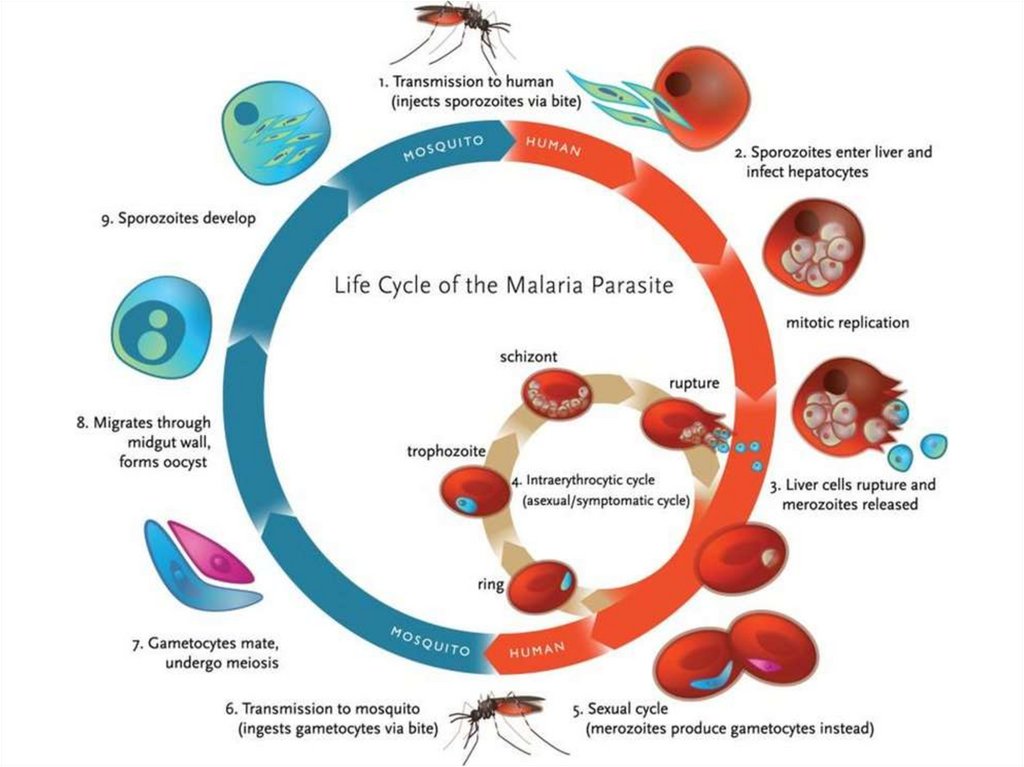
Malaria.Plasmodium species
Is a mosquito-borne infectious disease of humans and
other animals caused by parasitic protozoans belonging to
the Plasmodium species. There are four species
normally infecting humans, namely, Plasmodium
falciparum, Plasmodium vivax, Plasmodium ovale, and
Plasmodium malariae.
Definitive host: mosquito Anopheles
Intermediate host: human
Distribution:
Tropical and subtropical regions around the equator. SubSaharan Africa, Asia, Latin America.
63.
64.
Symptoms:Fever, fatigue, vomiting, headaches, anemia, hemoglobin in the
urine. In severe cases it can cause yellow skin, seizures, coma,
death.
Symptoms usually begin 10-15 days after being bitten. If not
properly treated, people have this disease some months or years.
The disease is most commonly transmitted by an infected
female Anopheles mosquito. The mosquito bite introduces the
parasites from mosquito’s saliva into a person’s blood. The
parasites travel to the liver where they mature and reproduce.
Diagnosis:
Microscopic examination of blood
Antigen-based rapid diagnostic tests (serology)
Detection DNA of parasites in blood samples (PCR)
Microscopy of the urine (hemoglobin in the urine)
65.
Treatment:Because chloroquine – resistant stains of P.falciparum are
present in many parts of the world, infection of P.falciparum may
be treated with other agents including mefloquine, quinine,
guanidine, pyrimethamine – sulfadoxine, and doxycycline. If the
laboratory reports a mixed infection involving P.falciparum and
P.vivax, the treatment must eradicate not only P.falciparum from
the erythrocytes but also the liver stages of P.vivax to avoid
relapses provided that the person no longer lives in a malaria
endemic area.
Prevention:
Mosquito control measures:
Using mosquito nets
Insecticides, insect repellents
66.
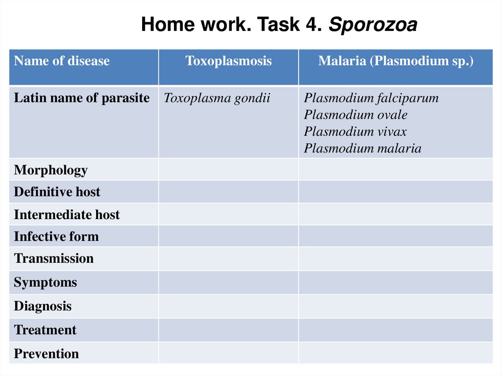
Home work. Task 4. SporozoaName of disease
Toxoplasmosis
Latin name of parasite Toxoplasma gondii
Morphology
Definitive host
Intermediate host
Infective form
Transmission
Symptoms
Diagnosis
Treatment
Prevention
Malaria (Plasmodium sp.)
Plasmodium falciparum
Plasmodium ovale
Plasmodium vivax
Plasmodium malaria
67.
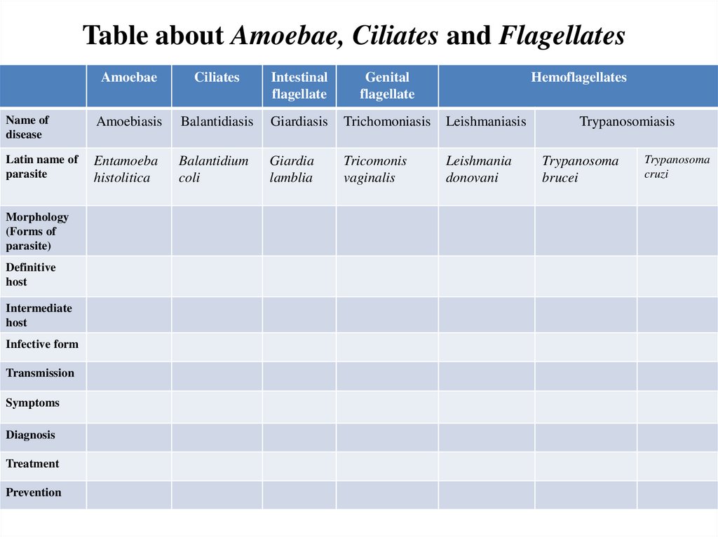
Table about Amoebae, Ciliates and FlagellatesAmoebae
Ciliates
Intestinal
flagellate
Genital
flagellate
Name of
disease
Amoebiasis
Balantidiasis
Giardiasis
Trichomoniasis
Leishmaniasis
Latin name of
parasite
Entamoeba
histolitica
Balantidium
coli
Giardia
lamblia
Tricomonis
vaginalis
Leishmania
donovani
Morphology
(Forms of
parasite)
Definitive
host
Intermediate
host
Infective form
Transmission
Symptoms
Diagnosis
Treatment
Prevention
Hemoflagellates
Trypanosomiasis
Trypanosoma
brucei
Trypanosoma
cruzi