

 medicine
Similar presentations:


")
")
Medical protozoology sarcodina and flagellata
1.
MEDICALPROTOZOOLOGY
SARCODINA AND FLAGELLATA
И ЖГУТИКОВЫЕ
2.
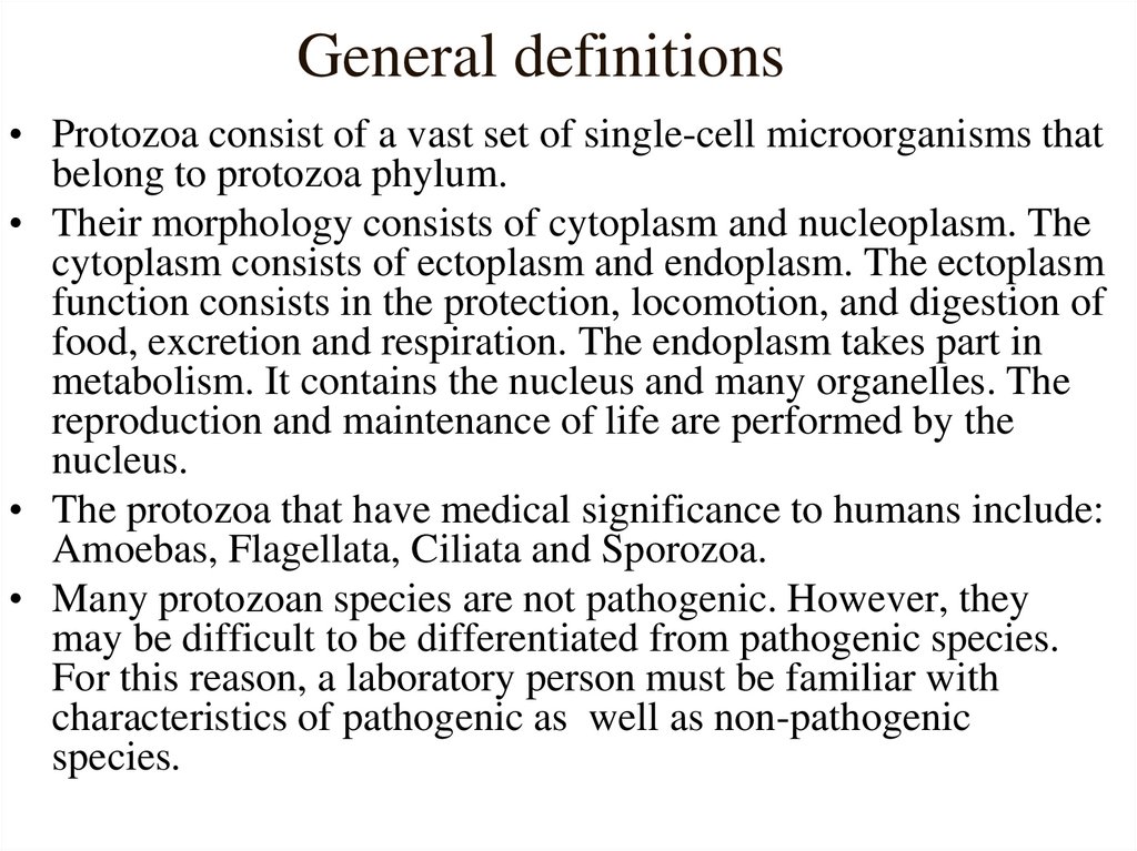
General definitions• Protozoa consist of a vast set of single-cell microorganisms that
belong to protozoa phylum.
• Their morphology consists of cytoplasm and nucleoplasm. The
cytoplasm consists of ectoplasm and endoplasm. The ectoplasm
function consists in the protection, locomotion, and digestion of
food, excretion and respiration. The endoplasm takes part in
metabolism. It contains the nucleus and many organelles. The
reproduction and maintenance of life are performed by the
nucleus.
• The protozoa that have medical significance to humans include:
Amoebas, Flagellata, Ciliata and Sporozoa.
• Many protozoan species are not pathogenic. However, they
may be difficult to be differentiated from pathogenic species.
For this reason, a laboratory person must be familiar with
characteristics of pathogenic as well as non-pathogenic
species.
3.
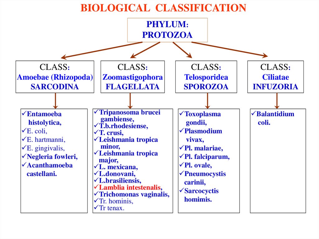
BIOLOGICAL CLASSIFICATIONPHYLUM:
PROTOZOA
CLASS:
CLASS:
CLASS:
CLASS:
Amoebae (Rhizopoda)
SARCODINA
Zoomastigophora
FLAGELLATA
Telosporidea
SPOROZOA
Ciliatae
INFUZORIA
Tripanosoma brucei
gambiense,
T.b.rhodesiense,
T. crusi,
Leishmania tropica
minor,
Leishmania tropica
major,
L. mexicana,
L.donovani,
L.brasiliensis,
Lamblia intestenalis,
Trichomonas vaginalis,
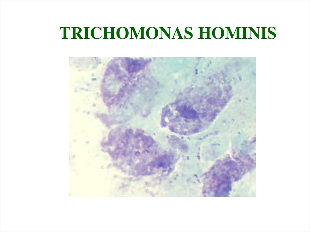
Tr. hominis,
Tr tenax.
Toxoplasma
gondii,
Plasmodium
vivax,
Pl. malariae,
Pl. falciparum,
Pl. ovale,
Pneumocystis
carinii,
Sarcocyctis
homimis.
Entamoeba
histolytica,
E. coli,
E. hartmanni,
E. gingivalis,
Negleria fowleri,
Acanthamoeba
castellani.
Balantidium
coli.
4.
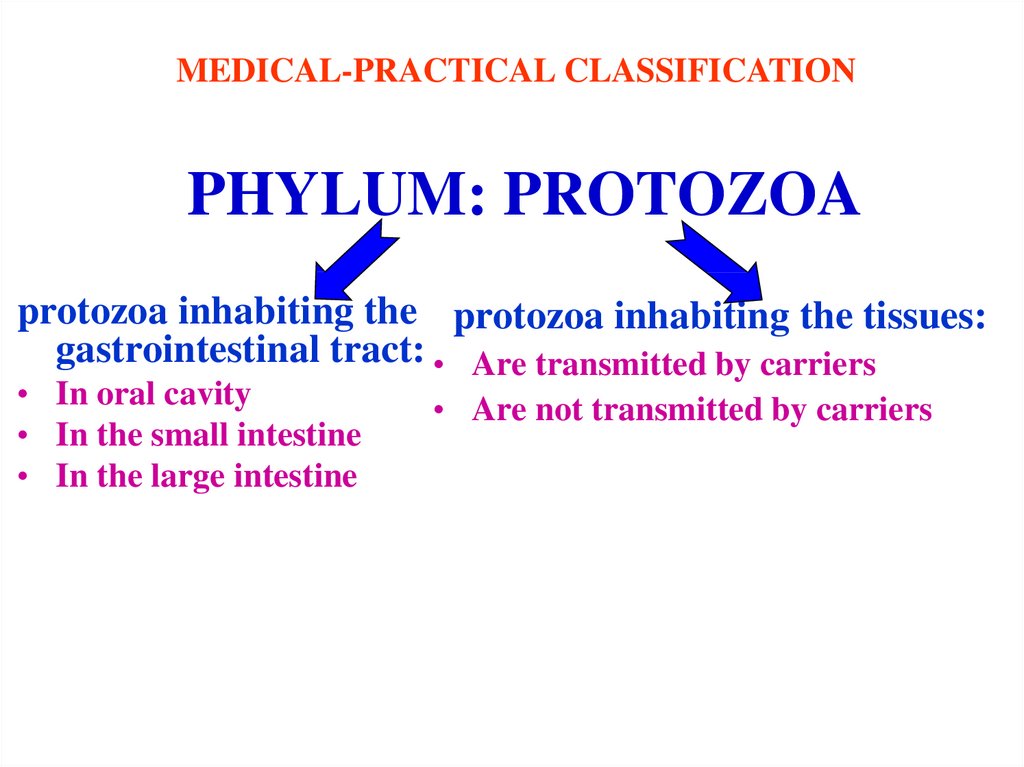
MEDICAL-PRACTICAL CLASSIFICATIONPHYLUM: PROTOZOA
protozoa inhabiting the protozoa inhabiting the tissues:
gastrointestinal tract: • Are transmitted by carriers
• In oral cavity
• In the small intestine
• In the large intestine
• Are not transmitted by carriers
5.
CLASSAmoebae (Rhizopoda)
SARCODINA
6.
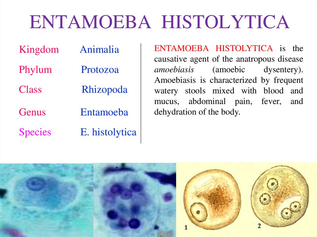
ENTAMOEBA HISTOLYTICAKingdom
Animalia
Phylum
Protozoa
Class
Rhizopoda
Genus
Entamoeba
Species
E. histolytica
ENTAMOEBA HISTOLYTICA is the
causative agent of the anatropous disease
amoebiasis
(amoebic
dysentery).
Amoebiasis is characterized by frequent
watery stools mixed with blood and
mucus, abdominal pain, fever, and
dehydration of the body.
7.
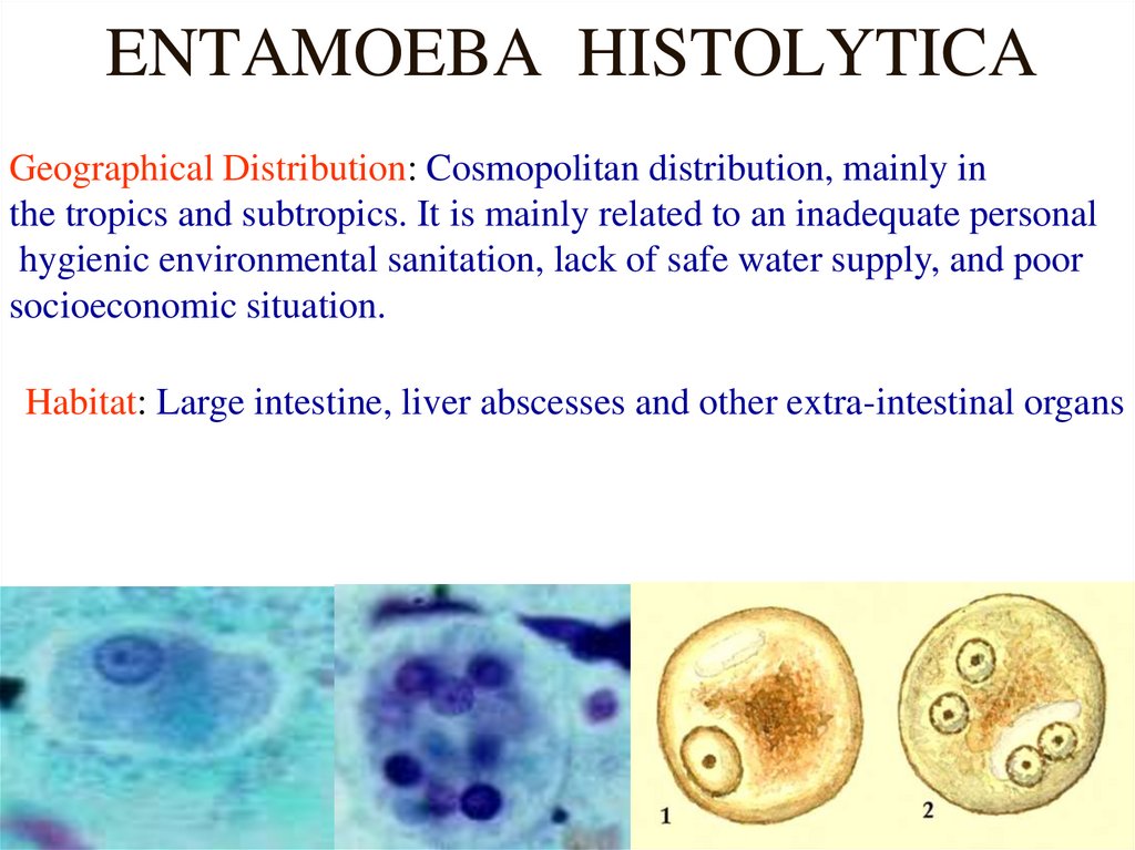
ENTAMOEBA HISTOLYTICAGeographical Distribution: Cosmopolitan distribution, mainly in
the tropics and subtropics. It is mainly related to an inadequate personal
hygienic environmental sanitation, lack of safe water supply, and poor
socioeconomic situation.
Habitat: Large intestine, liver abscesses and other extra-intestinal organs
8.
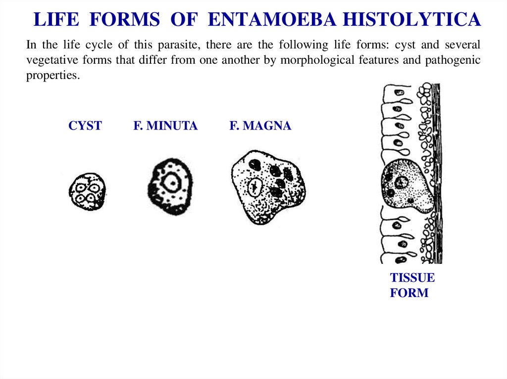
LIFE FORMS OF ENTAMOEBA HISTOLYTICAIn the life cycle of this parasite, there are the following life forms: cyst and several
vegetative forms that differ from one another by morphological features and pathogenic
properties.
CYST
F. MINUTA
F. MAGNA
TISSUE
FORM
9.
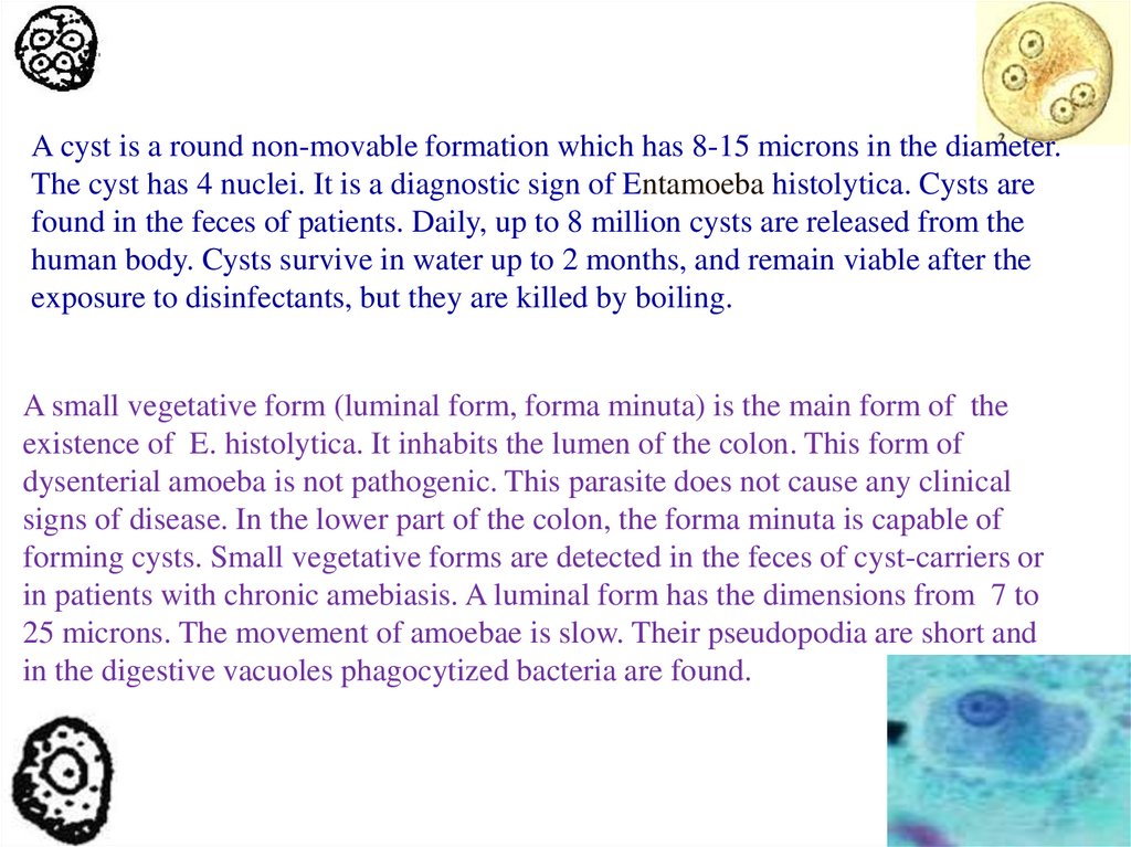
A cyst is a round non-movable formation which has 8-15 microns in the diameter.The cyst has 4 nuclei. It is a diagnostic sign of Entamoeba histolytica. Cysts are
found in the feces of patients. Daily, up to 8 million cysts are released from the
human body. Cysts survive in water up to 2 months, and remain viable after the
exposure to disinfectants, but they are killed by boiling.
A small vegetative form (luminal form, forma minuta) is the main form of the
existence of E. histolytica. It inhabits the lumen of the colon. This form of
dysenterial amoeba is not pathogenic. This parasite does not cause any clinical
signs of disease. In the lower part of the colon, the forma minuta is capable of
forming cysts. Small vegetative forms are detected in the feces of cyst-carriers or
in patients with chronic amebiasis. A luminal form has the dimensions from 7 to
25 microns. The movement of amoebae is slow. Their pseudopodia are short and
in the digestive vacuoles phagocytized bacteria are found.
10.
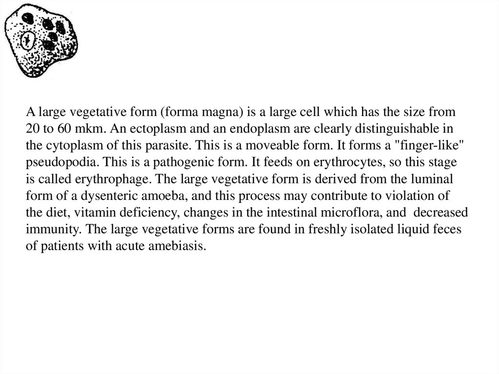
A large vegetative form (forma magna) is a large cell which has the size from20 to 60 mkm. An ectoplasm and an endoplasm are clearly distinguishable in
the cytoplasm of this parasite. This is a moveable form. It forms a "finger-like"
pseudopodia. This is a pathogenic form. It feeds on erythrocytes, so this stage
is called erythrophage. The large vegetative form is derived from the luminal
form of a dysenteric amoeba, and this process may contribute to violation of
the diet, vitamin deficiency, changes in the intestinal microflora, and decreased
immunity. The large vegetative forms are found in freshly isolated liquid feces
of patients with acute amebiasis.
11.
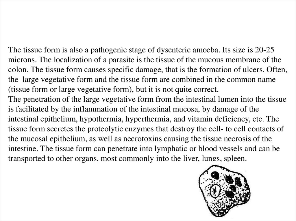
The tissue form is also a pathogenic stage of dysenteric amoeba. Its size is 20-25microns. The localization of a parasite is the tissue of the mucous membrane of the
colon. The tissue form causes specific damage, that is the formation of ulcers. Often,
the large vegetative form and the tissue form are combined in the common name
(tissue form or large vegetative form), but it is not quite correct.
The penetration of the large vegetative form from the intestinal lumen into the tissue
is facilitated by the inflammation of the intestinal mucosa, by damage of the
intestinal epithelium, hypothermia, hyperthermia, and vitamin deficiency, etc. The
tissue form secretes the proteolytic enzymes that destroy the cell- to cell contacts of
the mucosal epithelium, as well as necrotoxins causing the tissue necrosis of the
intestine. The tissue form can penetrate into lymphatic or blood vessels and can be
transported to other organs, most commonly into the liver, lungs, spleen.
12.
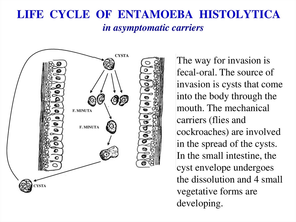
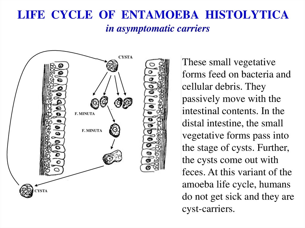
LIFE CYCLE OF ENTAMOEBA HISTOLYTICAin asymptomatic carriers
CYSTA
F. MINUTA
F. MINUTA
CYSTA
The way for invasion is
fecal-oral. The source of
invasion is cysts that come
into the body through the
mouth. The mechanical
carriers (flies and
cockroaches) are involved
in the spread of the cysts.
In the small intestine, the
cyst envelope undergoes
the dissolution and 4 small
vegetative forms are
developing.
13.
LIFE CYCLE OF ENTAMOEBA HISTOLYTICAin asymptomatic carriers
CYSTA
F. MINUTA
F. MINUTA
CYSTA
These small vegetative
forms feed on bacteria and
cellular debris. They
passively move with the
intestinal contents. In the
distal intestine, the small
vegetative forms pass into
the stage of cysts. Further,
the cysts come out with
feces. At this variant of the
amoeba life cycle, humans
do not get sick and they are
cyst-carriers.
14.
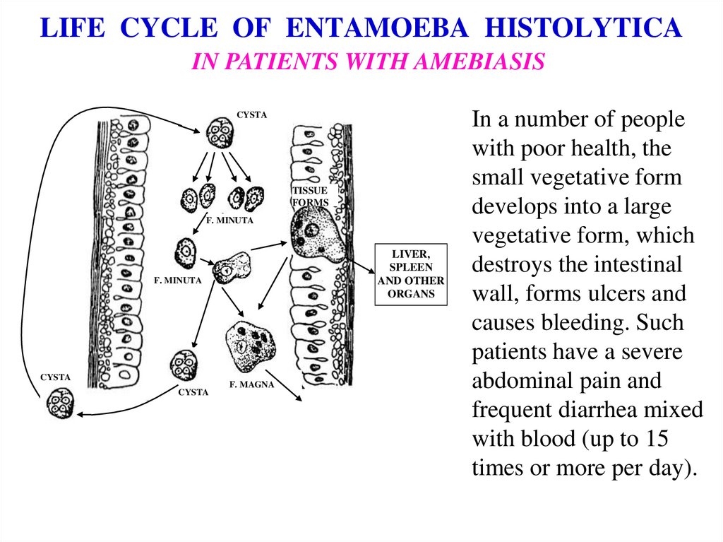
LIFE CYCLE OF ENTAMOEBA HISTOLYTICAIN PATIENTS WITH AMEBIASIS
CYSTA
TISSUE
FORMS
F. MINUTA
LIVER,
SPLEEN
AND OTHER
ORGANS
F. MINUTA
CYSTA
CYSTA
F. MAGNA
In a number of people
with poor health, the
small vegetative form
develops into a large
vegetative form, which
destroys the intestinal
wall, forms ulcers and
causes bleeding. Such
patients have a severe
abdominal pain and
frequent diarrhea mixed
with blood (up to 15
times or more per day).
15.
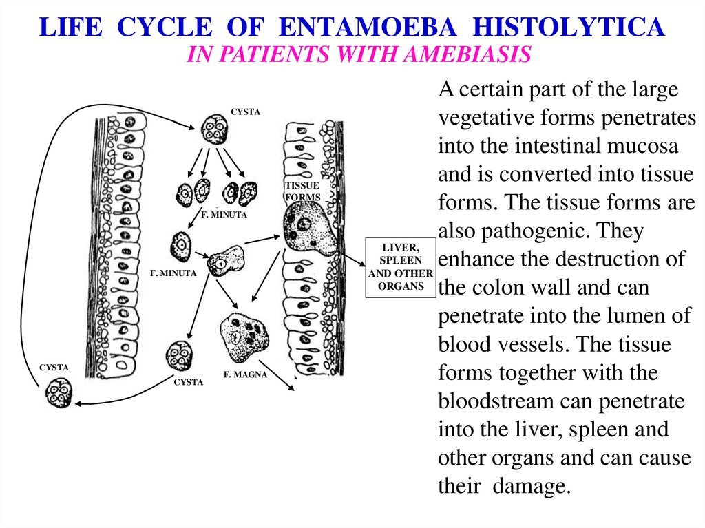
LIFE CYCLE OF ENTAMOEBA HISTOLYTICAIN PATIENTS WITH AMEBIASIS
A certain part of the large
vegetative forms penetrates
into the intestinal mucosa
and is converted into tissue
TISSUE
FORMS
forms. The tissue forms are
also pathogenic. They
LIVER,
SPLEEN enhance the destruction of
AND OTHER
ORGANS the colon wall and can
penetrate into the lumen of
blood vessels. The tissue
forms together with the
bloodstream can penetrate
into the liver, spleen and
other organs and can cause
their damage.
CYSTA
F. MINUTA
F. MINUTA
CYSTA
CYSTA
F. MAGNA
16.
• Patients with amoebic dysentery must behospitalized. In the absence of proper
treatment, such patients have a variety of
complications of the disease: anemia,
dehydration, disturbance of electrolytic
composition of the blood etc. These
abnormalities may cause death. Spontaneous
recovery rarely occurs.
17.
Diagnostics of amebiasis• During the acute form of the disease many forma magna
with ingested erythrocytes are found in patient's feces.
• In the chronic form of the disease many cysts and a little of
forma magna are found in patient's feces.
• During cysts-carriage many cysts are found in patient's
feces.
18.
PREVENTIONof amebiasis can be personal and public. The personal prevention is
the activities that each patient should carry out himself.
RECOMMENDATIONS FOR PERSONAL PREVENTION:
use boiled water,
wash hands before eating and after using the toilet,
scald fruits and vegetables,
protect products from flies and cockroaches.
19.
The public prevention is the measures which are carried out by asanitary doctor.
• RECOMMENDATIONS FOR PUBLIC
PREVENTION:
• closing of access to local water-sources,
• import of fresh water,
• identification and treatment of patients and
humans, that are carriers of cysts,
• disinfection of water closets,
• sanitary-educational work in the community.
20.
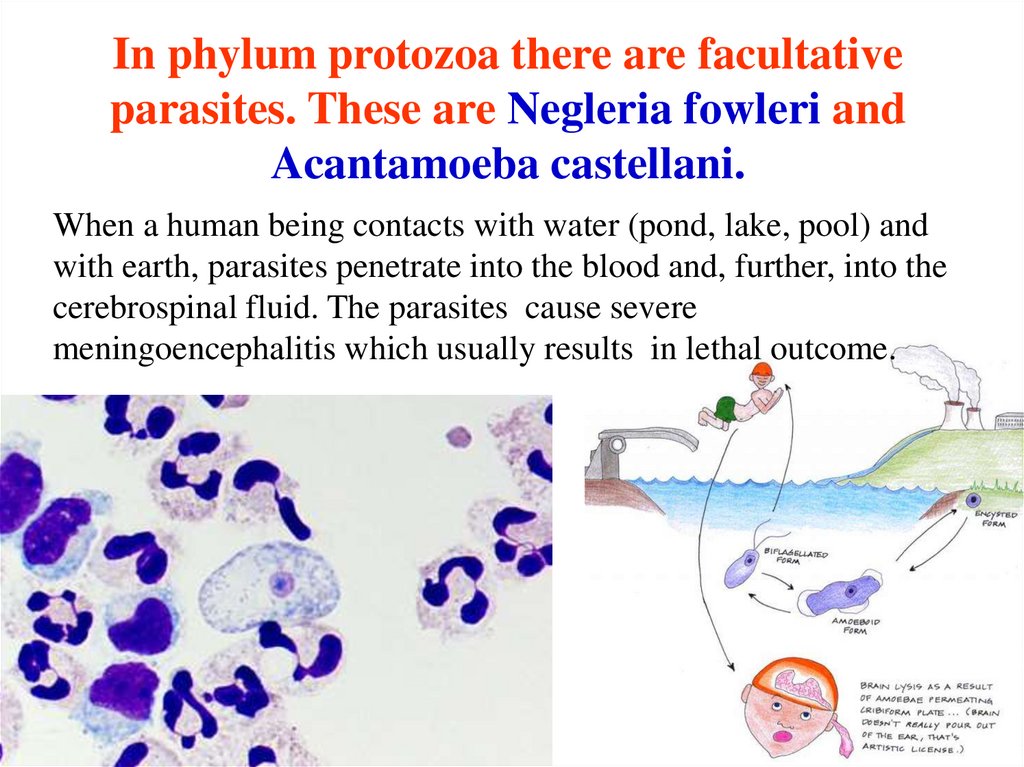
In phylum protozoa there are facultativeparasites. These are Negleria fowleri and
Acantamoeba castellani.
When a human being contacts with water (pond, lake, pool) and
with earth, parasites penetrate into the blood and, further, into the
cerebrospinal fluid. The parasites cause severe
meningoencephalitis which usually results in lethal outcome.
21.
CLASSZOOMASTIGOPHORA
(FLAGELLATA)
22.
• All members of the flagellata class can bedivided into two groups: parasites which have
one flagellum and parasites which have many
flagella.
• The parasites which have only one flagellum is
called also the oro-intestinal and urogenital
flagellata.
• The parasites which have many flagella are also
called hemo-somatic flagellata.
• Our acquaintance with the parasites of class
flagellata we will begin with oro-intestinal and
urogenital flagellata.
23.
Flagellata with one flagellum have different life forms.А
B
C
E
D
А- Metacyclic form; B- Trypomastigote (Trypanosomal)
form; C- Epimastigote (Crithidial) form; D- Promastigote
(Leptomonad) form; E- Amastigote (leishmania) form.
The different life forms of flagellates differ from one another by a cell
shape, the presence or absence of an undulating membrane and a
flagellum, as well as a kinetoplast localization (basal body).
24.
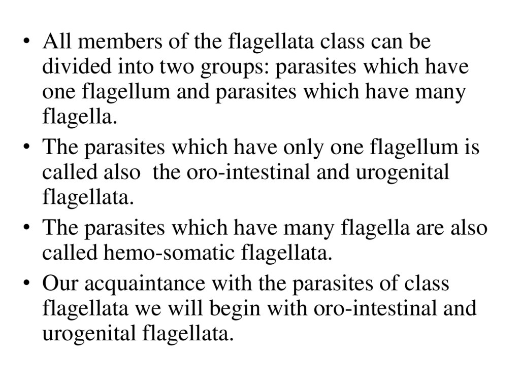
E - Amastigote (Leishmania) formА
B
C
D
E
The amastigote form is an intracellular spherical form. It has
no flagellum and has no undulating membrane. The amastigote
form is the intracellular form of all leishmania species and
Trypanosome cruzi.
25.
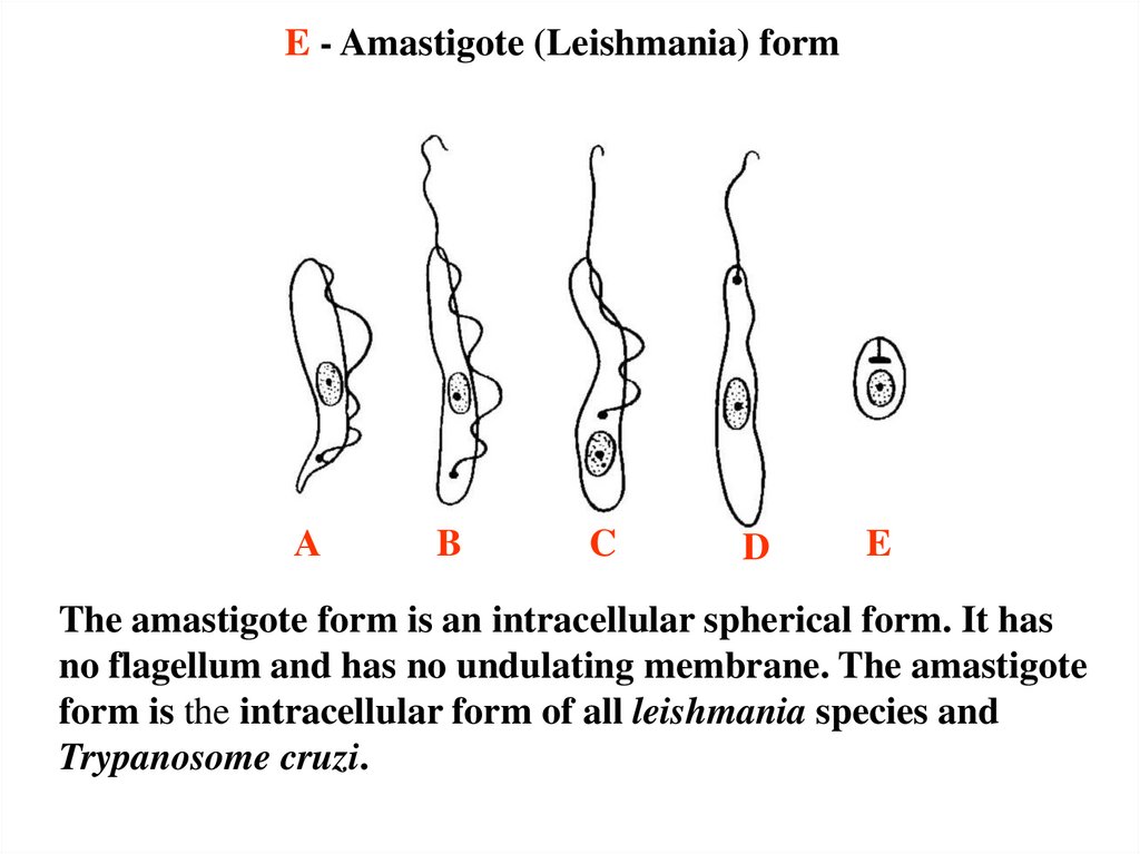
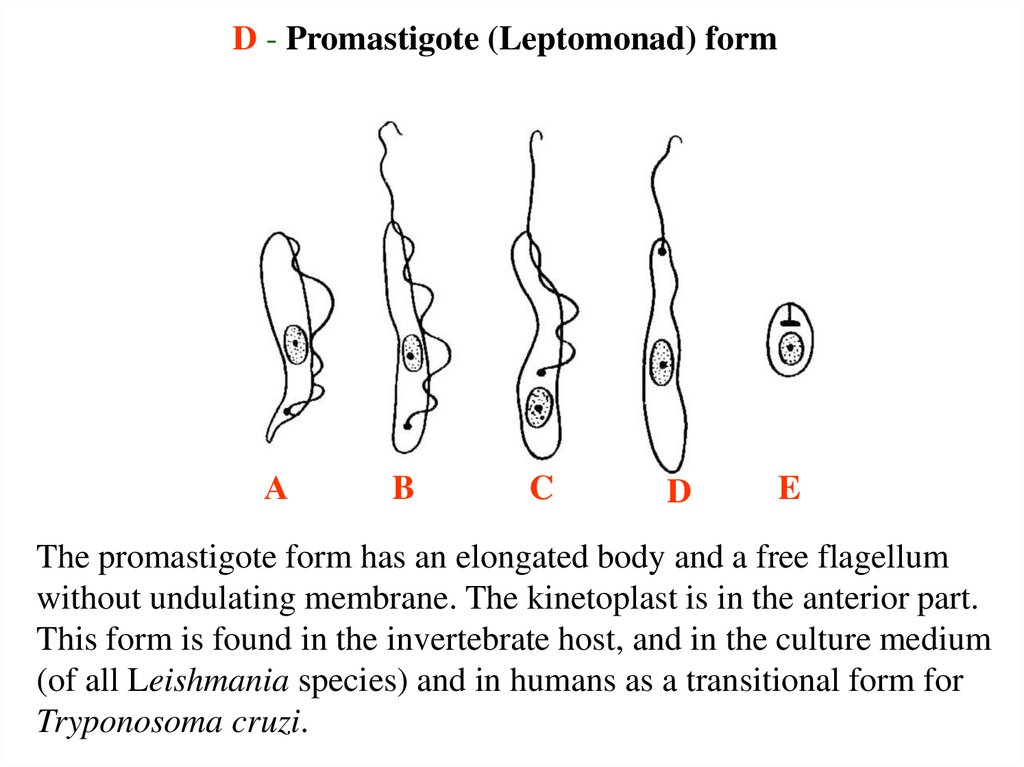
D - Promastigote (Leptomonad) formА
B
C
D
E
The promastigote form has an elongated body and a free flagellum
without undulating membrane. The kinetoplast is in the anterior part.
This form is found in the invertebrate host, and in the culture medium
(of all Leishmania species) and in humans as a transitional form for
Tryponosoma cruzi.
26.
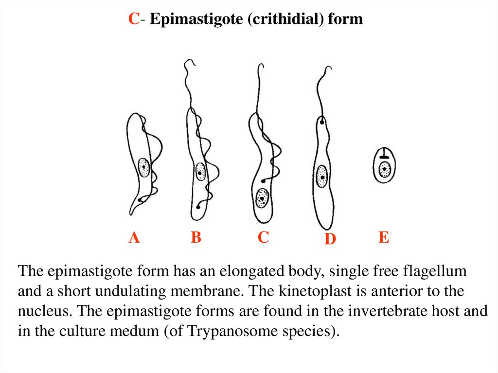
C- Epimastigote (crithidial) formА
B
C
D
E
The epimastigote form has an elongated body, single free flagellum
and a short undulating membrane. The kinetoplast is anterior to the
nucleus. The epimastigote forms are found in the invertebrate host and
in the culture medum (of Trypanosome species).
27.
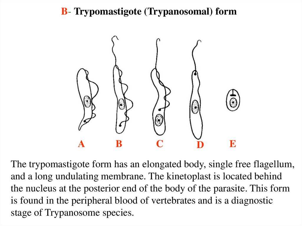
B- Trypomastigote (Trypanosomal) formА
B
C
D
E
The trypomastigote form has an elongated body, single free flagellum,
and a long undulating membrane. The kinetoplast is located behind
the nucleus at the posterior end of the body of the parasite. This form
is found in the peripheral blood of vertebrates and is a diagnostic
stage of Trypanosome species.
28.
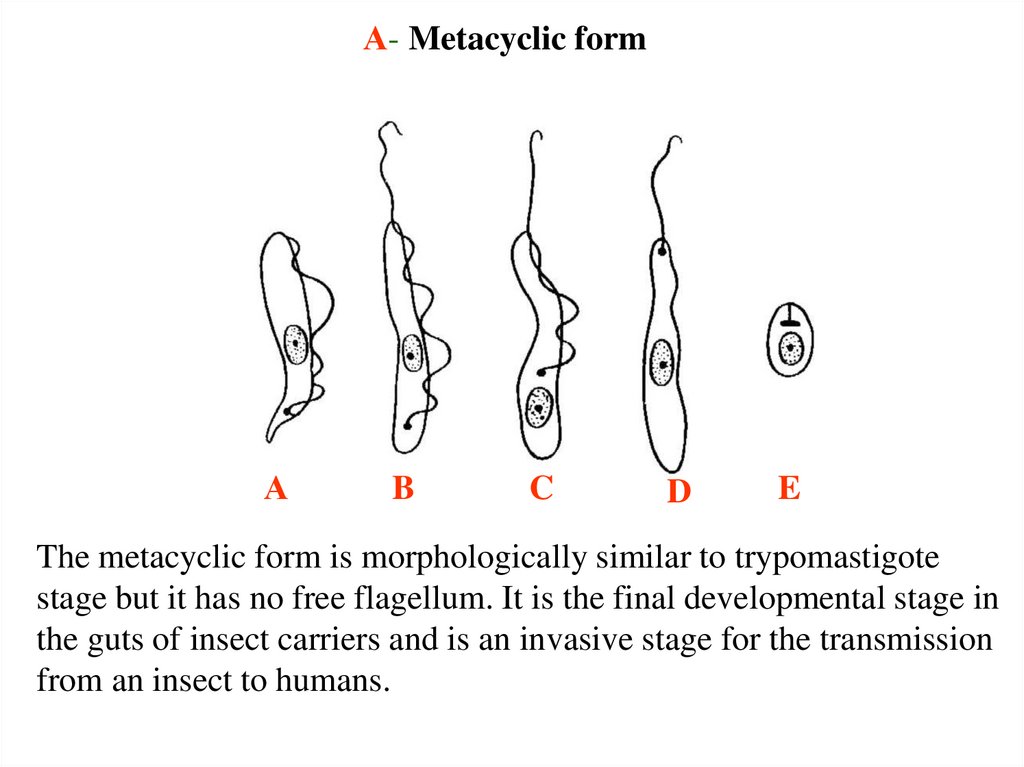
А- Metacyclic formА
B
C
D
E
The metacyclic form is morphologically similar to trypomastigote
stage but it has no free flagellum. It is the final developmental stage in
the guts of insect carriers and is an invasive stage for the transmission
from an insect to humans.
29.
The causative agents of leishmaniasis30.
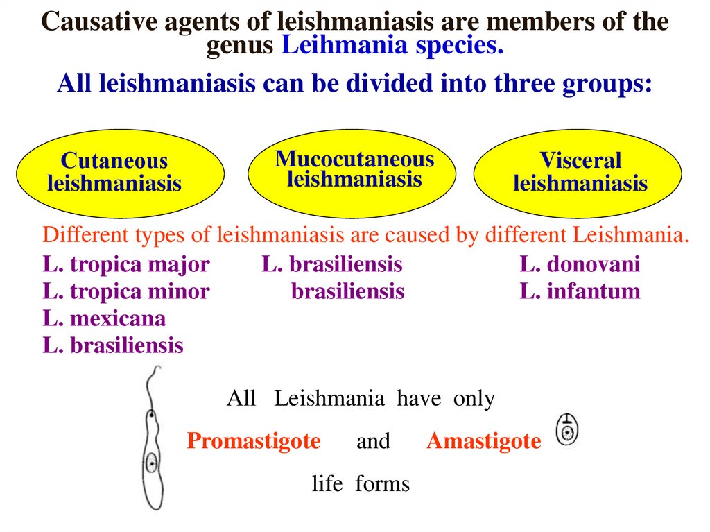
Causative agents of leishmaniasis are members of thegenus Leihmania species.
All leishmaniasis can be divided into three groups:
Сutaneous
leishmaniasis
Mucocutaneous
leishmaniasis
Visceral
leishmaniasis
Different types of leishmaniasis are caused by different Leishmania.
L. tropica major
L. brasiliensis
L. donovani
L. tropica minor
brasiliensis
L. infantum
L. mexicana
L. brasiliensis
All Leishmania have only
Promastigote
and
life forms
Amastigote
31.
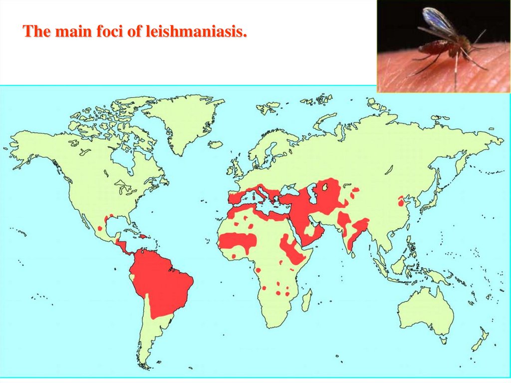
The main foci of leishmaniasis.32.
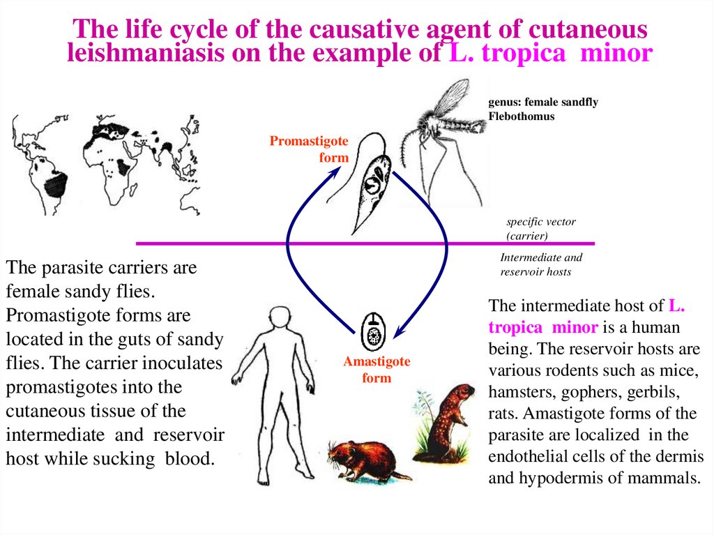
The life cycle of the causative agent of cutaneousleishmaniasis on the example of L. tropica minor
genus: female sandfly
Flebothomus
Promastigote
form
specific vector
(carrier)
The parasite carriers are
female sandy flies.
Promastigote forms are
located in the guts of sandy
flies. The carrier inoculates
promastigotes into the
cutaneous tissue of the
intermediate and reservoir
host while sucking blood.
Intermediate and
reservoir hosts
Amastigote
form
The intermediate host of L.
tropica minor is a human
being. The reservoir hosts are
various rodents such as mice,
hamsters, gophers, gerbils,
rats. Amastigote forms of the
parasite are localized in the
endothelial cells of the dermis
and hypodermis of mammals.
33.
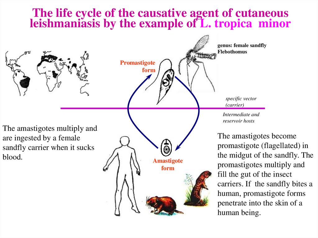
The life cycle of the causative agent of cutaneousleishmaniasis by the example of L. tropica minor
genus: female sandfly
Flebothomus
Promastigote
form
specific vector
(carrier)
Intermediate and
reservoir hosts
The amastigotes multiply and
are ingested by a female
sandfly carrier when it sucks
blood.
Amastigote
form
The amastigotes become
promastigote (flagellated) in
the midgut of the sandfly. The
promastigotes multiply and
fill the gut of the insect
carriers. If the sandfly bites a
human, promastigote forms
penetrate into the skin of a
human being.
34.
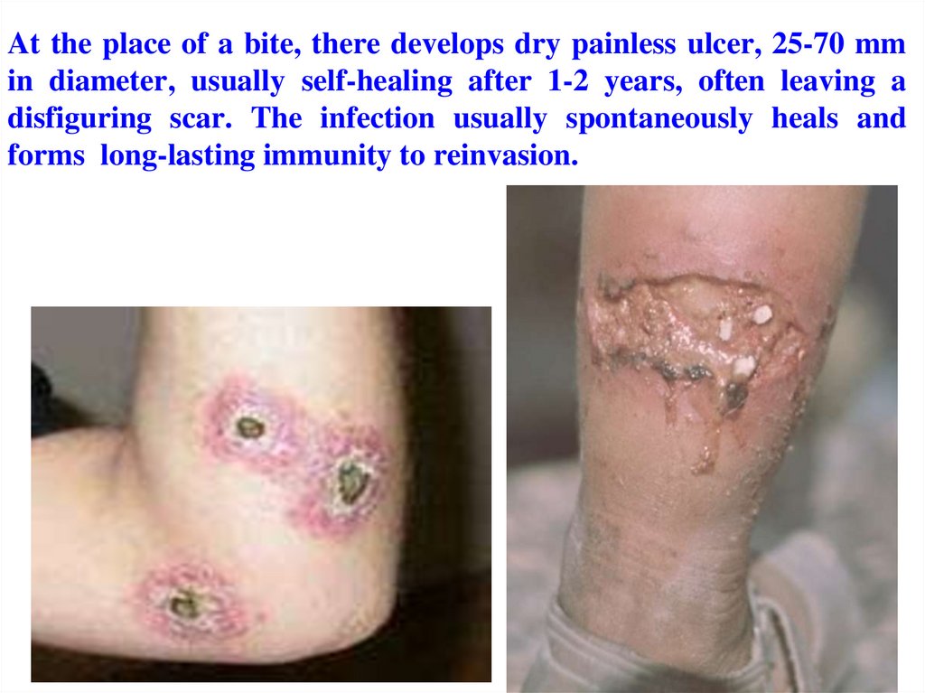
At the place of a bite, there develops dry painless ulcer, 25-70 mmin diameter, usually self-healing after 1-2 years, often leaving a
disfiguring scar. The infection usually spontaneously heals and
forms long-lasting immunity to reinvasion.
35.
THE CUTANEOUS LEISHMANIASIS36.
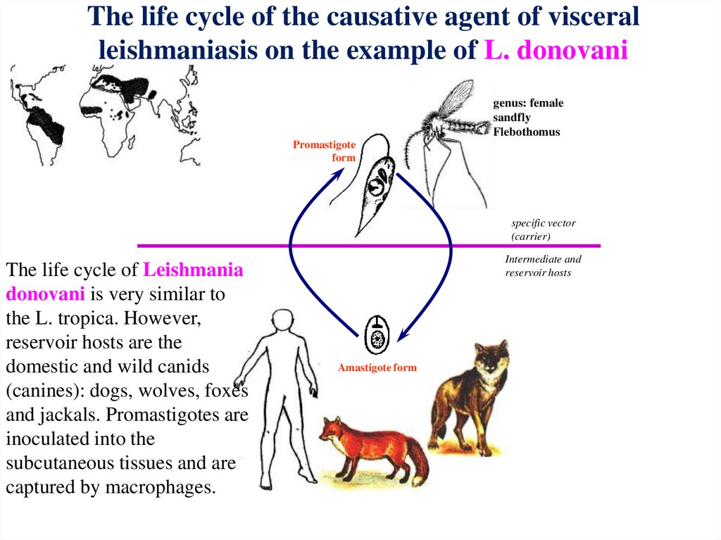
The life cycle of the causative agent of visceralleishmaniasis on the example of L. donovani
genus: female
sandfly
Flebothomus
Promastigote
form
specific vector
(carrier)
The life cycle of Leishmania
donovani is very similar to
the L. tropica. However,
reservoir hosts are the
domestic and wild canids
(canines): dogs, wolves, foxes
and jackals. Promastigotes are
inoculated into the
subcutaneous tissues and are
captured by macrophages.
Intermediate and
reservoir hosts
Amastigote form
37.
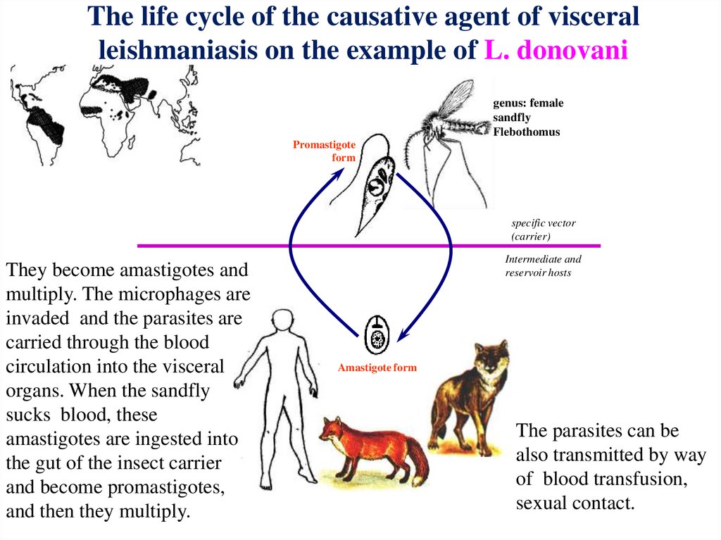
The life cycle of the causative agent of visceralleishmaniasis on the example of L. donovani
genus: female
sandfly
Flebothomus
Promastigote
form
specific vector
(carrier)
They become amastigotes and
multiply. The microphages are
invaded and the parasites are
carried through the blood
circulation into the visceral
organs. When the sandfly
sucks blood, these
amastigotes are ingested into
the gut of the insect carrier
and become promastigotes,
and then they multiply.
Intermediate and
reservoir hosts
Amastigote form
The parasites can be
also transmitted by way
of blood transfusion,
sexual contact.
38.
Leishmania braziliensis braziliensis• Geographical Distribution: Tropical forests of South
America and Central America.
• Reservoir hosts are rodents and some domestic animals.
• Habitat: Amastigote: in the reticulo-endothelial cells of
muco-cutaneous tissues of the nose, mouth, lips, larynx.
Promastigote: in the gut of Lutzomyia sandflies
• Life cycle: Lutzomyia sandflies are the main carriers, and
man acquires infection from an enzootic area.
• Pathology: Mucocutaneous leishmaniasis (espundia).
Chronic ulceration of the mucus membrane of the mouth
nose, throat, etc. with the destruction of bones and
cartilages.
39.
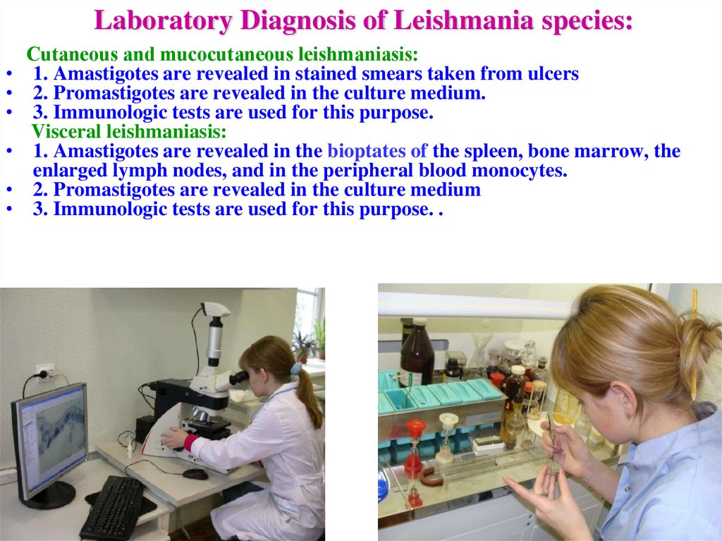
Laboratory Diagnosis of Leishmania species:Сutaneous and mucocutaneous leishmaniasis:
1. Amastigotes are revealed in stained smears taken from ulcers
2. Promastigotes are revealed in the culture medium.
3. Immunologic tests are used for this purpose.
Visceral leishmaniasis:
1. Amastigotes are revealed in the bioptates of the spleen, bone marrow, the
enlarged lymph nodes, and in the peripheral blood monocytes.
2. Promastigotes are revealed in the culture medium
3. Immunologic tests are used for this purpose. .
40.
PREVENTION• RECOMMENDATIONS FOR PUBLIC
PREVENTION :
• Treatment of infected individuals,
• Destruction of specific carriers,
• Destruction of reservoir hosts,
• Health education in the community
• RECOMMENDATIONS FOR PERSONAL
PREVENTION :
• Avoiding endemic areas,
• Avoiding insect bites.
41.
The causative agentsof African sleeping sickness
(African trypanosomiasis)
42.
Causative agents of African sleeping sickness are members ofthe species Tripanosoma brucei.
There are two subspecies of Tripanosoma brucei, which are
pathogenic for humans: Tripanosoma brucei gambiense and
Tripanosoma brucei rhodesiense.
Tr. b. gambiense – is the cause of the West African
variant (chronic trypanosomiasis or Gambian version)
Tr. b. rhodesiense - is the cause of the East African
variant (acute trypanosomiasis, Rhodesian version)
43.
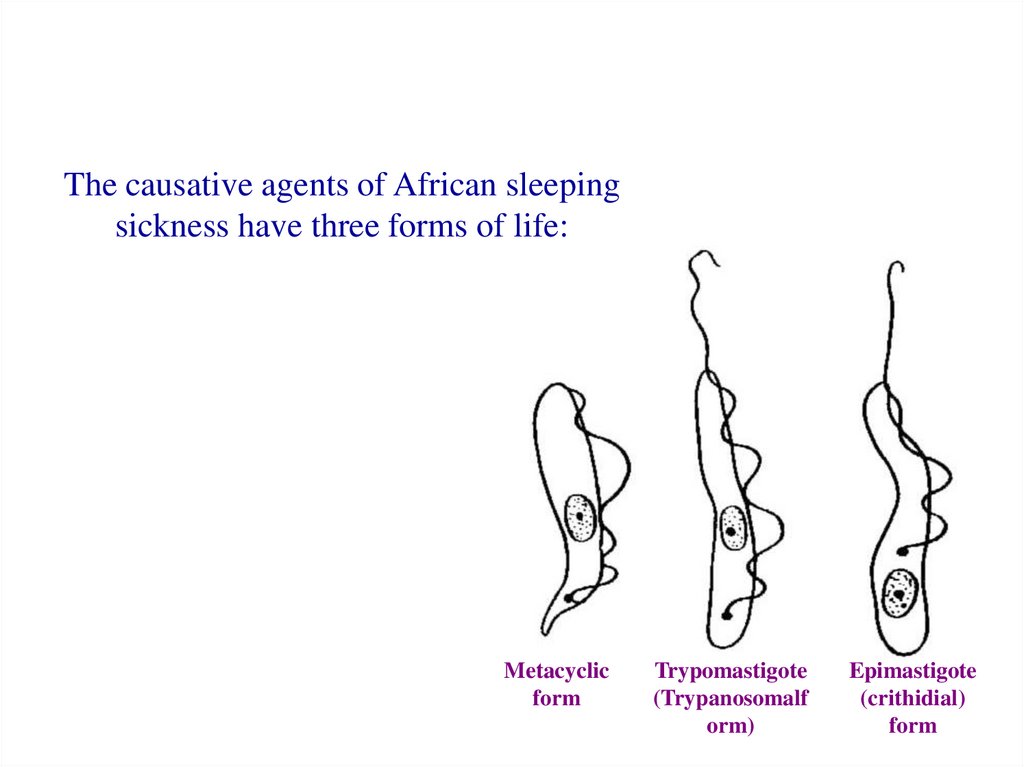
The causative agents of African sleepingsickness have three forms of life:
Metacyclic
form
Trypomastigote
(Trypanosomalf
orm)
Epimastigote
(crithidial)
form
44.
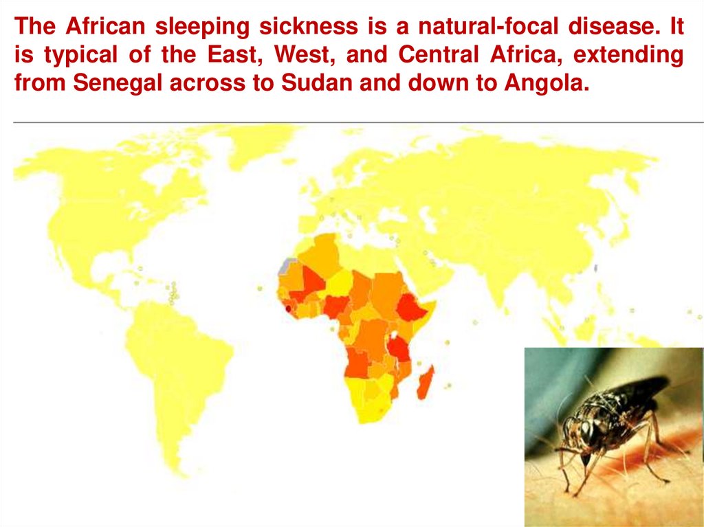
The African sleeping sickness is a natural-focal disease. Itis typical of the East, West, and Central Africa, extending
from Senegal across to Sudan and down to Angola.
45.
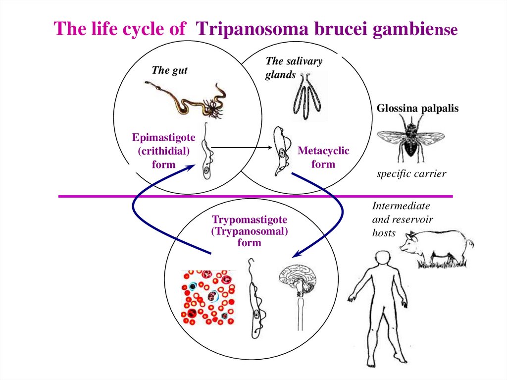
The life cycle of Tripanosoma brucei gambienseThe gut
The salivary
glands
Glossina palpalis
Epimastigote
(crithidial)
form
Metacyclic
form
Trypomastigote
(Trypanosomal)
form
specific carrier
Intermediate
and reservoir
hosts
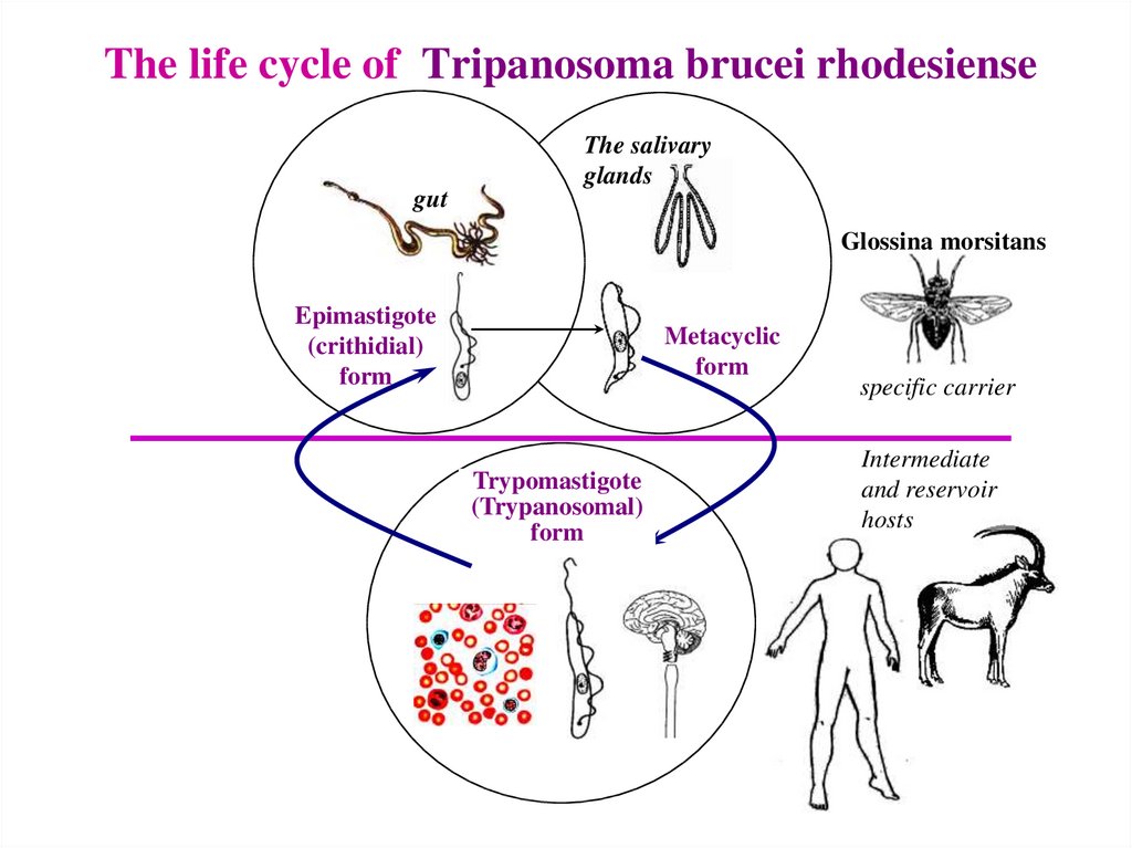
46.
The life cycle of Tripanosoma brucei rhodesienseThe salivary
glands
gut
Glossina morsitans
Epimastigote
(crithidial)
form
Metacyclic
form
Trypomastigote
(Trypanosomal)
form
specific carrier
Intermediate
and reservoir
hosts
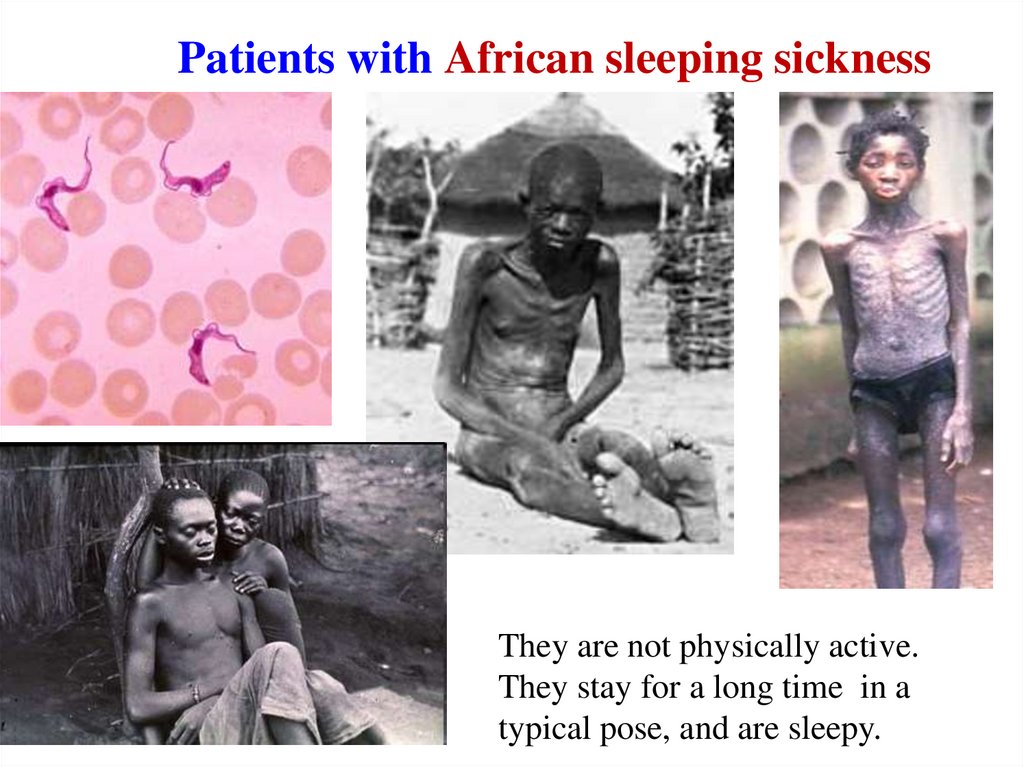
47.
Patients with African sleeping sicknessThey are not physically active.
They stay for a long time in a
typical pose, and are sleepy.
48.
African sleeping sickness49.
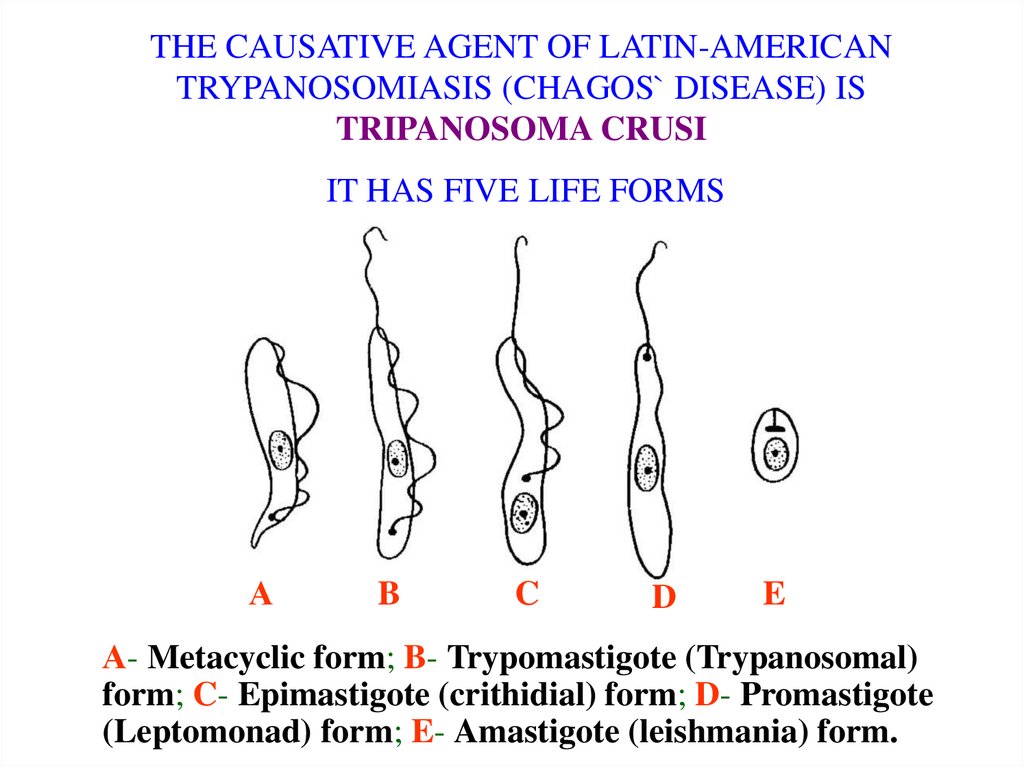
THE CAUSATIVE AGENT OF LATIN-AMERICANTRYPANOSOMIASIS (CHAGOS` DISEASE) IS
TRIPANOSOMA CRUSI
IT HAS FIVE LIFE FORMS
А
B
C
D
E
А- Metacyclic form; B- Trypomastigote (Trypanosomal)
form; C- Epimastigote (crithidial) form; D- Promastigote
(Leptomonad) form; E- Amastigote (leishmania) form.
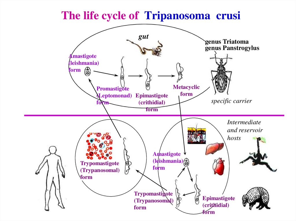
50.
The life cycle of Tripanosoma crusigut
genus Triatoma
genus Panstrogylus
Amastigote
(leishmania)
form
Metacyclic
Promastigote
form
(Leptomonad) Epimastigote
form
(crithidial)
form
specific carrier
Intermediate
and reservoir
hosts
Trypomastigote
(Trypanosomal)
form
Amastigote
(leishmania)
form
Trypomastigote
(Trypanosomal)
form
Epimastigote
(crithidial)
form
51.
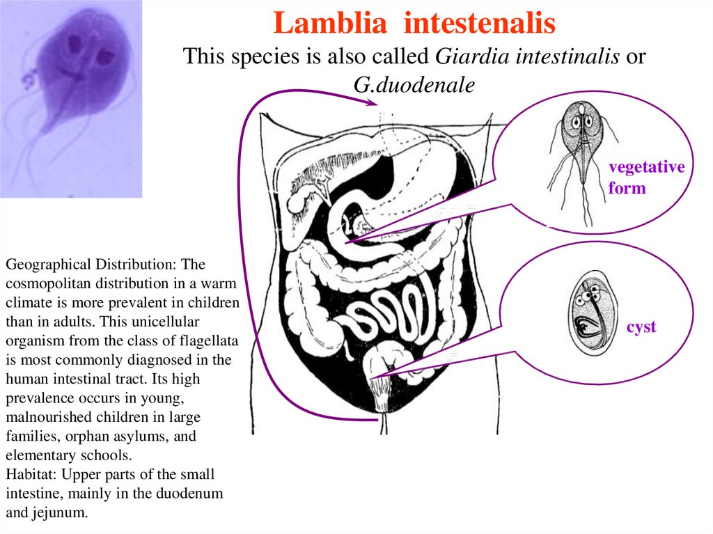
Lamblia intestenalisThis species is also called Giardia intestinalis or
G.duodenale
vegetative
form
Geographical Distribution: The
cosmopolitan distribution in a warm
climate is more prevalent in children
than in adults. This unicellular
organism from the class of flagellata
is most commonly diagnosed in the
human intestinal tract. Its high
prevalence occurs in young,
malnourished children in large
families, orphan asylums, and
elementary schools.
Habitat: Upper parts of the small
intestine, mainly in the duodenum
and jejunum.
cyst
52.
Manifestations of lambliosis53.
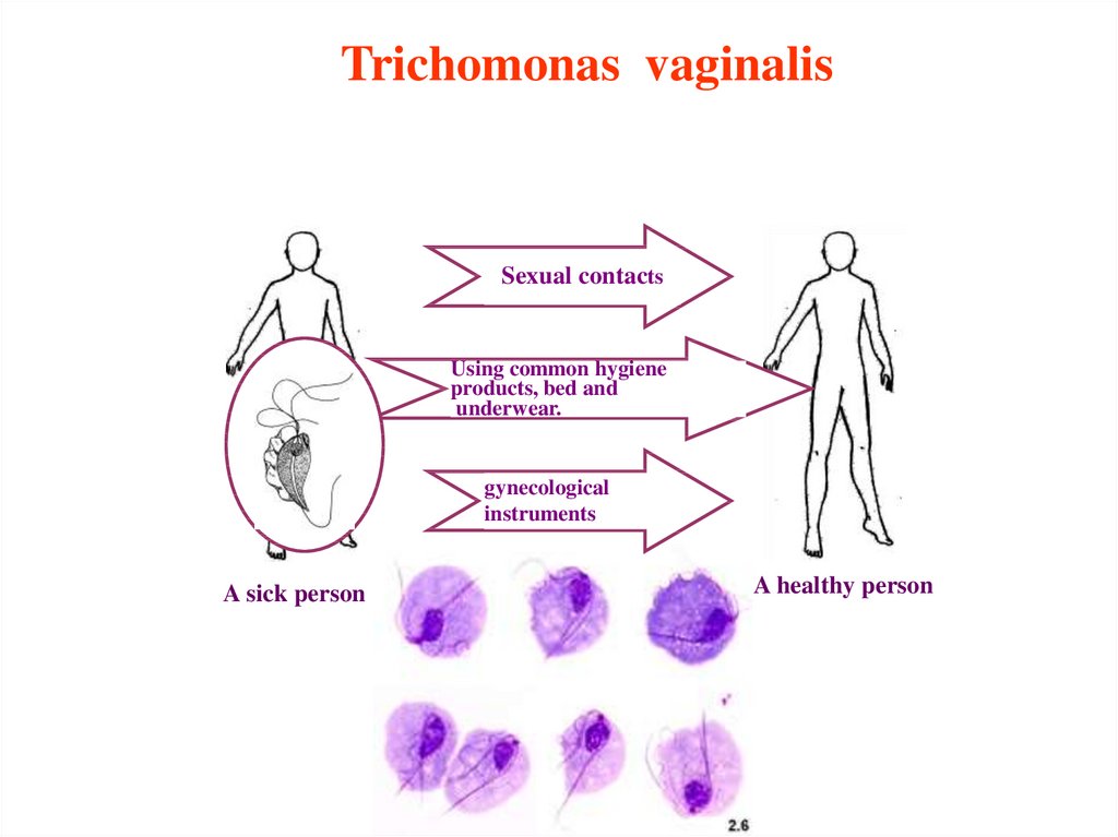
Trichomonas vaginalisSexual contacts
Using common hygiene
products, bed and
underwear.
gynecological
instruments
A sick person
A healthy person