


 medicine
Similar presentations:


Chronic pancreatitis and pancreonecro sis
1.
CHRONICPANCREATITIS
AND
PANCREONECRO
SIS
Prepared by: Tatayeva
Kh. 339GM
2.
ETIOLOGYPrimary pancreatitis :
➤ Misuse of alcohol (70-80% of
all diagnostic cases )
➤ the systematic eating of fatty
foods
➤ influence of drugs
(azathioprine , isoniazide ,
tetracycline , sulfonamides )
➤ protein deficiency
➤ Hereditary
➤ Ischemic (in lesions of
vascular , which supplies
blood pancreas )
➤ Idiopathic
Secondary pancreatitis :
➤
diseases of the biliary
tract (in 30-40%)
➤
disease of duodenum
➤ a primary (tumors,papillitis)
and a secondary
(dyskinesia of billiary tract)
➤
liver disease
➤
bowel disease
➤
viral infections (parotitis )
➤
allergic conditions
➤
hyperlipidemia
➤
hyperparathyroidism
➤
injury of the pancreas
3.
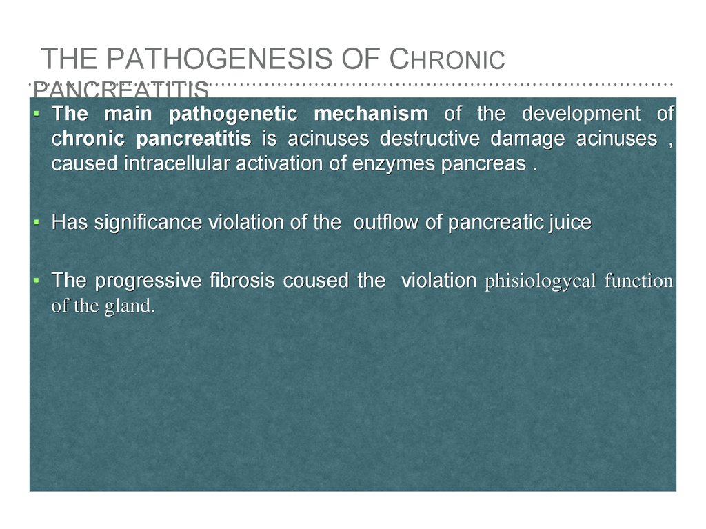
THE PATHOGENESIS OF CHRONICPANCREATITIS
▪ The main pathogenetic mechanism of the development of
chronic pancreatitis is acinuses destructive damage acinuses ,
caused intracellular activation of enzymes pancreas .
▪ Has significance violation of the outflow of pancreatic juice
▪ The progressive fibrosis coused the violation phisiologycal function
of the gland.
4.
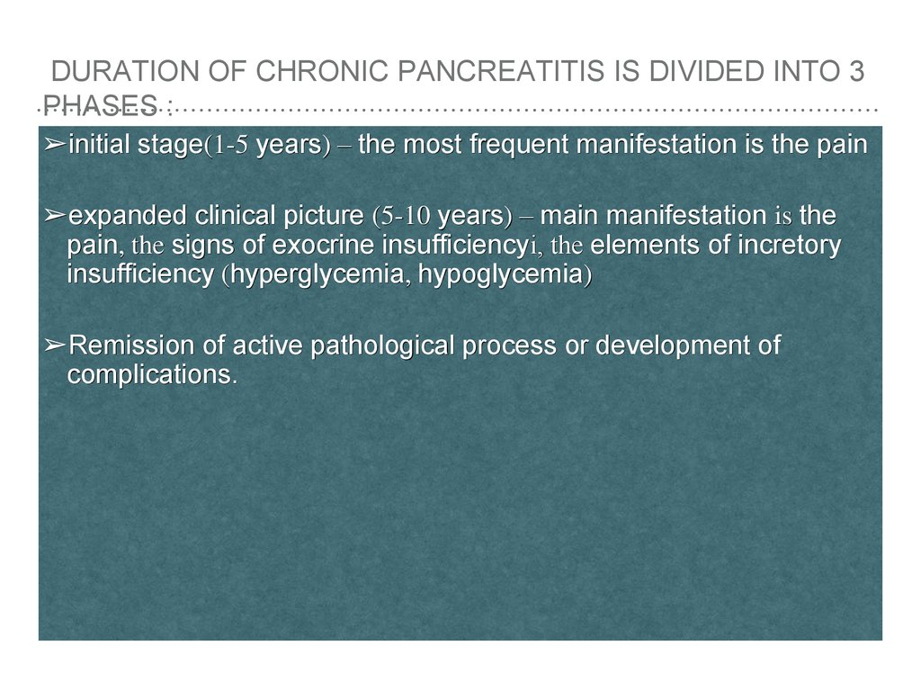
DURATION OF CHRONIC PANCREATITIS IS DIVIDED INTO 3PHASES :
➢initial stage(1-5 years) – the most frequent manifestation is the pain
➢expanded clinical picture (5-10 years) – main manifestation is the
pain, the signs of exocrine insufficiencyі, the elements of incretory
insufficiency (hyperglycemia, hypoglycemia)
➢Remission of active pathological process or development of
complications.
5.
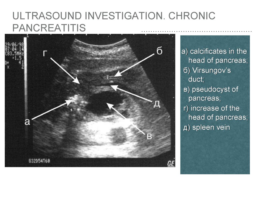
ULTRASOUND INVESTIGATION. CHRONICPANCREATITIS
The pancreas might appear atrophic, calcified or fibrotic (advanced
stages). Findings that may be present on ultrasound include:
• hyperechogenicity (often diffuse) often indicates fibrotic
changes
• pseudocysts
• pseudoaneurysms
• presence of ascites
Ultrasound may also assist to differentiate between the autoimmune
type vs. acquired:
• the pancreas is enlarged (either focally or diffusely) in the
autoimmune type
• calcifications are visible in acquired types
6.
ULTRASOUND INVESTIGATION. CHRONICPANCREATITIS
а) calcificates in the
head of pancreas;
б) Virsungov’s
duct;
в) pseudocyst of
pancreas;
г) increase of the
head of pancreas;
д) spleen vein
7.
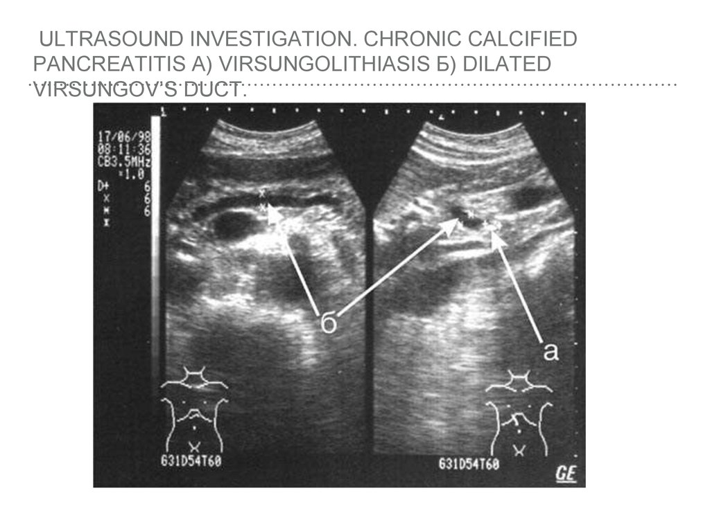
ULTRASOUND INVESTIGATION. CHRONIC CALCIFIEDPANCREATITIS А) VIRSUNGOLITHIASIS Б) DILATED
VIRSUNGOV’S DUCT.
8.
ENDOSCOPIC ULTRASOUND➤
has a vital diagnostic role because it is extremely
sensitive in detecting the early pathological changes of
chronic pancreatitis. Endoscopic ultrasound is the
investiga tion of choice if chronic pancreatitis is
suspected but not proven. Endoscopic ultrasoundguided neneedle aspiration cytology is useful for the
diagnosis of chronic pancreatitis and also for help ing
to exclude pancreatic cancer, although it may be dif
cult to obtain a good sample from an indurated gland.
9.
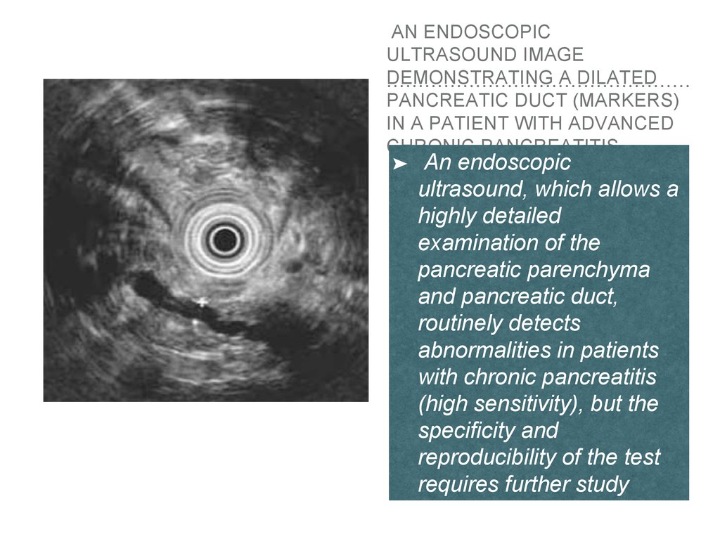
AN ENDOSCOPICULTRASOUND IMAGE
DEMONSTRATING A DILATED
PANCREATIC DUCT (MARKERS)
IN A PATIENT WITH ADVANCED
CHRONIC PANCREATITIS
➤
An endoscopic
ultrasound, which allows a
highly detailed
examination of the
pancreatic parenchyma
and pancreatic duct,
routinely detects
abnormalities in patients
with chronic pancreatitis
(high sensitivity), but the
specificity and
reproducibility of the test
requires further study
10.
COMPUTER TOMOGRAMPHYThe diagnostic information similar to ultrasound, is indicated for
suspected tumors and cysts of the pancreas;
CT features of chronic pancreatitis include:
➤ dilatation
of the main pancreatic duct
➤ pancreatic
➤ changes
calcification
in pancreatic size (i.e. atrophy), shape, and contour
➤ pancreatic
pseudocysts
11.
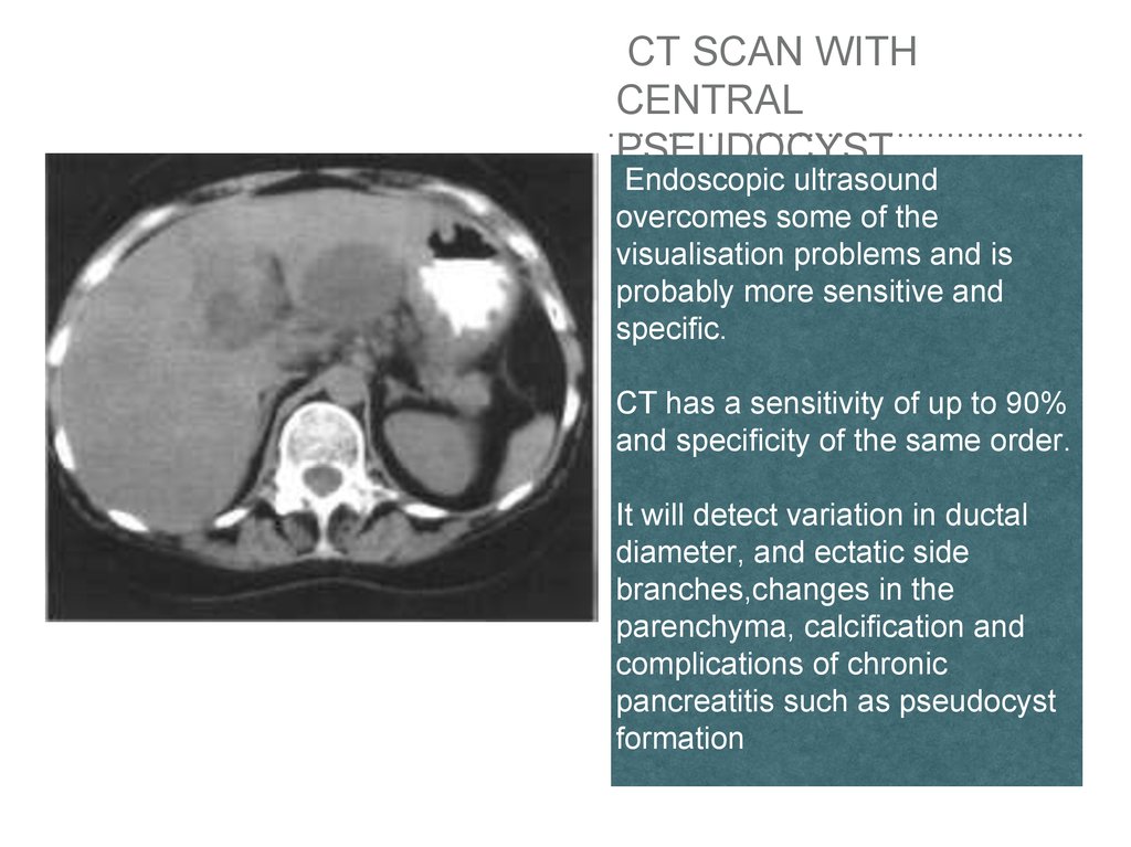
CT SCAN WITHCENTRAL
PSEUDOCYST
Endoscopic ultrasound
overcomes some of the
visualisation problems and is
probably more sensitive and
specific.
CT has a sensitivity of up to 90%
and specificity of the same order.
It will detect variation in ductal
diameter, and ectatic side
branches,changes in the
parenchyma, calcification and
complications of chronic
pancreatitis such as pseudocyst
formation
12.
ENDOSCOPIC RETROGRADE CHOLANGYIOPANKREATOGRAPHY
➤
reveals impaired patency of the main and secondary
ducts. “Chain of lakes" is a classic symptom of chronic
pancreatitis (areas of constriction and expansion of
virsunhov ducts).
➤
It is also possible the segmental or total obstruction of
a ductal system of pancreas.
13.
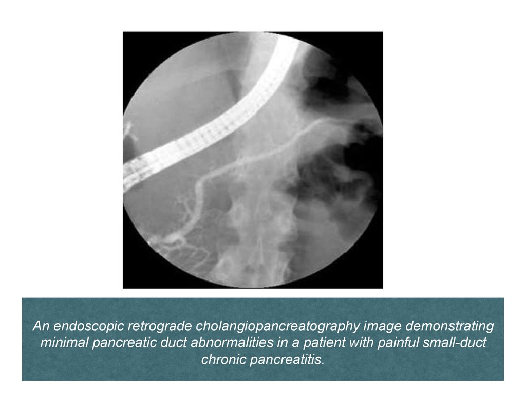
An endoscopic retrograde cholangiopancreatography image demonstratingminimal pancreatic duct abnormalities in a patient with painful small-duct
chronic pancreatitis.
14.
An endoscopic retrograde cholangiopancreatography imagedemonstrating massive pancreatic duct dilatation in a patient
with bigduct chronic pancreatitis.
15.
PANCREONECROSIS➤
➤
➤
➤
➤
➤
➤
Necrotizing Pancreatitis
Necrosis of pancreatic parenchyma or peripancreatic tissues
occurs in 10-15 % of patients.
It is characterized by a protracted clinical course, a high
incidence of local complications, and a high mortality rate.
There are 3 subtypes of necrotizing pancreatitis:
1 Necrosis of both pancreatic parenchyma and peripancreatic
tissues (most common).
2 Necrosis of only extrapancreatic tissue without necrosis of
pancreatic parenchyma (less common).
3 Necrosis of pancreatic parenchyma without surrounding
necrosis of peripancreatic tissue (very rare).
16.
➤➤
➤
➤
➤
➤
➤
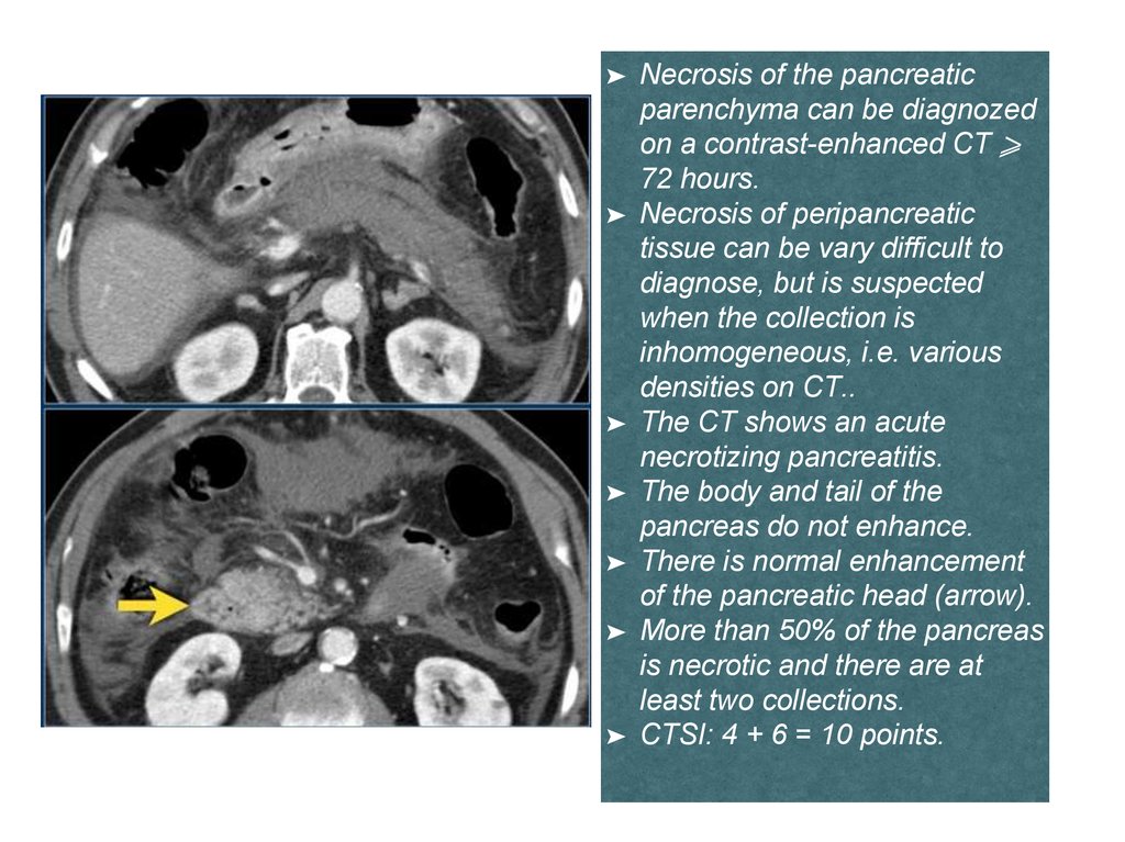
Necrosis of the pancreatic
parenchyma can be diagnozed
on a contrast-enhanced CT ⩾
72 hours.
Necrosis of peripancreatic
tissue can be vary difficult to
diagnose, but is suspected
when the collection is
inhomogeneous, i.e. various
densities on CT..
The CT shows an acute
necrotizing pancreatitis.
The body and tail of the
pancreas do not enhance.
There is normal enhancement
of the pancreatic head (arrow).
More than 50% of the pancreas
is necrotic and there are at
least two collections.
CTSI: 4 + 6 = 10 points.
17.
MRI➤
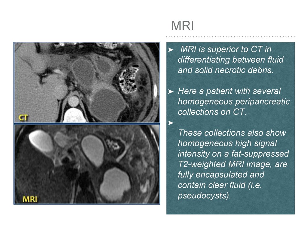
MRI is superior to CT in
differentiating between fluid
and solid necrotic debris.
➤
Here a patient with several
homogeneous peripancreatic
collections on CT.
➤
These collections also show
homogeneous high signal
intensity on a fat-suppressed
T2-weighted MRI image, are
fully encapsulated and
contain clear fluid (i.e.
pseudocysts).
18.
➤➤
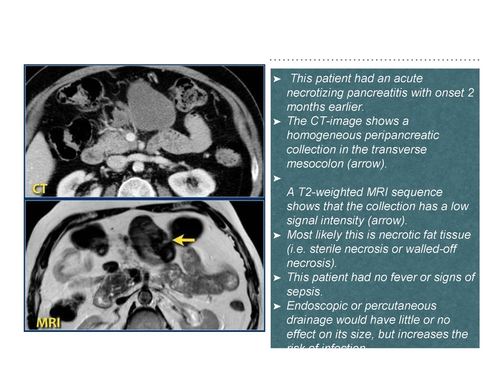
This patient had an acute
necrotizing pancreatitis with onset 2
months earlier.
The CT-image shows a
homogeneous peripancreatic
collection in the transverse
mesocolon (arrow).
➤
➤
➤
➤
A T2-weighted MRI sequence
shows that the collection has a low
signal intensity (arrow).
Most likely this is necrotic fat tissue
(i.e. sterile necrosis or walled-off
necrosis).
This patient had no fever or signs of
sepsis.
Endoscopic or percutaneous
drainage would have little or no
effect on its size, but increases the
risk of infection.
19.
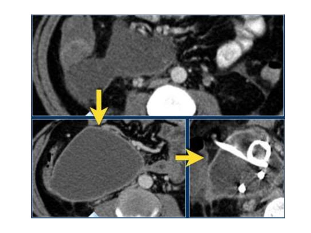
WALLED-OFF NECROSIS WON➤
➤
➤
➤
➤
➤
➤
On a follow-up scan the collection in the
right anterior pararenal space increased in
size.
It has fluid density and a thin enhancing
wall.
This can be a pseudocyst or walled-offnecrosis and it may or may not be infected.
The patient became septic and a
percutaneous drainage was performed.
After drainage the collection barely
diminished in size.
The patient underwent surgery and the
collection was found to consist of necrotic
debris, which was not appreciated on CT,
hence this was a walled-off-necrosis and
not a pseudocyst.
➤
The necrotic debris was too viscous for
successful percutaneous drainage.
Based on CT alone it is sometimes
impossible to determine whether a
collection contains fluid only or a mixture
of fluid and necrotic tissue.
➤
➤
➤
➤
➤
Consequently it is sometimes better to
describe these as 'indeterminate
peripancreatic collections'.
The images are of a patient with acute
pancreatitis.
On the upper image is a collection in the
area of the pancreatic head in the right
anterior pararenal space.
At this stage, it is not possible to
distinguish between an acute
peripancreatic fluid collection and acute
necrotic collection.
20.
21.
WALLED-OFFNECROSIS➤
➤
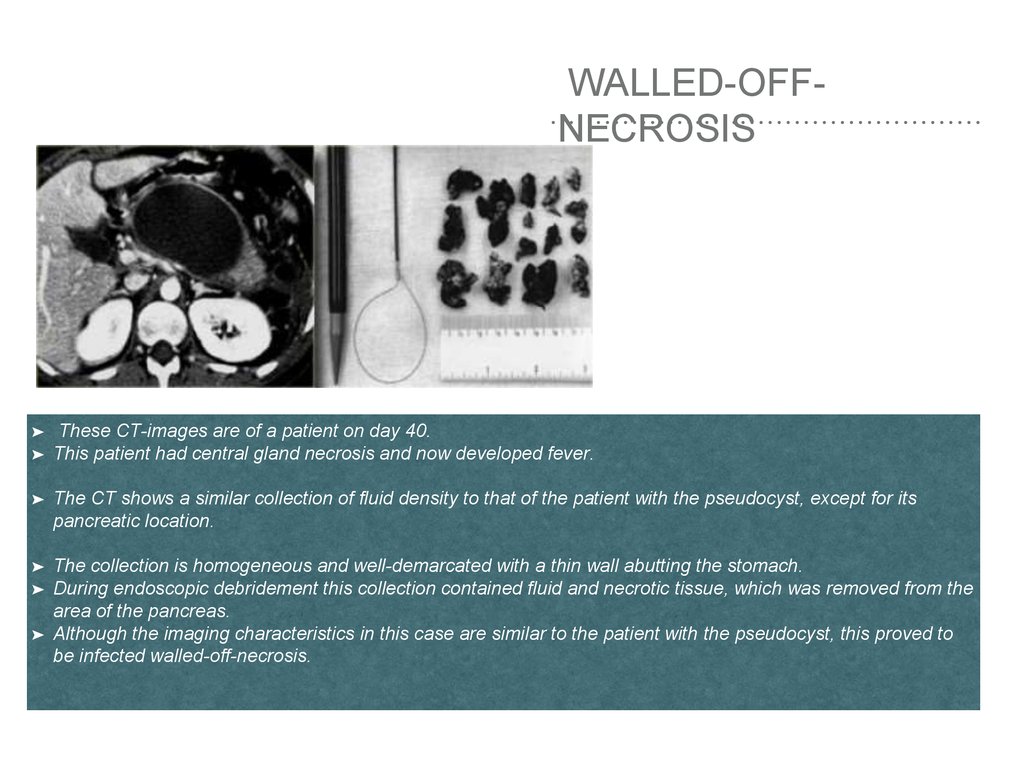
These CT-images are of a patient on day 40.
This patient had central gland necrosis and now developed fever.
➤
The CT shows a similar collection of fluid density to that of the patient with the pseudocyst, except for its
pancreatic location.
➤
The collection is homogeneous and well-demarcated with a thin wall abutting the stomach.
During endoscopic debridement this collection contained fluid and necrotic tissue, which was removed from the
area of the pancreas.
Although the imaging characteristics in this case are similar to the patient with the pseudocyst, this proved to
be infected walled-off-necrosis.
➤
➤
22.
WALLED-OFFNECROSIS➤
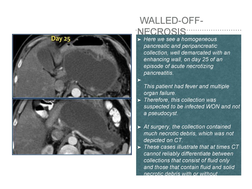
Here we see a homogeneous
pancreatic and peripancreatic
collection, well demarcated with an
enhancing wall, on day 25 of an
episode of acute necrotizing
pancreatitis.
➤
➤
➤
➤
This patient had fever and multiple
organ failure.
Therefore, this collection was
suspected to be infected WON and not
a pseudocyst.
At surgery, the collection contained
much necrotic debris, which was not
depicted on CT.
These cases illustrate that at times CT
cannot reliably differentiate between
collections that consist of fluid only
and those that contain fluid and solid
necrotic debris with or without
infection.
23.
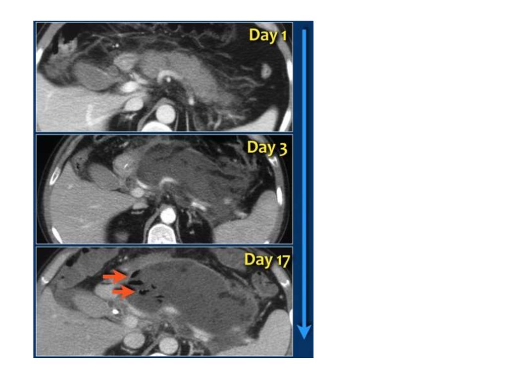
INFECTED NECROSIS➤
➤
➤
➤
➤
➤
➤
➤
➤
➤
➤
➤
➤
➤
Infected necrosis is:
• Infection of necrotic pancreatic parenchyma or extrapancreatic fatty tissue - i.e. infected ANC or
infected WON, depending on degree of encapsulation.
• Usually occurs in the 2nd-4th week and rarely in the first week.
• Most severe local complication of acute necrotizing pancreatitis.
• Most common cause of death in patients with acute pancreatitis.
• Diagnose infected necrosis when there are gas bubbles on CT (seen in 40%) or when FNA is positive
for bacteria.
This case is a typical example of infected pancreatic necrosis.
• On day 1 there is enhancement of the pancreas and it just looks like a mild interstitial pancreatitis.
On day 3 there is no enhancement of the pancreas, consistent with necrosis.
• The necrosis also involves the peripancreatic tissue.
• So this is an ANC - acute necrotic collection.
• On day 17 there are gas bubbles in the necrotic collection consistent with infected pancreatic and
peripancreatic necrosis.
• A wall surrounds the collection.
The term pancreatic abcess is no longer used, since a collection of pus without necrotic tissue is
extremely uncommon in acute pancreatitis.
24.
25.
CENTRAL GLANDNECROSIS
➤
Central gland necrosis is a specific
form of necrotizing pancreatitis,
representing full thickness necrosis
between the pancreatic head and tail
and is nearly always associated with
disruption of the pancreatic duct.
➤
This leads to persistent collections as
the viable pancreatic tail continues to
secrete pancreatic juices.
➤
These collections mayreact poorly to
endoscopic or percutaneous
drainage.
➤
Definitive treatment may require
distal pancreatectomy or long-term
endoscopic drainage.