


 medicine
Similar presentations:


")
")
Radionuclide examination of urinary tract
1.
Kazan State Medical Universitytopic :radionuclide examination of
urinary tract
sunny bhasal
Group 1317
2.
Radionuclide diagnostics methods▪ Noninvasive
▪ Are primarily physiologic
▪ Functional
▪ Does not provide the same anatomic details
▪ As morphologic method (X-ray,US,CT,MRI)
3.
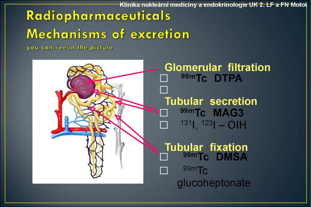
Klinika nukleární medicíny a endokrinologie UK 2. LF a FN MotolGlomerular filtration
⬜ 99mTc DTPA
⬜
Tubular secretion
⬜ 99mTc MAG3
⬜ 131I, 123I – OIH
Tubular fixation
⬜ 99mTc DMSA
⬜ 99mTc
glucoheptonate
4.
Klinika nukleární medicíny a endokrinologie UK 2. LF a FN Motol• Nuclear medicine is a medical specialty involving the application
of radioactive substances in the diagnosis and treatment
of disease.
• In nuclear medicine procedures, radionuclides are combined with
other elements to form chemical compounds, or else combined
with existing pharmaceutical compounds, to
form radiopharmaceuticals. These radiopharmaceuticals, once
administered to the patient, can localize to specific organs or
cellular receptors
• There are several techniques of diagnostic nuclear medicine.
• Scintigraphy
• PET
• SPECT
5.
Klinika nukleární medicíny a endokrinologie UK 2. LF a FN Motol6.
Klinika nukleární medicíny a endokrinologie UK 2. LF a FN Motol• Three basic classes of radionuclide are employed in nuclear
renography.
•Filtered agents
•Excreted agents
•Cortical imaging agents
7.
Klinika nukleární medicíny a endokrinologie UK 2. LF a FN Motol• DTPA and MAG3 are filtered through the glomerulus. This is
useful in evaluating:
• Perfusion
• Vascular supply
• Filtration
• Measuring renal function (glomerular filtration rate)
• Drainage
• Detects obstruction
8.
Klinika nukleární medicíny a endokrinologie UK 2. LF a FN Motol• MAG3 and Hipuran are excreted by the renal tubules. These
radionuclides are helpful in evaluating patients with:
• Diminished renal function
• Kidney transplants
• MAG3 is both filtered and excreted so some radiologists prefer it
to other radionuclides
9.
Klinika nukleární medicíny a endokrinologie UK 2. LF a FN Motol• DMSA and Glucoheptonate are accumulated in the cortex so
they are helpful in evaluating:
•Renal scarring from chronic infection
•Infarction
•Renal mass
•Differential renal mass (proportion of total renal
mass contributed by each kidney)
10.
Klinika nukleární medicíny a endokrinologie UK 2. LF a FN Motol• There are two main radionuclide techniques for studying the
kidneys:
• The Renogram which measures renal function. Scans of renal
morphology (DMSA scan). The advent of CT and ultrasound has
reduced the need for such scans. They are now used mainly for
evaluating renal scanning.
11.
Klinika nukleární medicíny a endokrinologie UK 2. LF a FN Motol12.
Klinika nukleární medicíny a endokrinologie UK 2. LF a FN MotolRadiopharmaceticals
99mTc-DTPA – Diethylentriamine pentaacetic acid
belongs to the group of chelate compounds
is excreted from kidneys through glomerular filtration
with a half-life of 70 minutes
it is the most suitable substance
for measuring glomerular filtration (GFR)
and good imaging of renal parenchyma
Vižďa J. a kol : Atlas of Renal Scintigraphy, 2002.
13.
Klinika nukleární medicíny a endokrinologie UK 2. LF a FN MotolRadiopharmaceticals
99mTc-MAG3 - Mercapto-acetyltriglycine
-is one of the newly developed radiopharmaceuticals
-is rapidly excreted by the kidneys via active tubular secretion
and minor part via glomerular filtration
-organic anions (which include MAG3) have a carboxyl group which
specifically binds to the receptors of tubular cells mediating the
active transport of MAG3 into the cells of the proximal tubulus
-with normaI renal function 70% of the administered activity of the
radiopharmaceutical (RP) is excreted within 30 minutes after the
application
14.
Klinika nukleární medicíny a endokrinologie UK 2. LF a FN MotolDynamic renal study
Radiopharmaceutical
99mTc - MAG3
Patient Preparation
adequately hydration prior to the examination
it is recommended to drink 100 ml of liquids per 10 kg
of the body weight 30 min prior the examination
empty bladder
p.are requested to void completely prior to the study
15.
Klinika nukleární medicíny a endokrinologie UK 2. LF a FN Motol16.
Klinika nukleární medicíny a endokrinologie UK 2. LF a FN Motol17.
Klinika nukleární medicíny a endokrinologie UK 2. LF a FN Motol18.
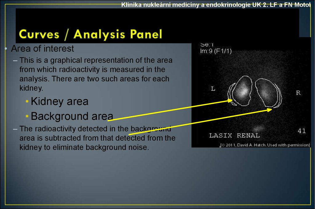
Klinika nukleární medicíny a endokrinologie UK 2. LF a FN Motol• Area of interest
– This is a graphical representation of the area
from which radioactivity is measured in the
analysis. There are two such areas for each
kidney.
• Kidney area
• Background area
– The radioactivity detected in the background
area is subtracted from that detected from the
kidney to eliminate background noise.
19.
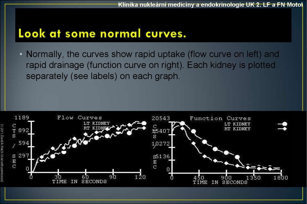
Klinika nukleární medicíny a endokrinologie UK 2. LF a FN Motol• Normally, the curves show rapid uptake (flow curve on left) and
rapid drainage (function curve on right). Each kidney is plotted
separately (see labels) on each graph.
20.
Klinika nukleární medicíny a endokrinologie UK 2. LF a FN MotolI. Vascular phase
II. Secretory
III. Excretory
A
II.
III.
I.
čas
21.
Klinika nukleární medicíny a endokrinologie UK 2. LF a FN MotolPatterns of renographic curves
A
obstructed pattern
impaired renal function
parenchymal lesion pattern
renal failure patern
normal
renal failure pattern
without measurable kidney uptake
čas
22.
Klinika nukleární medicíny a endokrinologie UK 2. LF a FN MotolNormal renal scan
ANT
RPO
POST
LPO
23.
Klinika nukleární medicíny a endokrinologie UK 2. LF a FN MotolEvaluation
- number of kidneys
- position
- size
- shape
- the size, number and location of areas cortical loss
- split renal function
Note!
Cortical „cold“ defect may be due to
different etiology :
tumor, abscess, cysts ….
alrealdy is necesarry to compare with US
24.
Klinika nukleární medicíny a endokrinologie UK 2. LF a FN MotolThank You!!!!