


 biology
Similar presentations:
 Everywhere!")


Bacteria. Overview
1. Bacteria
Copyright © 2005 Pearson Education, Inc. publishing as Benjamin Cummings2.
• Overview: They’re (Almost) Everywhere!• Most prokaryotes are microscopic
– But what they lack in size they more than
make up for in numbers
• The number of prokaryotes in a single handful
of fertile soil
– Is greater than the number of people who have
ever lived
Copyright © 2005 Pearson Education, Inc. publishing as Benjamin Cummings
3.
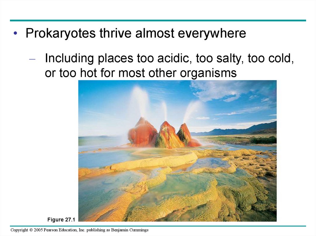
• Prokaryotes thrive almost everywhere– Including places too acidic, too salty, too cold,
or too hot for most other organisms
Figure 27.1
Copyright © 2005 Pearson Education, Inc. publishing as Benjamin Cummings
4.
• Biologists are discovering– That these organisms have an astonishing
genetic diversity
Copyright © 2005 Pearson Education, Inc. publishing as Benjamin Cummings
5.
• Structural, functional, and genetic adaptationscontribute to prokaryotic success
• Most prokaryotes are unicellular
– Although some species form colonies
Copyright © 2005 Pearson Education, Inc. publishing as Benjamin Cummings
6. Cell-Surface Structures
• One of the most important features of nearly allprokaryotic cells
– Is their cell wall, which maintains cell shape,
provides physical protection, and prevents the
cell from bursting in a hypotonic environment
Copyright © 2005 Pearson Education, Inc. publishing as Benjamin Cummings
7.
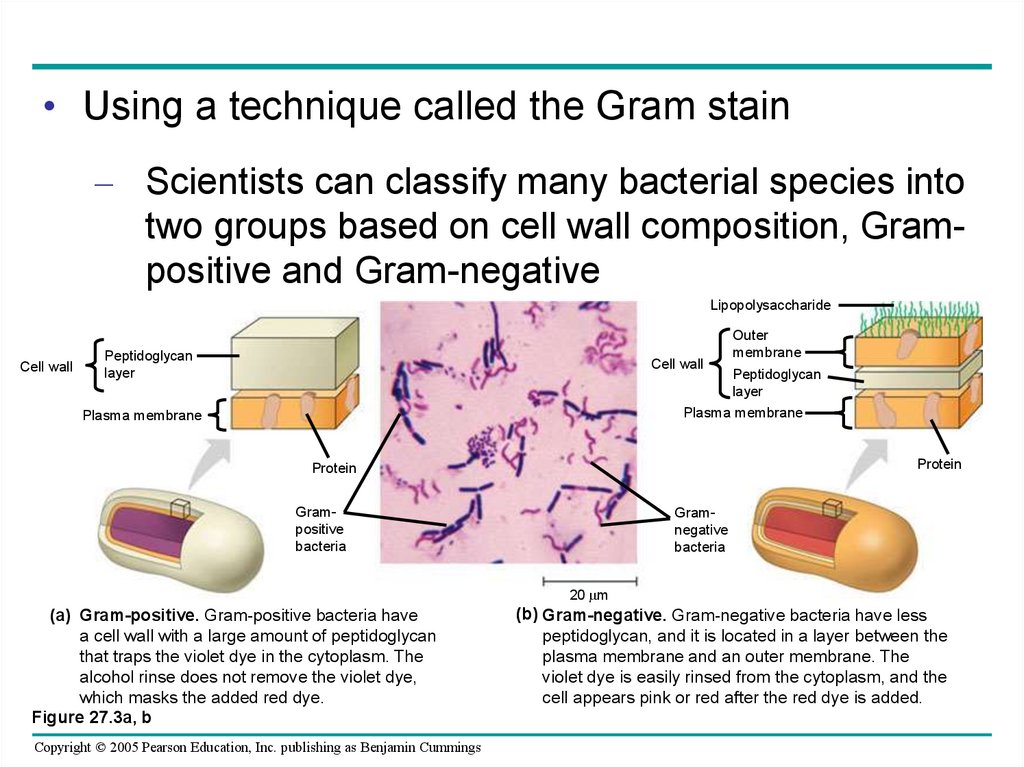
• Using a technique called the Gram stain– Scientists can classify many bacterial species into
two groups based on cell wall composition, Grampositive and Gram-negative
Lipopolysaccharide
Cell wall
Peptidoglycan
layer
Cell wall
Outer
membrane
Peptidoglycan
layer
Plasma membrane
Plasma membrane
Protein
Protein
Grampositive
bacteria
Gramnegative
bacteria
20 m
(a) Gram-positive. Gram-positive bacteria have
a cell wall with a large amount of peptidoglycan
that traps the violet dye in the cytoplasm. The
alcohol rinse does not remove the violet dye,
which masks the added red dye.
Figure 27.3a, b
Copyright © 2005 Pearson Education, Inc. publishing as Benjamin Cummings
(b) Gram-negative. Gram-negative bacteria have less
peptidoglycan, and it is located in a layer between the
plasma membrane and an outer membrane. The
violet dye is easily rinsed from the cytoplasm, and the
cell appears pink or red after the red dye is added.
8.
• The cell wall of many prokaryotes– Is covered by a capsule, a sticky layer of
polysaccharide or protein
200 nm
Capsule
Figure 27.4
Copyright © 2005 Pearson Education, Inc. publishing as Benjamin Cummings
9.
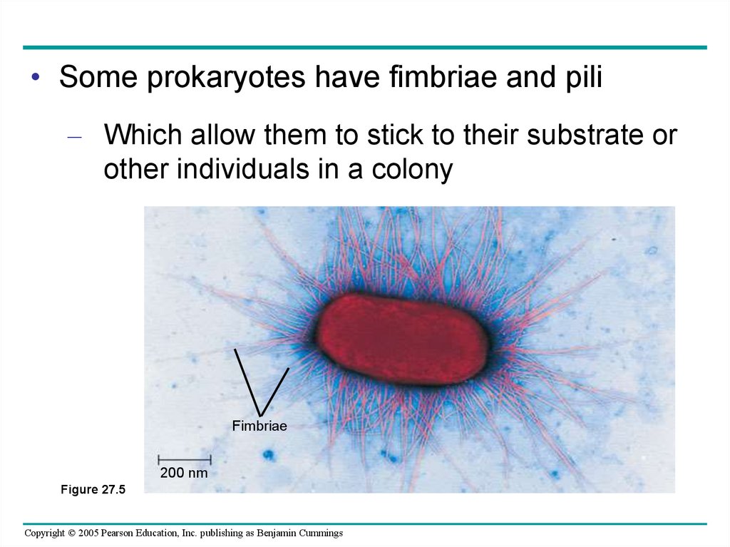
• Some prokaryotes have fimbriae and pili– Which allow them to stick to their substrate or
other individuals in a colony
Fimbriae
200 nm
Figure 27.5
Copyright © 2005 Pearson Education, Inc. publishing as Benjamin Cummings
10. Motility
• Most motile bacteria propel themselves by flagella– Which are structurally and functionally different
from eukaryotic flagella
Flagellum
Filament
50 nm
Cell wall
Hook
Basal apparatus
Figure 27.6
Plasma
membrane
Copyright © 2005 Pearson Education, Inc. publishing as Benjamin Cummings
11. Classification Systems in the Procaryotae
1.Microscopic morphology
2.
Macroscopic morphology – colony appearance
3.
Physiological / biochemical characteristics
4.
Chemical analysis
5.
Serological analysis
6.
Genetic and molecular analysis
G + C base composition
DNA analysis using genetic probes
Nucleic acid sequencing and rRNA analysis
Copyright © 2005 Pearson Education, Inc. publishing as Benjamin Cummings
12. Diagnostic Scheme for Medical Use
• Uses phenotypic qualities in identification–
restricted to bacterial disease agents
–
divides based on cell wall structure, shape,
arrangement, and physiological traits
Copyright © 2005 Pearson Education, Inc. publishing as Benjamin Cummings
13. Species and Subspecies
• Species–
collection of bacterial cells which share an overall similar
pattern of traits in contrast to other bacteria whose pattern
differs significantly
• Strain or variety
–
culture derived from a single parent that differs in
structure or metabolism from other cultures of that
species (biovars, morphovars)
• Type
–
subspecies that can show differences in antigenic
makeup (serotype or serovar), susceptibility to bacterial
viruses (phage type) and in pathogenicity (pathotype)
Copyright © 2005 Pearson Education, Inc. publishing as Benjamin Cummings
14.
• Prokaryotic cells have a variety of shapes– The three most common of which are spheres
(cocci), rods (bacilli), and spirals
1 m
Figure 27.2a–c (a) Spherical (cocci)
2 m
(b) Rod-shaped (bacilli)
Copyright © 2005 Pearson Education, Inc. publishing as Benjamin Cummings
5 m
(c) Spiral
15.
•Cocci (or coccus for a single cell) areround cells, sometimes slightly flattened
when they are adjacent to one another.
•Bacilli (or bacillus for a single cell) are
rod-shaped bacteria.
•Spirilla (or spirillum for a single cell) are
curved bacteria which can range from a
gently curved shape to a corkscrew-like
spiral. Many spirilla are rigid and capable
of movement. A special group of spirilla
known as spirochetes are long, slender,
and flexible.
Copyright © 2005 Pearson Education, Inc. publishing as Benjamin Cummings
16.
1. DiplococciThe cocci are arranged in pairs.
Examples: Streptococcus pneumoniae, Moraxella
catarrhalis, Neisseria gonorrhoeae, etc.
2. Streptococci
The cocci are arranged in chains, as the cells divide in one plane.
Examples: Streptococcus pyogenes, Streptococcus agalactiae
Copyright © 2005 Pearson Education, Inc. publishing as Benjamin Cummings
17.
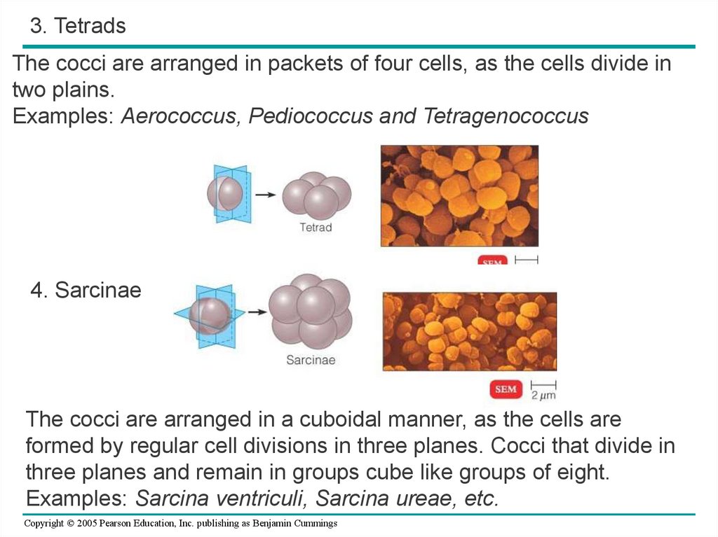
3. TetradsThe cocci are arranged in packets of four cells, as the cells divide in
two plains.
Examples: Aerococcus, Pediococcus and Tetragenococcus
4. Sarcinae
The cocci are arranged in a cuboidal manner, as the cells are
formed by regular cell divisions in three planes. Cocci that divide in
three planes and remain in groups cube like groups of eight.
Examples: Sarcina ventriculi, Sarcina ureae, etc.
Copyright © 2005 Pearson Education, Inc. publishing as Benjamin Cummings
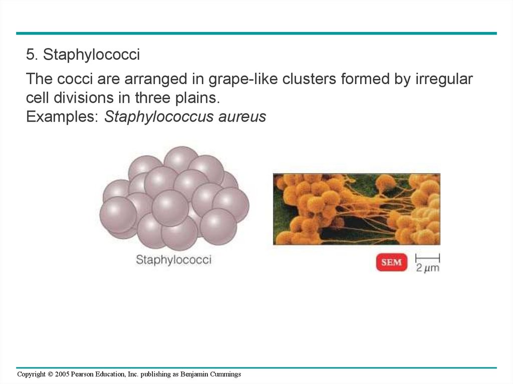
18.
5. StaphylococciThe cocci are arranged in grape-like clusters formed by irregular
cell divisions in three plains.
Examples: Staphylococcus aureus
Copyright © 2005 Pearson Education, Inc. publishing as Benjamin Cummings
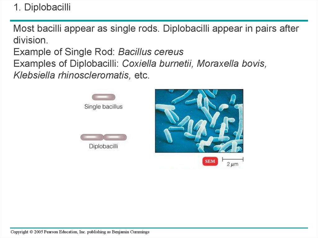
19.
1. DiplobacilliMost bacilli appear as single rods. Diplobacilli appear in pairs after
division.
Example of Single Rod: Bacillus cereus
Examples of Diplobacilli: Coxiella burnetii, Moraxella bovis,
Klebsiella rhinoscleromatis, etc.
Copyright © 2005 Pearson Education, Inc. publishing as Benjamin Cummings
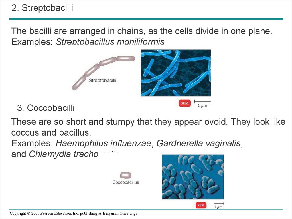
20.
2. StreptobacilliThe bacilli are arranged in chains, as the cells divide in one plane.
Examples: Streptobacillus moniliformis
3. Coccobacilli
These are so short and stumpy that they appear ovoid. They look like
coccus and bacillus.
Examples: Haemophilus influenzae, Gardnerella vaginalis,
and Chlamydia trachomatis
Copyright © 2005 Pearson Education, Inc. publishing as Benjamin Cummings
21.
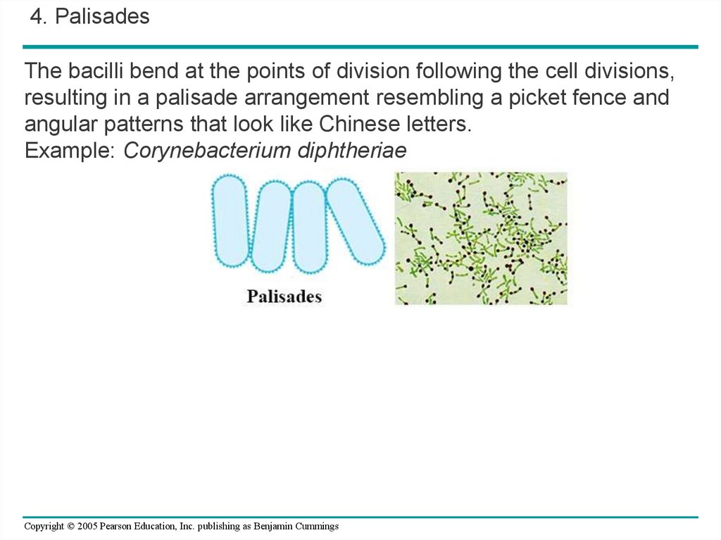
4. PalisadesThe bacilli bend at the points of division following the cell divisions,
resulting in a palisade arrangement resembling a picket fence and
angular patterns that look like Chinese letters.
Example: Corynebacterium diphtheriae
Copyright © 2005 Pearson Education, Inc. publishing as Benjamin Cummings
22.
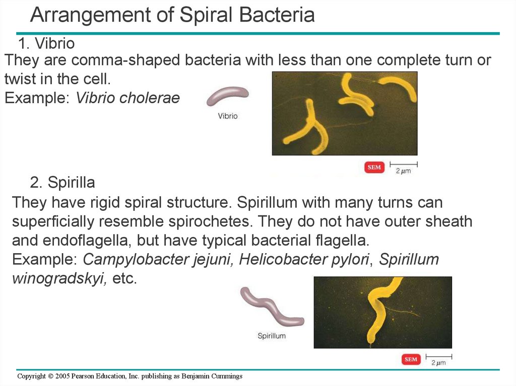
Arrangement of Spiral Bacteria1. Vibrio
They are comma-shaped bacteria with less than one complete turn or
twist in the cell.
Example: Vibrio cholerae
2. Spirilla
They have rigid spiral structure. Spirillum with many turns can
superficially resemble spirochetes. They do not have outer sheath
and endoflagella, but have typical bacterial flagella.
Example: Campylobacter jejuni, Helicobacter pylori, Spirillum
winogradskyi, etc.
Copyright © 2005 Pearson Education, Inc. publishing as Benjamin Cummings
23.
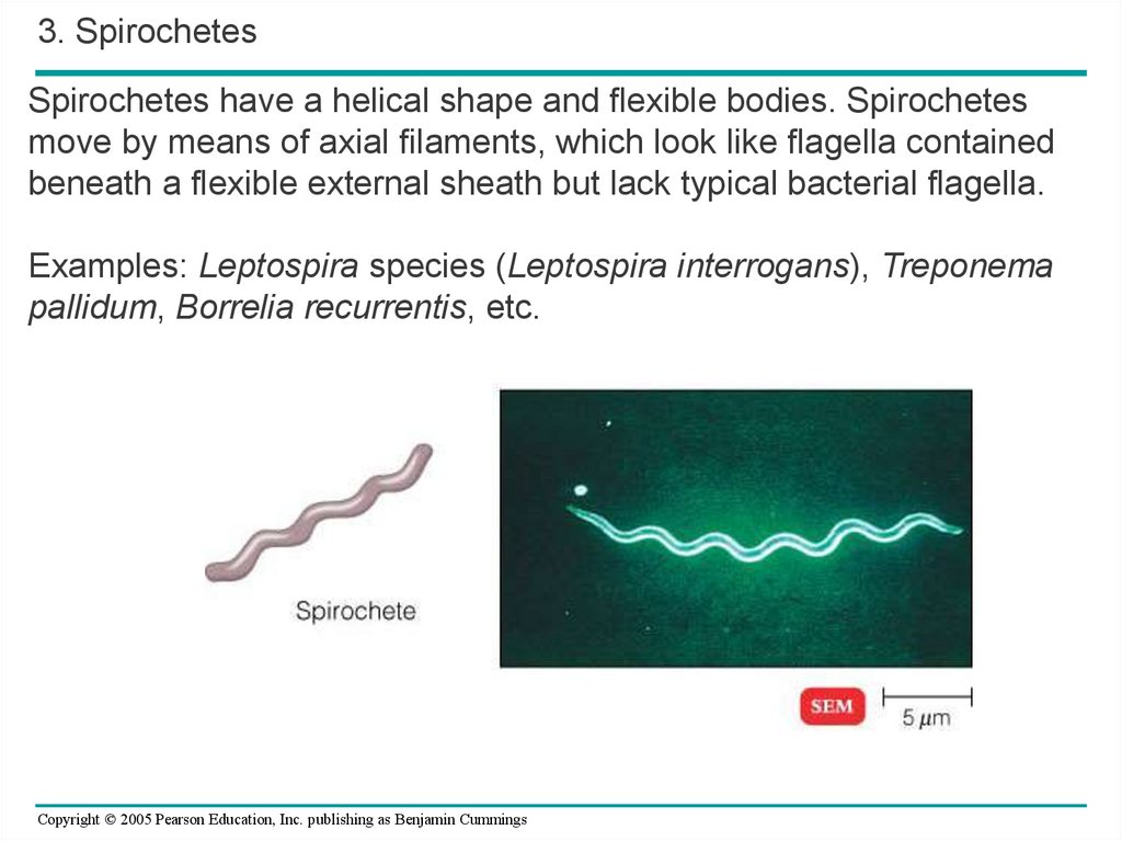
3. SpirochetesSpirochetes have a helical shape and flexible bodies. Spirochetes
move by means of axial filaments, which look like flagella contained
beneath a flexible external sheath but lack typical bacterial flagella.
Examples: Leptospira species (Leptospira interrogans), Treponema
pallidum, Borrelia recurrentis, etc.
Copyright © 2005 Pearson Education, Inc. publishing as Benjamin Cummings
24.
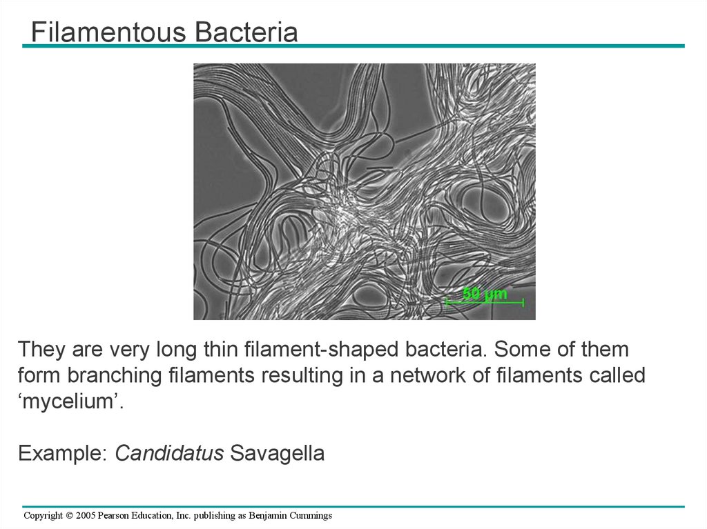
Filamentous BacteriaThey are very long thin filament-shaped bacteria. Some of them
form branching filaments resulting in a network of filaments called
‘mycelium’.
Example: Candidatus Savagella
Copyright © 2005 Pearson Education, Inc. publishing as Benjamin Cummings
25.
• In a heterogeneous environment, manybacteria exhibit taxis
– The ability to move toward or away from
certain stimuli
Copyright © 2005 Pearson Education, Inc. publishing as Benjamin Cummings
26. Internal and Genomic Organization
• Prokaryotic cells– Usually lack complex compartmentalization
Copyright © 2005 Pearson Education, Inc. publishing as Benjamin Cummings
27.
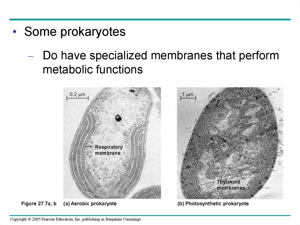
• Some prokaryotes– Do have specialized membranes that perform
metabolic functions
0.2 m
1 m
Respiratory
membrane
Thylakoid
membranes
Figure 27.7a, b
(a) Aerobic prokaryote
Copyright © 2005 Pearson Education, Inc. publishing as Benjamin Cummings
(b) Photosynthetic prokaryote
28.
Copyright © 2005 Pearson Education, Inc. publishing as Benjamin Cummings29.
Copyright © 2005 Pearson Education, Inc. publishing as Benjamin Cummings30.
Copyright © 2005 Pearson Education, Inc. publishing as Benjamin Cummings31.
Copyright © 2005 Pearson Education, Inc. publishing as Benjamin Cummings32.
Copyright © 2005 Pearson Education, Inc. publishing as Benjamin Cummings33.
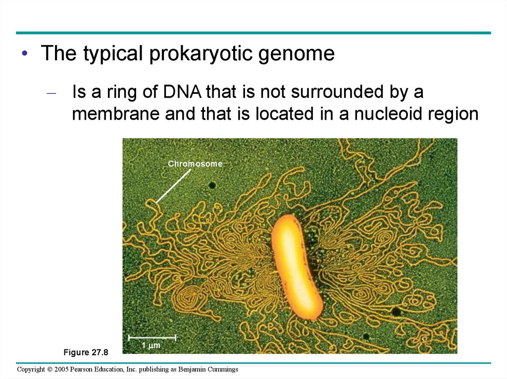
• The typical prokaryotic genome– Is a ring of DNA that is not surrounded by a
membrane and that is located in a nucleoid region
Chromosome
Figure 27.8
1 m
Copyright © 2005 Pearson Education, Inc. publishing as Benjamin Cummings
34.
• Some species of bacteria– Also have smaller rings of DNA called
plasmids
Copyright © 2005 Pearson Education, Inc. publishing as Benjamin Cummings
35. Reproduction and Adaptation
• Prokaryotes reproduce quickly by binary fission– And can divide every 1–3 hours
Copyright © 2005 Pearson Education, Inc. publishing as Benjamin Cummings
36.
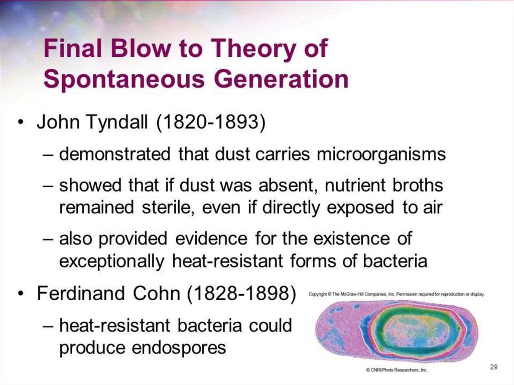
• Many prokaryotes form endospores– Which can remain viable in harsh conditions
for centuries
Endospore
0.3 m
Figure 27.9
Copyright © 2005 Pearson Education, Inc. publishing as Benjamin Cummings
37.
Copyright © 2005 Pearson Education, Inc. publishing as Benjamin Cummings38.
Copyright © 2005 Pearson Education, Inc. publishing as Benjamin Cummings39.
• Rapid reproduction and horizontal genetransfer
– Facilitate the evolution of prokaryotes to
changing environments
Copyright © 2005 Pearson Education, Inc. publishing as Benjamin Cummings
40.
• A great diversity of nutritional and metabolicadaptations have evolved in prokaryotes
• Examples of all four models of nutrition are
found among prokaryotes
– Photoautotrophy
– Chemoautotrophy
– Photoheterotrophy
– Chemoheterotrophy
Copyright © 2005 Pearson Education, Inc. publishing as Benjamin Cummings