


 Membrane")


")


 biology
 english
Similar presentations:


Morphology & structure of microorganisms
1. Microbiology
2. Morphology & structure of Microorganisms Lecture 1
Morphology & structure ofMicroorganisms
Lecture 1
3. Microbiology
Microbiology is the study of organisms that toosmall to be seen without magnification.
Microbiology derived from:
Micro: too small to be seen with naked eye.
Bio: life
Logy: study of
4. History of Microbiology
1. Robert Koch was notable for his discovery ofthe bacterium Bacillus anthracis in 1876 and such had
launched the new scientific field of bacteriology.
His
discoveries about microscopic techniques and
different pathogenic bacteria like the Bacillus
anthracis, Staphylococcus, Mycobacterium
tuberculosis, Vibrio cholerae, etc. ensued the “golden
age” of scientific discovery.
In honor of his phenomenal discoveries, Koch was
5.
2. Robert Hooke: Known for his discovery ofthe first ever compound microscope, Robert Hooke
is often called the “Father of Cytology“. He used
such invention to view and observe the most minute
and previously unknown structures called as the
cells.
Aside
from his discoveries in biology, Hooke also
has made several significant contributions to the
field of agriculture, physics, chemistry, and
6. Microorganisms Are Ubiquitous
Microorganisms include:Bacteria
Viruses
Fungi
Parasites (protozoa & helminthes)
Algae
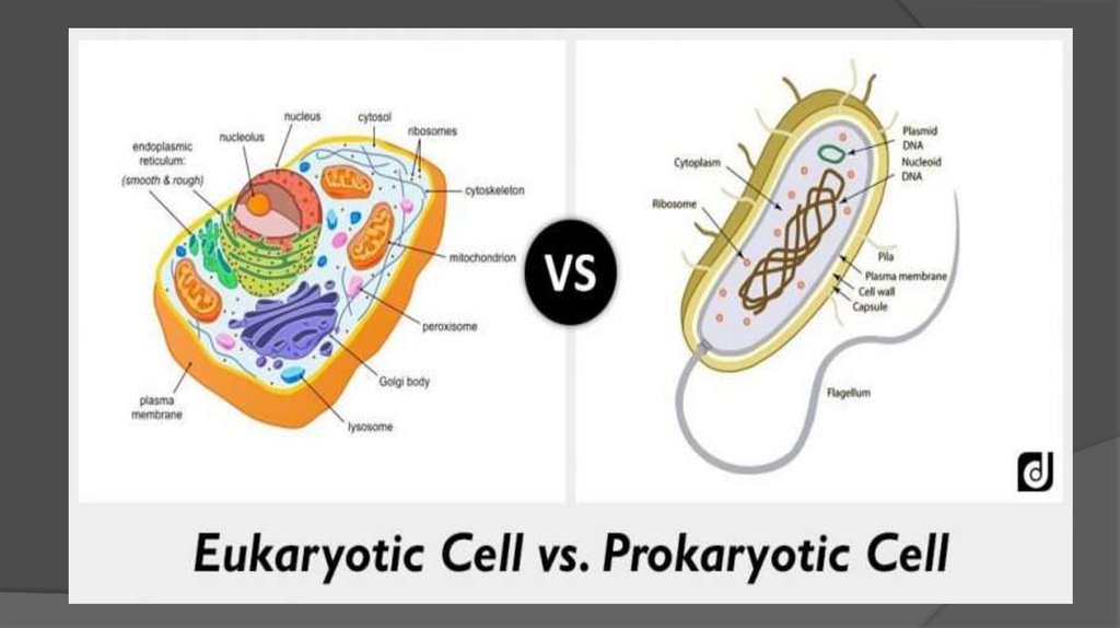
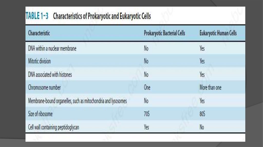
7. Introduction
Thereare several classes of living
organisms.
Based on the organization of their cellular
structures, all living cells can be divided
into two groups:
Eukaryotic cell: animals, plants, fungi,
protozoa and algae.
8.
9.
10. Prokaryotic Cells
Muchsmaller (microns) and more simple
than eukaryotes.
Prokaryotes are molecules surrounding by a
membrane and cell wall.
They lack true nucleus and don’t have
membrane
bound
organelles
like
mitochondria, Golgi complex, etc.
11.
12. Size of bacteria
Unitof measurement in bacteriology is
micron (micrometer) (µm) .
Bacteria of medical importance
0.2-1.5 µm in diameter
3-5 µm in length
13. Shape of Bacteria
Cocci:spherical / oval shaped (major
groups)
Bacilli: Rod shaped
Vibrios: comma shaped
Spirilla: rigid spiral forms
Spirochetes: flexible spiral forms
Actionomycetes: branching filamentous
14. Shape of Bacteria
15.
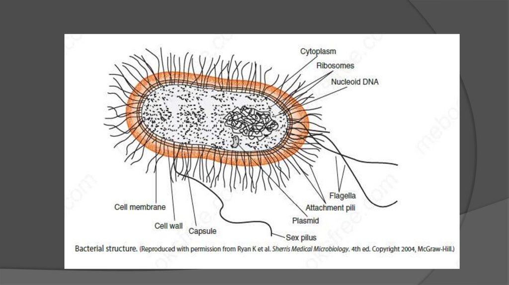
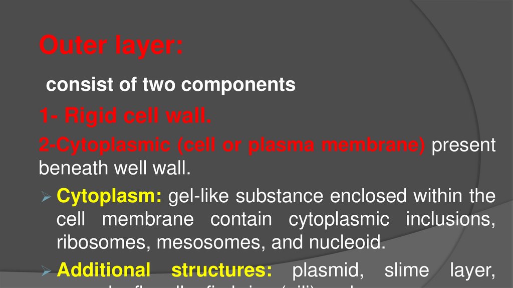
Outer layer:consist of two components
1- Rigid cell wall.
2-Cytoplasmic (cell or plasma membrane) present
beneath well wall.
Cytoplasm: gel-like substance enclosed within the
cell membrane contain cytoplasmic inclusions,
ribosomes, mesosomes, and nucleoid.
Additional structures: plasmid, slime layer,
16. Structure & Function of Cell Components
Structure & Functionof Cell Components
17.
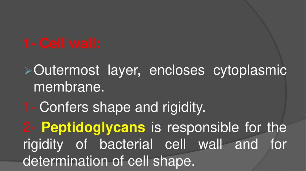
1- Cell wall:Outermost
layer, encloses cytoplasmic
membrane.
1- Confers shape and rigidity.
2- Peptidoglycans is responsible for the
rigidity of bacterial cell wall and for
determination of cell shape.
18.
4- Cell wall cannot be seen by direct lightmicroscope and do not stain with simple stain.
5- Carries bacterial antigens (important in
virulence & immunity)
6- Chemical nature of the cell wall helps to
divide bacteria into two broad groups (Gram
positive & Gram negative).
7- Gram +ve bacteria have simpler chemical
nature than –ve bacteria.
19.
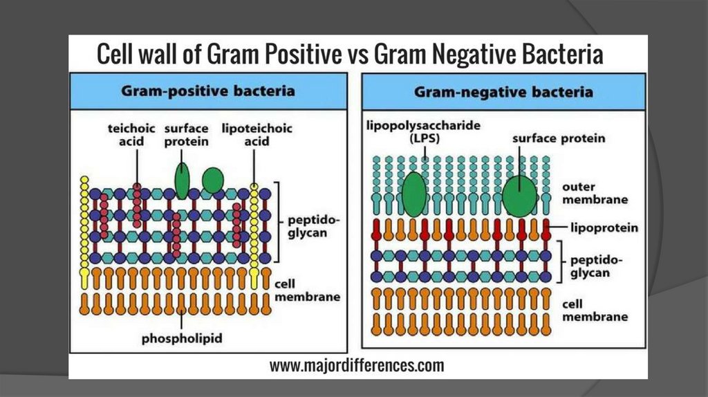
20. Gram Positive Cell Wall
TheGram +ve cell wall is composed of a
thick, multilayered Peptidoglycan sheath
outside of the cytoplasmic membrane.
Teichoic acids are linked to and embedded in
the peptidoglycan.
lipoteichoic acids extend into the cytoplasmic
membrane.
21. Gram negative Cell Wall
TheGram -ve cell wall is composed of an outer
membrane
linked
to
thin
single-layered
peptidoglycan by lipoproteins.
The peptidoglycan is located within the periplasmic
space that created between the outer and inner
membrane.
The outer membrane includes Porins, which allow
the passage of small hydrophilic molecules across
22.
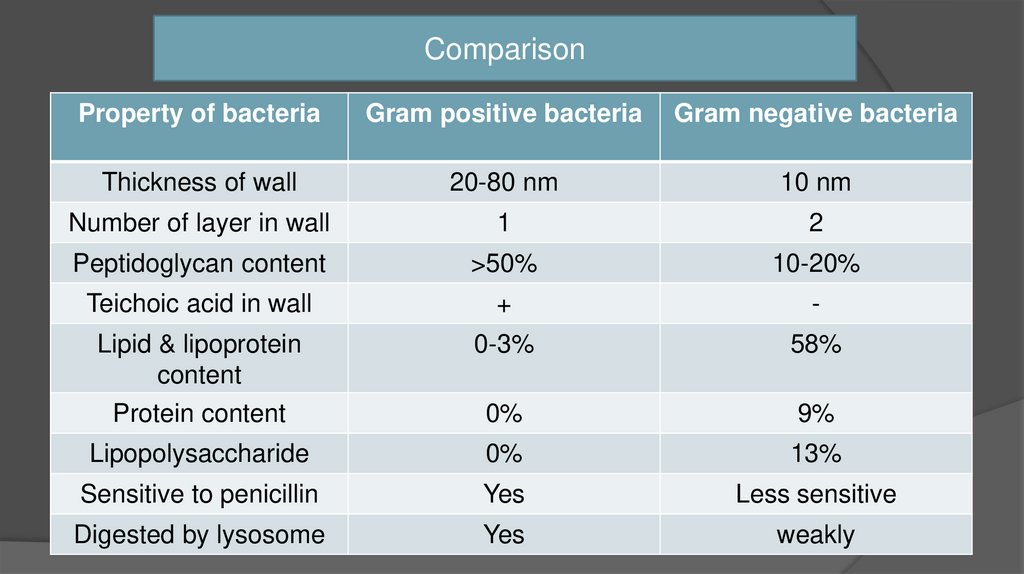
ComparisonProperty of bacteria
Gram positive bacteria
Gram negative bacteria
Thickness of wall
20-80 nm
10 nm
Number of layer in wall
1
2
Peptidoglycan content
>50%
10-20%
Teichoic acid in wall
+
-
Lipid & lipoprotein
content
Protein content
0-3%
58%
0%
9%
Lipopolysaccharide
0%
13%
Sensitive to penicillin
Yes
Less sensitive
Digested by lysosome
Yes
weakly
23. 2- Cytoplasmic (Plasma) Membrane
Thinlayer 5-10 nm, separates cell wall
from cytoplasm
Acts as a semi-permeable membrane:
controls the inflow and outflow of metabolites
Composed of lipoproteins with small amount
of carbohydrates
24. 3- Cytoplasm
Colloidalsystem of variety of organic
& inorganic solutes in viscous watery
solution.
- Cytoplasmic components
1- Ribosomes: function
synthesis (70s).
in
protein
25.
2- Mesosomes1- Multi-laminated structures formed as invaginations
of plasma membrane.
2- Principle sites of respiratory enzyme.
3- coordinate nuclear & cytoplasmic division during
binary fission.
4- more prominent G+ve bacteria
26.
4- NucleusNo
nucleolus
No nuclear membrane
Oval or elongated bodies generally 1 per
cell
Genome- single, circular bodies double
stranded DNA.
27. Additional Organelles
1-Plasmid:Extra-nuclear
genetic elements consisting of DNA.
Transmitted to daughter cells during binary fission
May ne transferred from one bacterium to another
Not essential for life of cell.
Confer certain properties e.g. drug resistant gene.
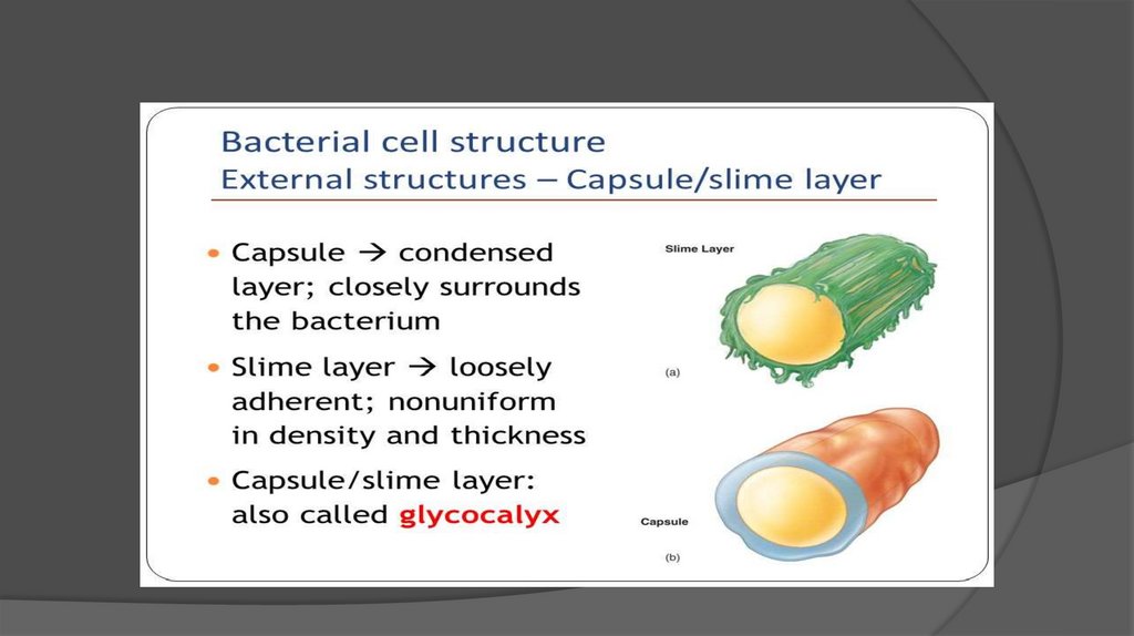
28. 2-Capsule & Slime Layer
2-Capsule & Slime LayerViscous
layer secreted around the cell wall.
Polysaccharide or polypeptide in nature..
1- Capsule:
Sharply defined structure, antigenic in nature.
Protects bacteria from lytic enzyme.
Inhibit phagocytosis.
Stained by negative staining using India
29.
B- Slime layer:Is loosely associated with bacterium and can be
easily washed off, whereas a capsule is
attached tightly to the bacterium and has
definite boundaries.
30.
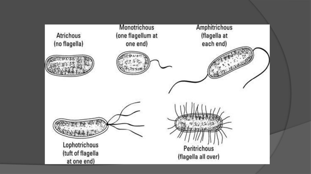
31. 3- Flagella
Long(3-12µm)
filamentous
surface
appendages.
Organs of locomotion.
Composed of protein called flagellins
The number and distribution of flagella on
the bacterial surface are characteristic for
given species –hence are useful in
identifying and classifying bacteria.
32.
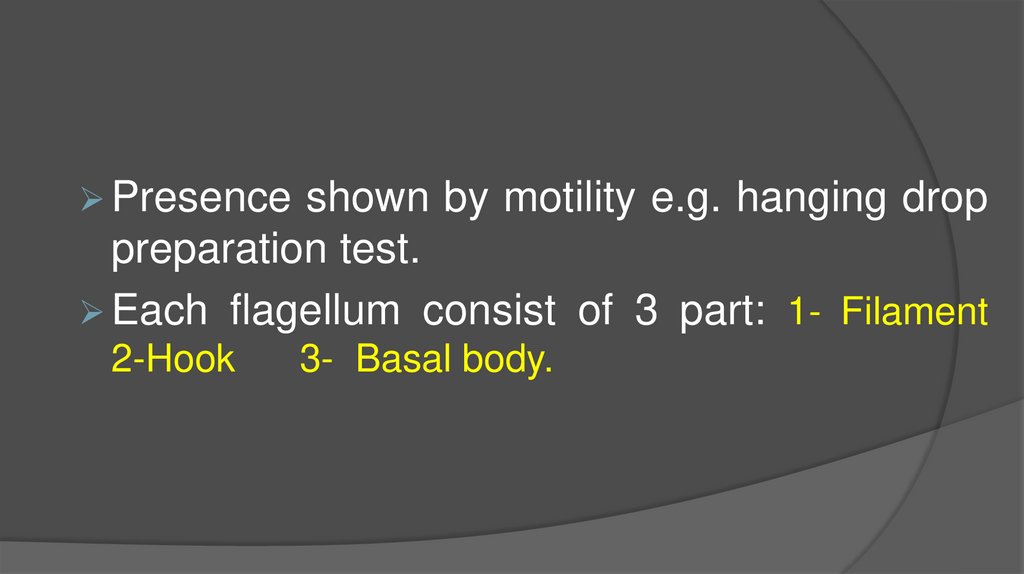
Presenceshown by motility e.g. hanging drop
preparation test.
Each flagellum consist of 3 part: 1- Filament
2-Hook
3- Basal body.
33.
34. 4- Fimbriae (Pili)
Thin,hair like appendages on the surface of many
Gram –ve bacteria
10-20µ long, acts as organs of adhesion
(attachment)-allowing
bacteria
to
colonize
environmental surface or cells and resist flushing.
Made up of proteins called Pilins.
Pili can be of two types:
A.
Common pili- short & abundant.
35. 5- Spores
Highlyresistant resting stages formed during
adverse
environmental
(depletion
of
nutrients).
Formed inside the parent cell, hence called
endospores.
Very
resistant to heat, radiation and drying
and can remain dormant for hundreds of
36.
37.
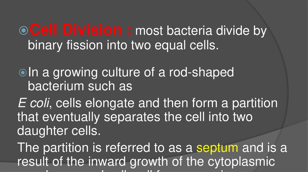
Cell Division : most bacteria divide bybinary fission into two equal cells.
In
a growing culture of a rod-shaped
bacterium such as
E coli, cells elongate and then form a partition
that eventually separates the cell into two
daughter cells.
The partition is referred to as a septum and is a
result of the inward growth of the cytoplasmic
38.
Growth is the orderly increase in the sumof all the components of an organism. The
increase in size that results when a cell takes
up water or deposits lipid or polysaccharide is
not true growth.
Cell multiplication is a consequence of cell
division of unicellular organisms, growth leads
to an increase in the number of single
bacteria making up a population, referred to
39.
Death means the irreversible loss of theability to reproduce (grow and divide).
The
empirical test of death is the culture of
cells on solid media: A cell is considered
dead if it fails to give rise to a colony on
any medium.