


 biology
Similar presentations:


")

. Occurrence and classification")


Nematodes
1.
BASHKIR STATE MEDICALUNIVERSITY
NEMATODES
BIOLOGY DEPARTMENT
BY Dr. MOHIT
2.
• Biohelminth nematodes include Trichinella, Dracunculus, Wuhireria,Dirofilaria, Onchocercus, and Loa-Loa.
• Thread like elongated body.
• Life cycle of biohelminthes have no eggs: females after fertilization give birth
to live larvae.
• most widespread: trichinellosis, dracunculiosis, vuchereriosis, onchocercosis,
loaosis, and dirofilariosis.
3.
Trichinella spiralis• sexually mature forms are in the
intestine.( in final host)
• larvae are in muscles( in
intermediate host)
• reach puberty after 2nd - 3rd day.
• give birth to live larvae.
• larvae migrate into the muscles by
hematogenous route.
• the larvae(with cyst) can remain in
the muscles for years, they remain
viable even after the death of the
host and decomposition of the
corpse.
4.
TRICHINELLOSIS• 1 - parasite- Trichinella spiralis.
• 2 - Systematic position (Phylum, Class) – Kingdom
Animalia, Subkingdom Metotozoa, Phylum
Nemathelminthes, Class Nematoda (biohelmint)
• 3 - Structure, organ systems (figure)
• Spindle-shaped body shape. The size of sexually mature
individuals varies from 2 mm (males) to 4 mm (females)
• 4 - Disease name – trichinellosis.
• 5 - Geographical distribution – common everywhere.
• 6 - Sources of invasion - infected carnivorous and
omnivorous animals (pigs, wild boars, cats, dogs, mice,
rats, bears, etc.).
• 7 - The way of penetration – alimentary.
• 8 - Factors of invasion – meat (pork, meat of wild boars,
bears, etc.) contaminated by spiral larvae.
5.
• 9 - Invasion stage for human – cyst withlarva.
• 10 - Place (organ) of localization in the
human body - small intestine, diaphragm,
muscles
• 11 - Pathogenic effects - toxic-allergic effects
• 12 - Clinical symptoms –The disease begins
acutely,38 - 39 ° C temp., there are swelling
of the eyelids and face, myalgia, heartache.
• 13 - Laboratory diagnosis - based on the
anamnesis, muscle biopsies (2 to 5 weeks
after infection). immunological methods and
PCR are mainly used to confirm the
diagnosis.
• 14 - Preventive measures - Personal – (heat
processing of meat does not kill larvae);
public.
6.
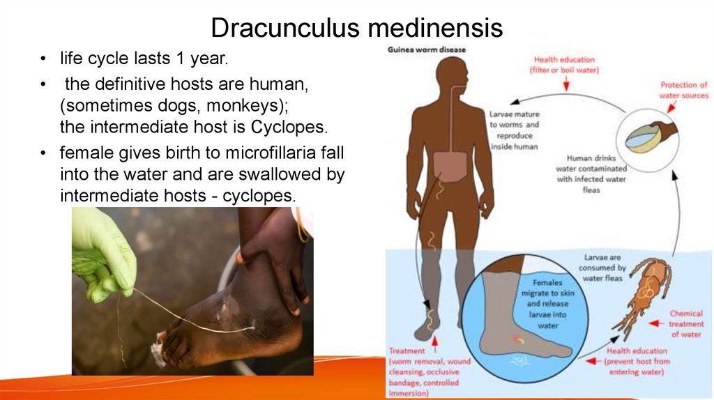
Dracunculus medinensis• life cycle lasts 1 year.
• the definitive hosts are human,
(sometimes dogs, monkeys);
the intermediate host is Cyclopes.
• female gives birth to microfillaria fall
into the water and are swallowed by
intermediate hosts - cyclopes.
7.
DRACUNCULOSIS:• 1 - parasite - Dracunculus medinensis.
• 2 - Systematic position (Phylum, Class) –
Kingdom Animalia, Subkingdom
Metotozoa, Phylum Nemathelminthes,
Class Nematoda
• 3 - Structure, organ systems -Female up to
150 cm long, male up to 3 cm.
• 4 - Disease name – dracunculiasis.
• 5 - Geographical distribution – tropical and
subtropical climate.
• 6 - Sources of invasion - infected human.
• 7 - The way of penetration – alimentary.
• 8 - Factors of invasion – water
contaminated by cyclops (water flea)
infected with larva (microfilaria).
8.
• 9 - Invasion stage for human – microfillariawithin cyclops.
• 10 - Place (organ) of localization in the
human body – subcutaneous tissues,
especially of the legs, arms and back.
• 11 - Pathogenic effects - toxic-allergic
effects and local mechanic effect such as
injury of subcutaneous tissues of the legs,
arms and back.
• 12 - Clinical symptoms – leg pain, itching
and burning in the area of the formed
subcutaneous bladder.
• 13 - Laboratory diagnosis - detection of
parasites under the skin of the lower
extremities (the worm can be detected
visually). Immunological methods and PCR
are mainly used to confirm the diagnosis.
9.
• 14 - Preventive measures -. Personal –filtering and boiling drinking water taken
from river and lake; public - detection and
treatment infected people; guarding places
of water intake, prohibition to bathe in
water intakes elimination of cyclopes.
• The old method of treatment is the
gradual extraction of the worm with
winding of a few centimeters on a stick
every day, over 3-4 weeks. The worm
can also be removed surgically.
10.
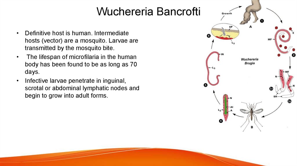
Wuchereria Bancrofti• Definitive host is human. Intermediate
hosts (vector) are a mosquito. Larvae are
transmitted by the mosquito bite.
• The lifespan of microfilaria in the human
body has been found to be as long as 70
days.
• Infective larvae penetrate in inguinal,
scrotal or abdominal lymphatic nodes and
begin to grow into adult forms.
11.
VUCHERERIOSIS:• 1 - Name of the parasite (in Latin) Wuchereria Bancrofti.
• 2 - Systematic position (Phylum, Class) –
Kingdom Animalia, Subkingdom
Metotozoa, Phylum Nemathelminthes,
Class Nematoda
• 3 - Structure, organ systems
• These are long hair-like transparent
nematodes. The male measures 2 to 4 cm
in length. Its tail-end is curved ventrally
and contains two spicules of unequal
length. The female measures 8 to 10 cm in
length. Its tail-end is narrow and abruptly
pointed.
• The lifespan of the adult worms is long,
probably several years (5 to 10 years).
12.
• 4 - Disease name – vuchereriosis.• 5 - Geographical distribution – tropical and
subtropical counries.
• 6 - Sources of invasion - infected human.
• 7 - The way of penetration – transmissible
(bite of mosquito).
• 8 - Factors of invasion – saliva of
mosquito.
• 9 - Invasion stage for human –microfillaria.
• 10 - Place (organ) – lymph nodes,
peripheral blood.
• 11 - Pathogenic effects - toxic-allergic
effects , enlarged of lymph nodes,
blockage of the lymphatic ducts, lymph
congestion in the limbs, genitals.
• 12 - Clinical symptoms – stagnation of
lymph, lymphedema, elephantiasis.
13.
• 13 - Laboratory diagnosis - laboratory diagnosis of vuchereriosis is based on detection ofmicrofilaria in the peripheral blood (at night), lymph and detection of adult worms in the
biopsied lymph node. Immunological methods and PCR are mainly used to confirm the
diagnosis.
• Microfilariae are released into the blood in the evening and at night - a period of maximum
activity of mosquitoes.
• 14 - Preventive measures - Personal – prevention of mosquito bites by using the insecticides
(repellents); public - detection and treatment infected people; elimination of mosquitoes;
personal and public health education.
14.
Onchocerca volvulus• two hosts. Definitive
host - humans.
Intermediate hosts are
midge( genu silunium).
• disease is also known as
RIVER BLINDNESS ,
because the vector
breeds in fast flowing
river.
• The adult worm lives in
the human host for about
15 years and the
microfilariae for about 1
year.
15.
ONCHOCERCOSIS• 1 - parasite - Onchocerca volvulus.
• 2 - Systematic position (Phylum, Class) –
Kingdom Animalia, Subkingdom
Metotozoa, Phylum Nemathelminthes,
Class Nematoda
• 3 - Structure, organ systems -The adult
worms are whitish, opalescent, with
transverse striations on the cuticle the
posterior end is curved, hence the name
Onchocerca, which means "curved tail''.
The male worm measures about 30 mm in
length and the female measures 50 cm.
• 4 - Disease name – onchocercosis.
16.
• 5 - Geographical distribution – tropical and subtropical climate mainly in tropical Africa, butalso in Central and South America. A small focus of infection exists in Yemen and South
Arabia.
• 6 - Sources of invasion - infected human.
• 7 - The way of penetration – transmissible - bite of midge (day-biting female black flies of the
genus Simulium).
• 8 - Factors of invasion – saliva of midge 9 - Invasion stage for human –microfillaria.
• 10 - Place (organ) of localization in the human body – subcutaneous connective tissue of
infected persons.
• 11 - Pathogenic effects - toxic-allergic effects and local mechanic effect such as injury of
subcutaneous tissues.
17.
• 12 - Clinical symptoms – the subcutaneousnodule or onchocercoma is a
circumscribed, firm, non tender tumor,
formed as a result of fibroblastic reaction
around the worms. 1 nodules vary in size
from a few mm to about 10 cm. They tend
to occur over anatomical sites where the
bones are superficial, such as the scalp,
scapulae, ribs, elbows, iliac crest, sacrum
and knees. The nodules are painless and
cause no trouble except for their unsightly
appearance Microfilariae cause lesions in
the skin and eyes.
18.
• 13 - Laboratory diagnosis - detection of microfilariae which may be found inconjunctival biopsies microscopically. Adult worms can be detected in the
biopsy material of the the biopsy material of the subcutaneous nodule;
immunoassay and PCR analysis.
• 14 - Preventive measures - Personal – midge (flies of the genius Simulium)
bites protection by using the insecticides (repellents); public - detection and
treatment infected people; elimination of midge (black flies of the genius
Simulium).
19.
Loa-Loa• Definitive host is human. Intermediate host or
vectors - horsefly of the genus Chrysops in
which the microfilariae develop into the
infective third-stage larvae.
• Infection is transmitted to man through the
bite of infected Chrysops during their blood
meal.
• develop into mature adult worm over 6-12
months and migrate in subcutaneous tissues.
• Development in Chrysops is completed in
about 10 days.
20.
LOAOSIS• 1 parasite - Loa-Loa.
• 2 - Systematic position – Kingdom Animalia, Subkingdom Metotozoa, Phylum
Nemathelminthes, Class Nematoda
• 3 - Structure, organ systemsThe adult worm is thin and transparent, measuring about 30-70
mm in length and 0.3-0.5 mm in thickness. Adults live for 4-7 years. The microfilariae are
sheathed with column of nuclei extending completely to the tip of the tail. They appear in
peripheral circulation only during the day from 12 noon to 2 pm diurnal periodicity).
• 4 - Disease name – loaosis.
• 5 - Geographical distribution – tropical and subtropical climate mainly in tropical Africa.
21.
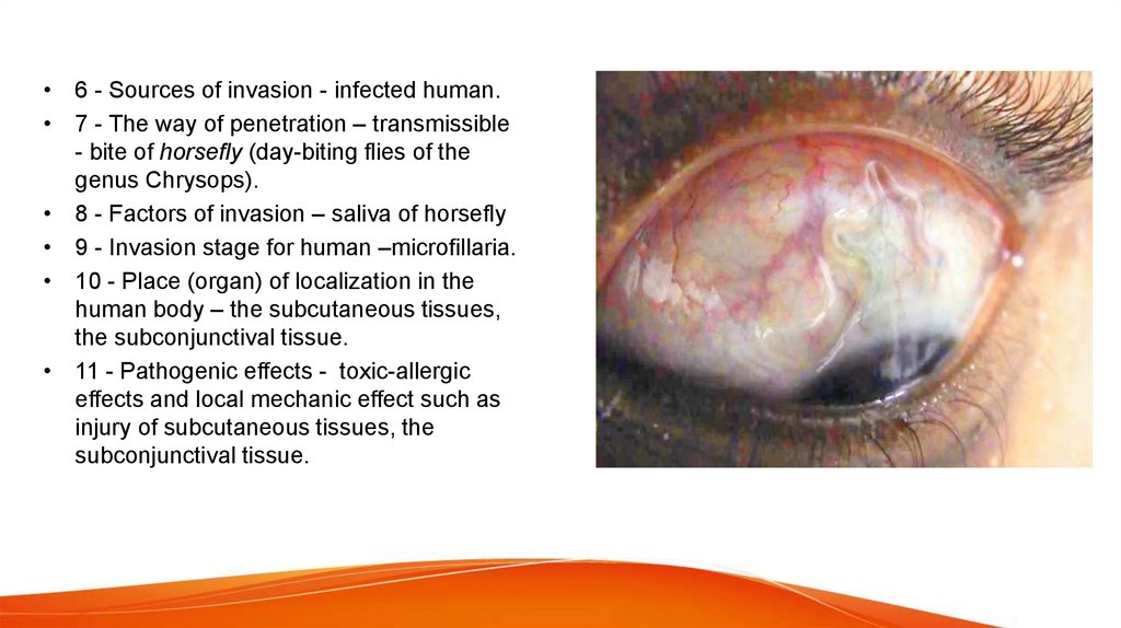
• 6 - Sources of invasion - infected human.• 7 - The way of penetration – transmissible
- bite of horsefly (day-biting flies of the
genus Chrysops).
• 8 - Factors of invasion – saliva of horsefly
• 9 - Invasion stage for human –microfillaria.
• 10 - Place (organ) of localization in the
human body – the subcutaneous tissues,
the subconjunctival tissue.
• 11 - Pathogenic effects - toxic-allergic
effects and local mechanic effect such as
injury of subcutaneous tissues, the
subconjunctival tissue.
22.
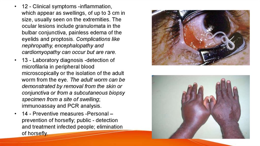
• 12 - Clinical symptoms -inflammation,which appear as swellings, of up to 3 cm in
size, usually seen on the extremities. The
ocular lesions include granulomata in the
bulbar conjunctiva, painless edema of the
eyelids and proptosis. Complications like
nephropathy, encephalopathy and
cardiomyopathy can occur but are rare.
• 13 - Laboratory diagnosis -detection of
microfilaria in peripheral blood
microscopically or the isolation of the adult
worm from the eye. The adult worm can be
demonstrated by removal from the skin or
conjunctiva or from a subcutaneous biopsy
specimen from a site of swelling;
immunoassay and PCR analysis.
• 14 - Preventive measures -Personal –
prevention of horsefly; public - detection
and treatment infected people; elimination
of horsefly.