


")


 biology
Similar presentations:


")
Phyllum plathyhelminthes
1. LECTURE PHYLLUM PLATHYHELMINTHES
ZAPOROZHYE STATE MEDICAL UNIVERSITYDEPARTMENT OF MEDICAL BIOLOGY
LECTURE
PHYLLUM PLATHYHELMINTHES
Composed by
Doctor of Philosophy
Popovich A. P.
madbio@zsmu.zp.ua
Zaporozhye - 2016
2. QUESTIONS
-Plathyhelmintes in general-Class Trematoda: main features and life
cycles of some parasites
-Class Cestodea: main features and life
cycles of some parasites
3.
Plathyhelminthes are known as flatworms, because theyare much flattened dorso-ventrally. They are
characterized by the following features:
*They are bilatary symmetrical animals. Many organs
of flatworms become pair-ed with respect to a single
plane that divides the animal into equal right and left
halves.
*They are acoelomates. There is no coelom. Their
primary body cavity is invaded by mesoderm cells.
*They are triploblastic. A third germ layer called
mesoderm is formed between ecto-derm and
endoderm. The mesoderm produces a new tissue
and organs. Much of the mesoderm remains
undifferentiated. It forms a packing tissue
known as parenchyma.
4.
--
-
-
-
*Body dorsoventrally flattened, leaf like or tape
like and segmented.
*First animals to have organ system
organization.
*Digestive system incomplete, anus is absent.
*Nervous system – a pair of ganglia with
longitudinal nerve cords
*Excretory system has one or two canals with
branches, the finer branches end in flame cells.
*Mostly hermaphrodite.
5. Classification of Plathyhelminthes
Phylum PlathyhelminthesClass Turbellaria
Free – living, Planaria
Class Trematoda
flukes: Fasciola,
Clonorchis,
Schistosomes
Class Cestoda
tapeworms: Taenia,
Echinoccocus,
Diphillobothrium latum
6. Сlass Trematoda.
General:Trematodes have flat and oval body, ranging from
1 mm to 3 cm in length.
Organs of attachment (suckers) and reproduction
are highly developed, but organs of lost entirely
Trematodes have a complicated life cycles with the
production of the following stages: the adult flukethe egg- the ciliated miracidium, the sporocyst, the
redia, the adult in the defenitive host.
Metacercaria is an infective stage.
7. Fasciola hepatica
8.
9. Schistosomes or Blood Flukes
They are much differ from the other Trematodes:they are flukes with sexual dimorphism
the larger males carry the more slender females in
the gynaecophoric canal
their eggs have spine
in the life-cycle no redia stage
10.
11. Life – history of Schistosomes
12. CESTODES (TAPEWORMS)
I. GENERAL1. The tapeworms are hermaphroditic worms,
which as adults parasitize the
gastrointestinal tract of vertebrates.
2. They are segmented /flat shape
A. TAPEWORM ANATOMY:
1. HEAD or SCOLEX, with adhesive organs, at
anterior end of worm.
Attachment to the intestinal mucosa is
accomplished by the scolex.
13.
2. PROGLOTTIDS -the multiple, hermaphroditic, eggproducing units.These are the flattened segments of the worm body.
Medically important species of cestode.
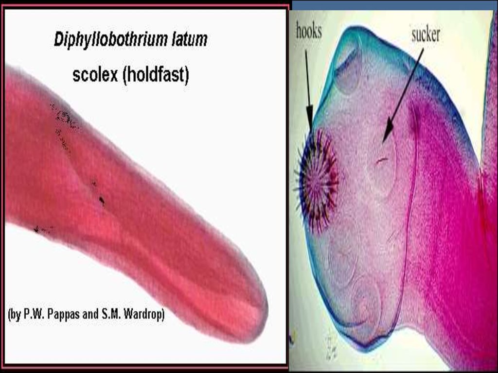
a. Diphyllobothrium latum has a SCOLEX with
elongated, slit-like attachment organs - Fish tape
worm
b. Taenia saginata has four muscular SUCKERSBeef tape worm.
c. Taenia solium has similar muscular SUCKERSPork tape worm.
d. Hymenolepis nana _ Dwarf tape worm.
e. Echinococcus granulosis _ Dog tape worm.
14.
15. TAPEWORM LIFE CYCLE:
The DEFINITIVE HOST ingests the larval form. Wormsmature rom larval
forms in the intestine of definitive host. The definitive host
harbors ADULT
WORMS in the intestine. EGGS are passed in the stool.
Eggs are ingested by the INTERMEDIATE HOST. LARVAE
develop from
eggs in the intermediate host and penetrate the host
intestinal mucosa.
Larvae develop into ENCYSTED FORMS in tissues of
intermediate host.
The CYSTICERCUS is the encysted form of the Taenia
species. The HYDATID is the encysted form of the
Echinococcus.
NOTE: Diphyllobothrium latum has two intermediate
hosts.
16. HUMAN DISEASE CAUSED BY TAPEWORMS
1. ADULT (WORM) STAGE:a. Taenia saginata, Taenia solium, Diphyllobothrium latum.
b. The presence of adult tapeworms in the human GI tract only rarely causes
symptomatic disease. People usually only become aware of the infection
when proglottids are passed in the feces.
NOTE: T. solium causes human disease in both adult and larval
stages.
2. LARVAL (CYST) STAGE (i.e., Cysticercosis):
a. Echinococcus granulosus, Echinococcus multilocularis, Taenia solium
b. The presence of the cyst stage of the tapeworm in extraintestinal tissues
causes signs and symptoms relative to the site of the expanding cyst.
LABORATORY IDENTIFICATION OF TAPEWORMS:
Morphology of the proglottid (degree of branching, configuration, size) is
important in distinguishing T. solium, T. saginata, and sometimes D. latum.
Eggs of T. solium and T. saginata are morphologically indistinguishable,
but the eggs of D. latum are operculated.
17. Taenia solium: TAENIASIS
Mode of transmission: Infection by ingestion of poorly cookedpork containing encysted larvae. Pathology: Adult worm
inhabits the human jejunum and sheds eggs which pass in
the stool. Pigs ingest the eggs which release embryos in the
GI tract. The embryos travel to systemic tissue where they
transform into encysted larvae (i.e., cysticerci). Humans are
infected by eating undercooked meat containing the
cysticerci.
Laboratory diagnosis: Examine STOOL for proglottids or eggs;
T. solium can be distinguished from T. saginata by the
proglottid branching. T. solium have proglottids with 5-10
primary uterine branches, but T. saginata proglottids have
15-20.
18. Taenia solium: CYSTICERCOSIS
Mode of transmission: Humans accidentally become the intermediatehost by ingestion of fecally contaminated food or water (most
common) or autoinfection (eggs from anus to hand to mouth) or
reverse peristalsis (rare).
Clinical manifestations: Clinical manifestations reflect the organ
system affected by the cyst(s). The most common clinical
manifestation results from CNS involvement. Symptoms include
headache, seizures, paresis. (Cysticercosis is a common cause of
childhood seizures in Mexico.)
19.
Pathology: Embryos emerge from the ingested eggand travel through the human body where cysticerci
develop. Common sites of encystment include the CNS,
eye, heart, muscle, and skin. Symptoms result from
mass effects of the expanding cyst(s) or from the host
inflammatory response to degenerating cysts.
Laboratory diagnosis: Radiological tests (especially
CT or NMR of the head with CNS symptoms shows ringenhancing lesions), serologic tests, sometimes
examination of cyst at surgery.
20. Echinococcus granulosus
21. Echinococcus granulosus:
Epidemiology: people (e.g., herders orhunters) who have close contact with dogs
that may feed contaminated wild herd
animals.
Mode of transmission: Disease has a
zoonotic pattern of transmission. Humans
ingest eggs shed by dogs that have fed on
infected domestic or wild herbivores.
22. Life cycle Echinococcus granulosus
23.
Pathology: Dogs & other canines are definitive hosts.The adult worm in dogs is very small, with only
three (3) proglottids. Eggs are
shed in feces. Sheep and cattle (and wild herbivores
in sylvatic form) are intermediate hosts which ingest
the eggs and harbor the cysts. In
humans, the larvae penetrate the intestinal mucosa,
invade submucosal venules, and are distributed to
tissue where HYDATID forms. Common hydatid
sites: liver, lung, bone. Hydatid cysts contain
multiple BROOD CAPSULES containing
SCOLICES. If cysts rupture either spontaneously or
during surgical removal, ANAPHYLAXIS can occur.
24.
Laboratory diagnosis: The diagnosis isusually suggested by the patient history or
by radiological findings (e.g., large hepatic
cyst).
Patients usually have eosinophilia. Diagnosis
usually confirmed serology and/or
examination of material removed at
surgery.
Treatment: Surgical excision is the treatment
of choice.