


 biology
Similar presentations:


Brucella (Brucellosis, Bang’s Disease). Occurrence and classification
1.
2. Why are you late?
3.
Brucella (Brucellosis, Bang’s Disease)Occurrence and classification.
The genus Brucella includes three medically relevant species—B.
abortus, B. melitensis, and B. suis – besides a number of others.
These three species are the causative organisms of classic zoonoses in
livestock and wild animals, specifically in cattle (B. abortus), goats
(B. melitensis), and pigs (B. suis). These bacteria can also be
transmitted from diseased animals to humans, causing a uniform
clinical picture, so-called undulant fever or Bang’s disease.
4.
Morphology and cultureBrucellae are slight, coccoid, Gram-negative rods with no flagella.
They only reproduce aerobically. In the initial isolation the
atmosphere must contain 5-10% CO2. Enriched mediums such as
blood agar are required to grow them in cultures.
5.
Pathogenesis and clinical pictureHuman brucellosis infections result from direct contact with
diseased animals or indirectly by way of contaminated foods, in
particular unpasteurized milk and dairy products. The bacteria
invade the body either through the mucosa of the upper intestinal
and respiratory tracts or through lesions in the skin, then enter the
subserosa or subcutis. From there they are transported by
microphages or macrophages, in which they can survive, to the
lymph nodes, where a lymphadenitis develops. The pathogens then
disseminate from the affected lymph nodes, at first lymphogenously
and then hematogenously, finally reaching the liver, spleen, bone
marrow, and other RES tissues, in the cells of which they can
survive and even multiply.
6.
DiagnosisThis is best achieved by isolating the pathogen from blood or
biopsies in cultures, which must be incubated for up to four weeks.
The laboratory must therefore be informed of the tentative
diagnosis. Brucellae are identified based on various metabolic
properties and the presence of surface antigens, which are detected
using a polyvalent Brucella-antiserum in a slide agglutination
reaction. Special laboratories are also equipped to differentiate the
three Brucella species. Antibody detection is done using the
agglutination reaction according to Gruber-Widal in a standardized
method. In doubtful cases, the сomplement binding reaction and
direct Coombs test can be applied to obtain a serological
diagnosis.
7.
Epidemiology and preventionBrucellosis is a zoonosis that affects animals all over the world.
Infections with B. melitensis occur most frequently in
Mediterranean countries, in Latin America, and in Asia. The
melitensis brucelloses seen in Europe are either caused by milk
products imported from these countries or occur in travelers. B.
abortus infections used to be frequent in central Europe, but the
disease has now practically disappeared there thanks to the
elimination of Brucella-infested cattle herds. Although control of
brucellosis infections focuses on prevention of exposure to the
pathogen, it is not necessary to isolate infected persons since the
infection is not communicable between humans. There is no
vaccine.
8.
Bordetella (Whooping Cough, Pertussis)The genus Bordetella, among others, includes the species B.
pertussis, B. parapertussis, and B. bronchiseptica. Of the three, the
pathogen responsible for whooping cough, B. pertussis, is of
greatest concern for humans. The other two species are occasionally
observed as human pathogens in lower respiratory tract infections.
Morphology and culture. B. pertussis bacteria are small, coccoid,
non-motile, Gram-negative rods that can be grown aerobically on
special culture mediums at 37°C for three to four days.
Pathogenesis. Pertussis bacteria are transmitted by aerosol
droplets. They are able to attach themselves to the cells of the
ciliated epithelium in the bronchi. They rarely invade the
epithelium. The infection results in (sub-) epithelial inflammations
and necroses.
9.
10.
DiagnosisThe pathogen can only be isolated and identified during the
catarrhal and early paroxysmal phases. Specimen material is
taken from the nasopharynx through the nose using a special
swabbing technique. A special medium is then carefully
inoculated or the specimen is transported to the laboratory
using a suitable transport medium. B. pertussis can also be
identified in nasopharyngeal secretion using the direct
immunofluorescence technique. Cultures must be aerobically
incubated for three to four days. Antibodies cannot be detected
by EIA until two weeks after onset at the earliest. Only a
seroconversion is conclusive.
11.
Therapy. Antibiotic treatment can only be expected to beeffective during the catarrhal and early paroxysmal phases before
the virulence factors are bound to the corresponding cell
receptors. Macrolides are the agents of choice.
Epidemiology and prevention. Pertussis occurs worldwide.
Humans are the only hosts. Sources of infection are infected
persons during the catarrhal phase, who cough out the pathogens
in droplets. There are no healthy carriers. The most important
preventive measure is the active vaccination (see vaccination
schedule). Although a whole-cell vaccine is available, various
acellular vaccines are now preferred.
12.
Treponema (Syphilis, Yaws, Pinta)Morphology and culture. These organisms are slender bacteria, 0.2
μm wide and 5–15 μm long; they feature 10–20 primary windings
and move by rotating around their lengthwise axis. Their small
width makes it difficult to render them visible by staining. They can
be observed in vivo using dark field microscopy. In-vitro culturing
has not yet been achieved.
Pathogenesis and clinical picture. Syphilis affects only humans.
The disease is normally transmitted by sexual intercourse. Infection
comes about because of direct contact with lesions containing the
pathogens, which then invade the host through microtraumata in the
skin or mucosa. The incubation period is two to four weeks.
13.
Left untreated, the disease manifests in several stages:Stage I (primary syphilis). Hard, indolent (painless) lesion, later
infiltration and ulcerous disintegration, called hard chancre.
Accompanied by regional lymphadenitis, also painless. Treponemes
can be detected in the ulcer.
Stage II (secondary syphilis). Generalization of the disease occurs
four to eight weeks after primary syphilis. Frequent clinical
symptoms include micropolylymphadenopathy and macular or
papulosquamous exanthem, broad condylomas, and enanthem.
Numerous organisms can be detected in seeping surface
efflorescences.
Latent syphilis. Stage of the disease in which no clinical symptoms
are manifested, but the pathogens are present in the body and serum
antibody tests are positive. Divided into early latency (less than four
years) and late latency (more than four years).
14.
Stage III (tertiary or late syphilis). Late gummatous syphilis:manifestations in skin, mucosa, and various organs. Tissue
disintegration is frequent. Lesions are hardly infectious or not at
all. Cardiovascular syphilis: endarteritis obliterans, syphilitic
aortitis. Neurosyphilis: two major clinical categories are
observed: meningovascular syphilis, i.e., endarteritis obliterans of
small blood vessels of the meninges, brain, and spinal cord;
parenchymatous syphilis, i.e., destruction of nerve cells in the
cerebral cortex (paresis) and spinal cord (tabes dorsalis). A great
deal of overlap occurs.
Syphilis connata. Transmission of the pathogen from mother to
fetus after the fourth month of pregnancy. Leads to miscarriage or
birth of severely diseased infant with numerous treponemes in its
organs.
15.
Serous transudate from moistmucocutaneous primary chancre. Direct
immunofluorescence.
16.
Therapy.Penicillin G is the antibiotic agent of choice. Dosage and duration
of therapy depend on the stage of the disease and the galenic
formulation of the penicillin used.
Epidemiology and prevention.
Syphilis is known all over the world. Annual prevalence levels in
Europe and the US are 10–30 cases per 100 000 inhabitants. The
primary preventive measure is to avoid any contact with syphilitic
efflorescences. When diagnosing a case, the physician must try to
determine the first-degree contact person, who must then be
examined immediately and provided with penicillin therapy as
required. National laws governing venereal disease management in
individual countries regulate the measures taken to diagnose,
prevent, and heal this disease. There is no vaccine.
17.
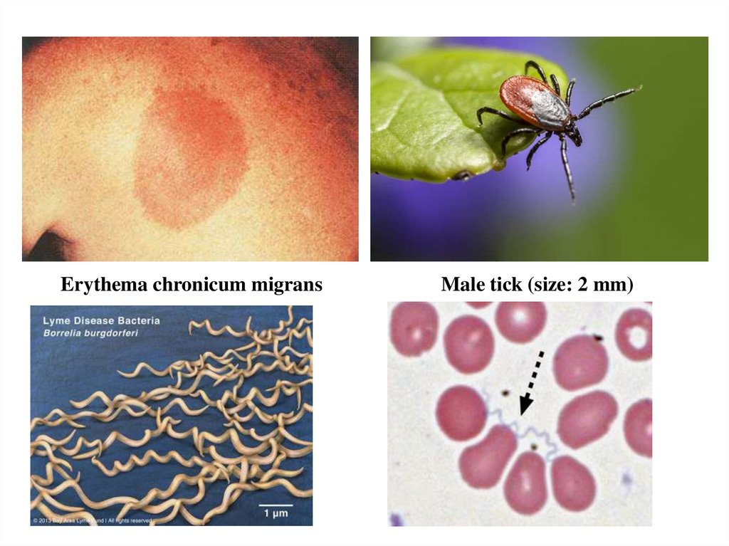
Borrelia (Relapsing Fever, Lyme Disease)Borrelia burgdorferi (Lyme Disease)
Classification.
The etiology of an increase in the incidence of acute cases of arthritis
among youths in the Lyme area of Connecticut in 1977 was at first
unclear. The illness was termed Lyme arthritis. It was not until 1981
that hitherto unknown borreliae were found to be responsible for the
disease. They were classified as B. burgdorferi in 1984 after their
discoverer. Analysis of the genome of various isolates has recently
resulted in a proposal to subclassify B. burgdorferi sensu lato in
three species: B. burgdorferi sensu stricto, B. garinii, B. afzelii.
18.
Morphology and cultureThese are thin, flexible, helically wound, highly motile
spirochetes. They can be rendered visible with Giemsa staining
or by means of dark field or phase contrast microscopy methods.
These borreliae can be grown in special culture mediums at 35°C
for five to 10 days, although culturing these organisms is
difficult and often unsuccessful.
Pathogenesis and clinical picture
The pathogens are transmitted by the bite of various tick species.
The incubation period varies from three to 30 days. Left
untreated, the disease goes through three stages, though
individual courses often deviate from the classic pattern. The
presenting symptom in stage I is the erythema chronicum
migrans.
19.
Erythema chronicum migransMale tick (size: 2 mm)
20.
DiagnosisDirect detection and identification of the pathogen by means
of microscopy and culturing techniques is possible, but laden
with uncertainties. In a recent development, the polymerase
chain reaction (PCR) is used for direct detection of pathogenspecific DNA. However, the method of choice is still the
antibody test (EIA or indirect immunofluorescence,Western
blotting if the result is positive).
Therapy
Stages I and II: amoxicillin, cefuroxime, doxycycline, or a
macrolide. Stage III: ceftriaxone.
21.
Epidemiology and preventionLyme disease occurs throughout the northern hemisphere. There are
some endemic foci where the infection is more frequent. The disease
is transmitted by various species of ticks, in Europe mostly by
Ixodes ricinus (sheep tick). In endemic areas of Germany,
approximately 3–7% of the larvae and 10–34% of nymphs and adult
ticks are infected with B. burgdorferi sensu lato. The annual
incidence of acute Lyme disease (stage I) in central Europe is 20–50
cases per 100 000 inhabitants. Wild animals from rodents on up to
deer are the natural reservoir of the Lyme disease Borrelia, although
these species seldom come down with the disease. The ticks obtain
their blood meals from these animals.
22.
Leptospira (Leptospirosis, Weil Disease)Classification. Leptospirae belong to the family Leptospiraceae. The
genus Leptospira comprises two species. L. biflexa includes all
apathogenic leptospirae and L. interrogans represents the pathogenic
species. Based on its specific surface antigen variety, L. interrogans is
subclassified in over 100 serovars in 19 serogroups. Some of the most
important serogroups are: icterohemorrhagiae, canicola, pomona,
australis, grippotyphosa, hyos, and sejroe.
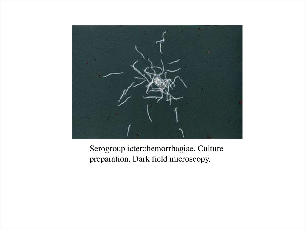
Morphology and culture. Leptospirae are fine spirochetes, 10–20
μm long, and 0.1–0.2 μm thick. They possess no flagella, but rather
derive their motility from rotating motions of the cell corpus.
Visualization of leptospirae is best done using dark field or phase
contrast microscopy. Leptospirae can be grown in special culture
mediums under aerobic conditions at temperatures between 27–30°C
23.
Serogroup icterohemorrhagiae. Culturepreparation. Dark field microscopy.
24.
Pathogenesis and clinical picture. Leptospirae invade the humanorganism through microinjuries in the skin or the intact
conjunctival mucosa. There are no signs of inflammation in
evidence at the portal of entry. The organisms spread to all parts of
the body, including the central nervous system, hematogenously.
Leptospirosis is actually a generalized vasculitis. The pathogens
damage mainly the endothelial cells of the capillaries, leading to
greater permeability and hemorrhage and interrupting the O2
supply to the tissues. Jaundice is caused by a nonnecrotic
hepatocellular dysfunction. Disturbances of renal function result
from hypoxic tubular damage. A clinical distinction is drawn
between anicteric leptospirosis, which has a milder course, and the
severe clinical picture of icteric leptospirosis (Weil disease). In
principle, any of the serovars could potentially cause either of these
two clinical courses. In practice, however, the serogroup
icterohemorrhagiae is isolated more frequently in Weil disease.
25.
DiagnosisDetection and identification of leptospirae are accomplished by
growing the organisms in cultures. Blood, cerebrospinal fluid,
urine, or organ biopsies, which must not be contaminated with
other bacteria, are incubated in special mediums at 27–30°C for
three to four weeks. A microscope check (dark field) is carried out
every week to see if any leptospirae are proliferating. The
Leptospirae are typed serologically in a lysis-agglutination
reaction with specific test sera.
The method of choice for a laboratory diagnosis is an antibody
assay. The antibodies produced after the first week of the
infection are detected in patient serum using a quantitative lysisagglutination test. Viable culture strains of the regionally endemic
serovars provide the test antigens. The reaction is read off under
the microscope.
26.
Therapy. The agent of choice is penicillin G.Epidemiology and prevention. Leptospiroses are typical
zoonotic infections. They are reported from every continent in
both humans and animals. The most important sources of
infection are rodents and domestic animals, mainly pigs. The
animals excrete the pathogen with urine. Leptospirae show little
resistance to drying out so that infections only occur because of
contact with a moist milieu contaminated with urine. The persons
most at risk are farmers, butchers, sewage treatment workers, and
zoo staff.
Prevention of these infections involves mainly avoiding contact
with material containing the pathogens, control of Muridae
rodents and successful treatment of domestic livestock. It is not
necessary to isolate infected persons or their contacts. There is no
commercially available vaccine.
27.
Rickettsia, Coxiella, Orientia, and Ehrlichia(Typhus, Spotted Fever, Q Fever, Ehrlichioses)
The genera of the Rickettsiaceae and Coxelliaceae contain short,
coccoid, small rods that can only reproduce in host cells. With
the exception of Coxiella (aerogenic transmission), they are
transmitted to humans via the vectors lice, ticks, fleas, or mites.
R. prowazekii and R. typhi cause typhus, a disease characterized
by high fever and a spotty exanthem. Several rickettsiae species
cause spotted fever, a milder typhuslike disease. Orientia
tsutsugamushi is transmitted by mite larvae to cause
tsutsugamushi fever. This disease occurs only in Asia. Coxiella
burnetii is responsible for Q fever, an infection characterized by
a pneumonia with an atypical clinical course.
28.
Several species of Ehrlichiaceae cause ehrlichiosis in animals andhumans. The method of choice for laboratory diagnosis of the
various rickettsioses and ehrlichioses is antibody assay by any of
several methods, in most cases indirect immunofluorescence.
Tetracyclines represent the antibiotic of choice for all of these
infections. Typhus and spotted fever no longer occur in Europe.
Q fever infections are reported from all over the world. Sources of
infection include diseased sheep, goats, and cattle. The prognosis
for the rare chronic form of Q fever (syn. Q fever endocarditis) is
poor. Ehrlichiosis infects mainly animals, but in rare cases humans
as well.
29.
The bacteria of this group belong to the families Rickettsiaceae(Rickettsia and Orientia), Coxelliaceae (Coxiella), and
Ehrlichiaceae (Ehrlichia, Anaplasma, Neorickettsia). Some of these
organisms can cause mild, selflimiting infections in humans, others
severe disease. Arthropods are the transmitting vectors in many
cases.
Morphology and culture. These obligate cell parasites are
coccoid, short rods measuring 0.3–0.5 lm that take gram staining
weakly, but Giemsa staining well. They reproduce by intracellular,
transverse fission only. They can be cultured in hen embryo yolk
sacs, in suitable experimental animals (mouse, rat, guinea pig) or in
cell cultures.
30.
Pathogenesis and clinical pictures.With the exception of C. burnetii, the organisms are transmitted
by arthropods. In most cases, the arthropods excrete them with
their feces and ticks transmit them with their saliva while
sucking blood. The organisms invade the host organism through
skin injuries. C. burnetii is transmitted exclusively by inhalation
of dust containing the pathogens. Once inside the body,
rickettsiae reproduce mainly in the vascular endothelial cells.
These cells then die, releasing increasing numbers of organisms
into the bloodstream. Numerous inflammatory lesions are caused
locally around the destroyed endothelia. Ehrlichiae reproduce in
the monocytes or granulocytes of membrane-enclosed
cytoplasmic vacuoles. The characteristic morulae clusters
comprise several such vacuoles stuck together.
31.
DiagnosisDirect detection and identification of these organisms in cell
cultures, embryonated hen eggs, or experimental animals is
unreliable and is also not to be recommended due to the risk of
laboratory infections. Special laboratories use the polymerase chain
reaction to identify pathogen-specific DNA sequences. However,
the method of choice is currently still the antibody assay, whereby
the immunofluorescence test is considered the gold standard among
the various methods. The Weil-Felix agglutination test is no longer
used today due to low sensitivity and specificity.
Therapy
Tetracyclines lower the fever within one to two days and are the
antibiotics of choice.
32.
Epidemiology and preventionThe epidemic form of typhus, and earlier scourge of eastern
Europe and Russia in particular, has now disappeared from
Europe and occurs only occasionally in other parts of theworld.
Murine typhus, on the other hand, is still a widespread disease in
the tropics and subtropics. Spotted fevers (e.g., Rocky Mountain
spotted fever) occur with increased frequency in certain
geographic regions, especially in the spring. Tsutsugamushi
fever occurs only in Japan and Southeast Asia.
33.
BartonellaClassification. The genus Bartonella includes, among others, the
species B. bacilliformis, B. quintana, B. henselae, and B.
clarridgeia.
Morphology and culture. Bartonella bacteria are small (0.6–1
μm), Gram-negative, frequently pleomorphic rods. Bartonellae
can be grown on culture mediums enriched with blood or serum.
34.
35.
Diagnosis. Special staining techniques are used to renderbartonellae visible under the microscope in tissue specimens.
Growth in cultures more than seven days. Amplification of specific
DNA in tissue samples or blood, followed by sequencing. Antibody
assay with IF or EIA.
Therapy. Tetracyclines, macrolides. Epidemiology and prevention.
Oroya fever (also known as Carrion disease) is observed only in
humans and is restricted to mountain valleys with elevations
above 800 m in the western and central Cordilleras in South
America because an essential vector, the sand fly, lives only there.
Cat scratch disease, on the other hand, is known all over the world.
It is transmitted directly from cats to humans or indirectly by cat
fleas. The cats involved are usually not sick.
36.
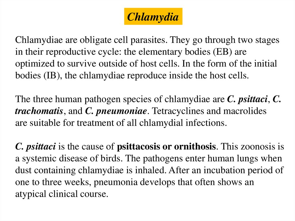
ChlamydiaChlamydiae are obligate cell parasites. They go through two stages
in their reproductive cycle: the elementary bodies (EB) are
optimized to survive outside of host cells. In the form of the initial
bodies (IB), the chlamydiae reproduce inside the host cells.
The three human pathogen species of chlamydiae are C. psittaci, C.
trachomatis, and C. pneumoniae. Tetracyclines and macrolides
are suitable for treatment of all chlamydial infections.
C. psittaci is the cause of psittacosis or ornithosis. This zoonosis is
a systemic disease of birds. The pathogens enter human lungs when
dust containing chlamydiae is inhaled. After an incubation period of
one to three weeks, pneumonia develops that often shows an
atypical clinical course.
37.
The bacteria in the taxonomic family Chlamydiaceaeare small (0.3–1 μm) obligate cell parasites with a Gram-negative
cell wall. The reproductive cycle of the chlamydiae comprises two
developmental stages: The elementary bodies are optimally adapted
to survival outside of host cells. The initial bodies, also known as
reticulate bodies, are the form in which the chlamydiae reproduce
inside the host cells by means of transverse fission.
38.
Two morphologically and functionally distinct forms are knownElementary bodies. The round to oval, optically dense elementary
bodies have a diameter of approximately 300 nm. They represent the
infectious form of the pathogen and are specialized for the demands
of existence outside the host cells. Once the elementary bodies have
attached themselves to specific host cell receptors, they invade the
cells by means of endocytosis. Inside the cell, they are enclosed in
an endocytotic membrane vesicle or inclusion, in which they
transform themselves into the other form - initial bodies - within a
matter of hours.
Initial bodies. Chlamydiae in this spherical to oval form are also
known as reticular bodies. They have a diameter of approximately
1000 nm. The initial bodies reproduce by means of transverse
fission and are not infectious while in this stage. At the end of the
cycle, the initial bodies are transformed back into elementary
bodies. The cell breaks open and releases the elementary bodies to
continue the cycle by attaching themselves to new host cells.
39.
Chlamydia psittaci (Ornithosis, Psittacosis)Pathogenesis and clinical picture
The natural hosts of C. psittaci are birds. This species causes
infections of the respiratory organs, the intestinal tract, the genital
tract, and the conjunctiva of parrots and other birds. Humans are
infected by inhalation of dust (from bird excrements) containing
the pathogens, more rarely by inhalation of infectious aerosols.
After an incubation period of one to three weeks, ornithosis
presents with fever, headache, and a pneumonia that often takes an
atypical clinical course. The infection may, however, also show no
more than the symptoms of a common cold, or even remain
clinically silent. Infected persons are not usually sources of
infection.
40.
Diagnosis. The pathogen can be grown from sputum in special cellcultures. Direct detection in the culture is difficult and only
possible in specially equipped laboratories. The complement
binding reaction can be used to identify antibodies to a generic
antigen common to all chlamydiae, so that this test would also have
a positive result in the presence of other chlamydial infections. The
antibody test of choice is indirect microimmunofluorescence.
Therapy. Tetracyclines (doxycycline) and macrolides.
Epidemiology and prevention. Ornithosis affects birds
worldwide. It is also observed in poultry. Diagnosis of an
ornithosis in a human patient necessitates a search for and
elimination of the source, especially if the birds in question are
household pets.
41.
Chlamydia trachomatis (Trachoma, Lymphogranuloma venereum)C. trachomatis is a pathogen that infects only humans. Trachoma is
a follicular keratoconjunctivitis. The disease occurs in all climatic
zones, although it is more frequent in warmer, less-developed
countries. It is estimated that 400 million people carry this chronic
infection and that it has caused blindness in six million. The
pathogen is transmitted by direct contact and indirectly via objects
in daily use. Left untreated, the initially acute inflammation can
develop a chronic course lasting months or years and leading
to formation of a corneal scar, which can then cause blindness. The
laboratory diagnostics procedure involves detection of C.
trachomatis in conjunctival smears using direct
immunofluorescence microscopy. The fluorochrome-marked
monoclonal antibodies are directed against the MOMP (major outer
membrane protein) of C. trachomatis.
42.
Lymphogranuloma venereum. This venereal disease (syn.Lymphogranuloma inguinale, lymphopathia venerea (FavreDurand-Nicolas disease) not to be confused with granuloma
inguinale) is frequently observed in the inhabitants of warm
climatic zones. A herpetiform primary lesion develops at the site
of invasion in the genital area, which then becomes an ulcus with
accompanying lymphadenitis. Laboratory diagnosis is based on
isolating the proliferating pathogen in cell cultures from purulent
material obtained from the ulcus or from matted lymph nodes. The
antibodies can be identified using the complement binding
reaction or the icroimmunofluorescence
test. Tetracyclines and macrolides are the potentially useful
antibiotic types.
43.
Chlamydia pneumoniaeThis new chlamydial species (formerly TWAR chlamydiae) causes infections
of the respiratory organs in humans that usually run a mild course:
influenzalike infections, sinusitis, pharyngitis, bronchitis, pneumonias
(atypical). Clinically silent infections are frequent. C. pneumoniae is
pathogenic in humans only. The pathogen is transmitted by aerosol droplets.
These infections are probably among the most frequent human chlamydial
infections. Serological studies have demonstrated antibodies to C. pneumoniae
in 60% of adults. Specific laboratory diagnosis is difficult. Special laboratories
can grow and identify the pathogen in cultures and detect it under the
microscope using marked antibodies to the LPS (although this test is positive
for all chlamydial infections). C. pneumoniae-specific antibodies can be
identified with the microimmunofluorescence method. In a primary infection, a
measurable titer does not develop for some weeks and is also quite low. The
antibiotics of choice are tetracyclines or macrolides. There is a growing body
of evidence supporting a causal contribution by C. pneumoniae to
atherosclerotic plaque in the coronary arteries, and thus to the pathogenesis of
coronary heart
disease.
44.
MycoplasmaMycoplasmas are bacteria that do not possess rigid cell walls for
lack of a murein layer. These bacteria take on many different
forms. They can only be rendered visible in their native state with
phase contrast or dark field microscopy. Mycoplasmas can be
grown on culture mediums with high osmotic pressure levels. M.
pneumoniae frequently causes pneumonias that run atypical
courses, especially in youths. Ten to twenty percent of
pneumonias contracted outside of hospitals are caused by this
pathogen. M. hominis and Ureaplasma urealyticum contribute to
nonspecific infections of the urogenital tract. Infections caused
by Mycoplasmataceae can be diagnosed by culture growth or
antibody assays. The antibiotics of choice are tetracyclines and
macrolides (macrolides not for M. hominis). Mycoplasmas show
high levels of natural resistance to all betalactam antibiotics.