


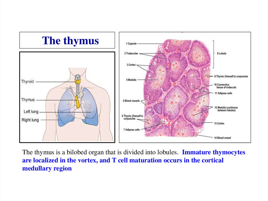

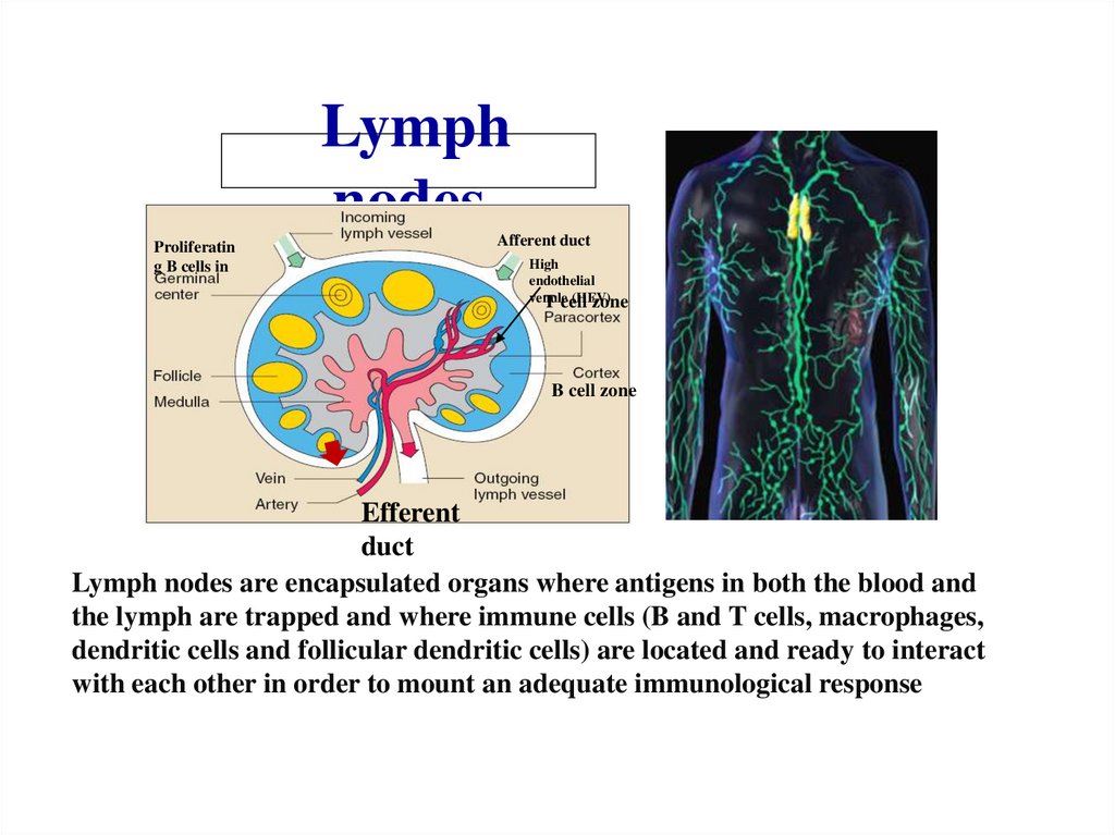

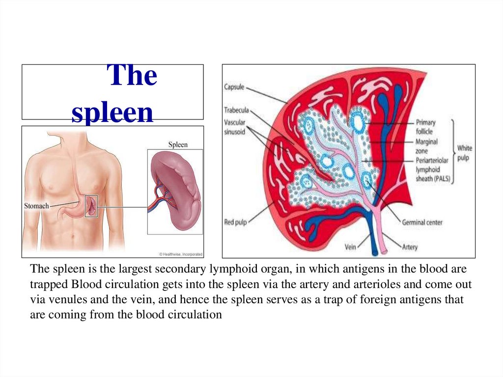
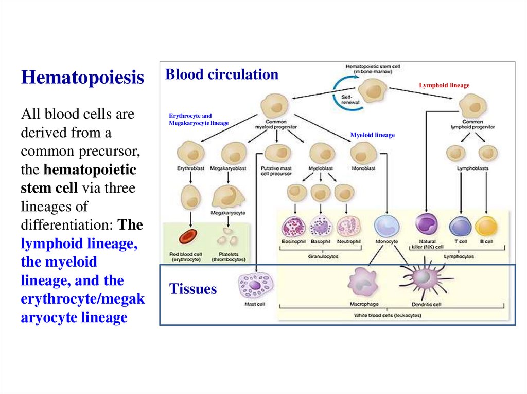


 biology
Similar presentations:


Electron transport system
1.
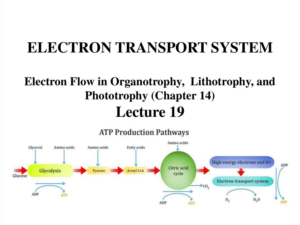
ELECTRON TRANSPORT SYSTEMElectron Flow in Organotrophy, Lithotrophy, and
Phototrophy (Chapter 14)
Lecture 19
2.
Energy, redox Reactions,and Enzymes
https://ecampusontario.pressbooks.pub
/microbio/chapter/energy-matter-andenzymes/
3.
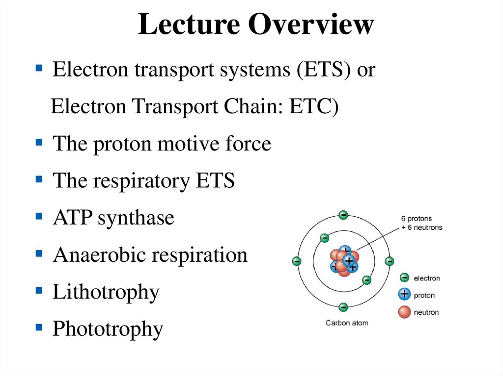
Lecture OverviewElectron transport systems (ETS) or
Electron Transport Chain: ETC)
The proton motive force
The respiratory ETS
ATP synthase
Anaerobic respiration
Lithotrophy
Phototrophy
4.
Introduction• We have learned previously how microorganisms can
catabolize nutrients and obtain energy in the form of energy
carriers
• ATP and GTP can produce energy by hydrolysis and cleavage
of the phosphate bond(s).
• However, NADH and FABH2 need to be transformed into ATP.
• Mot of the energy yield comes from successive redox
(coupled reduction and oxidation) steps within an electron
transport system (ETS).
• The types of metabolisms that use an ETS include
organotrophy (organic electron donors), lithotrophy (inorganic
electron donors), and phototrophy (electrons are excited by
light absorption).
• Our focus will mainly be on ETS organotrophy.
5.
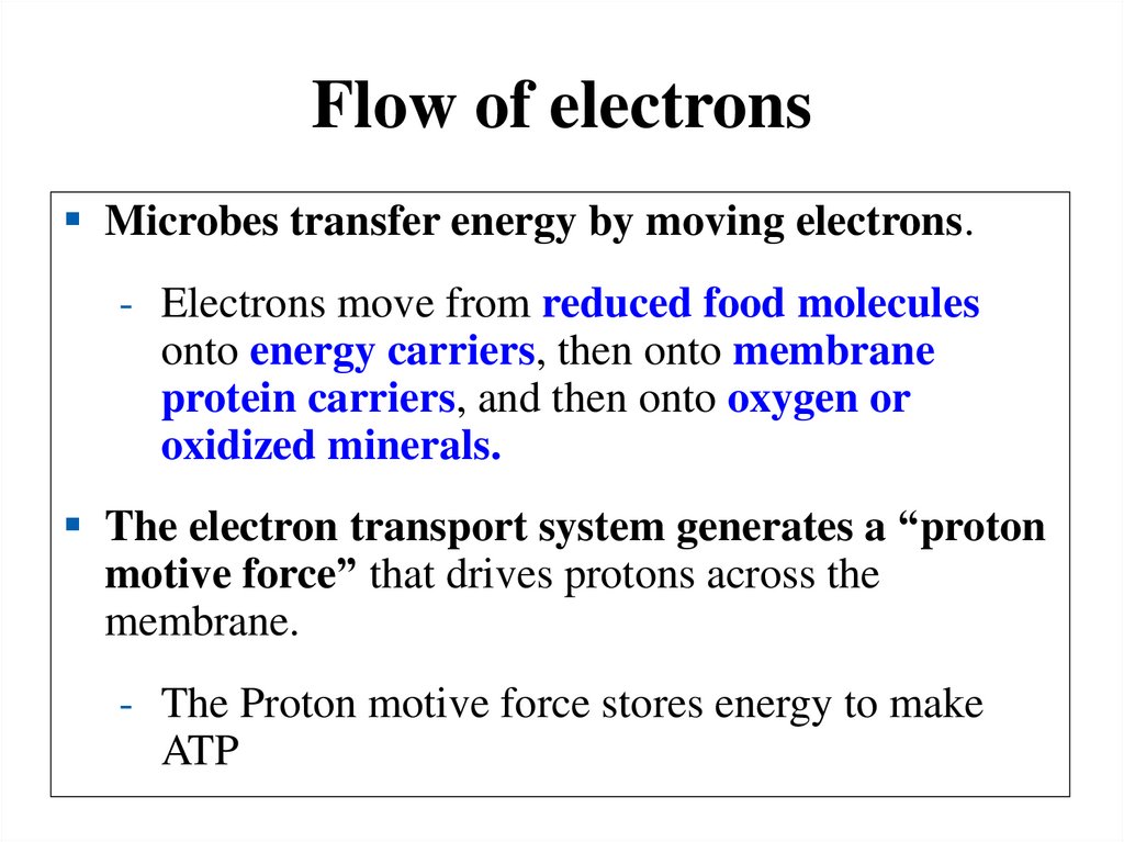
Flow of electronsMicrobes transfer energy by moving electrons.
- Electrons move from reduced food molecules
onto energy carriers, then onto membrane
protein carriers, and then onto oxygen or
oxidized minerals.
The electron transport system generates a “proton
motive force” that drives protons across the
membrane.
- The Proton motive force stores energy to make
ATP
6.
Energy transfer pathwayFlow of electron from
Donor to acceptor
Reduced food
molecules
eEnergy carriers
e-
eCytochrome
Oxygen or
Oxydized
minerals
7.
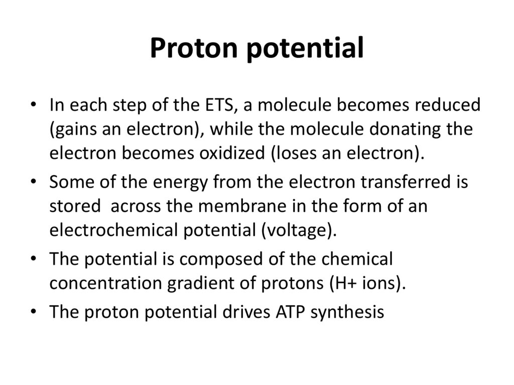
Proton potential• In each step of the ETS, a molecule becomes reduced
(gains an electron), while the molecule donating the
electron becomes oxidized (loses an electron).
• Some of the energy from the electron transferred is
stored across the membrane in the form of an
electrochemical potential (voltage).
• The potential is composed of the chemical
concentration gradient of protons (H+ ions).
• The proton potential drives ATP synthesis
8.
Electron transport systemThe electron transport system (ETS) or electron
transport chain (ETC) generates a proton motive
force that drives protons across the membrane
The proton motive force stores energy to
make ATP
A similar process occurs in mitochondria of
animals and photosynthetic membrane of plant
chloroplasts.
9.
The ETS is embedded in the membraneThe ETS can convert its energy into an ion
potential or electrochemical potential between
two compartments separated by a membrane
The ion potential is most commonly a proton
(H+) potential or proton motive force (PMF)
PMF drives essential cell processes such as
synthesis of ATP
10.
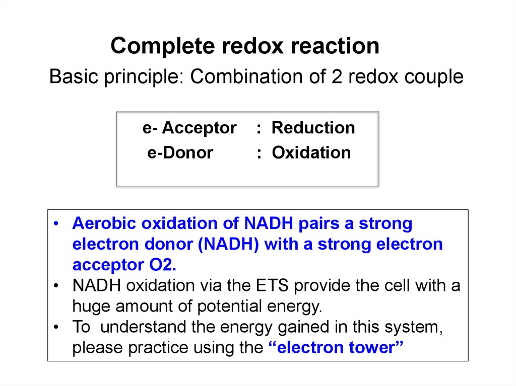
Complete redox reactionBasic principle: Combination of 2 redox couple
e- Acceptor
e-Donor
: Reduction
: Oxidation
• Aerobic oxidation of NADH pairs a strong
electron donor (NADH) with a strong electron
acceptor O2.
• NADH oxidation via the ETS provide the cell with a
huge amount of potential energy.
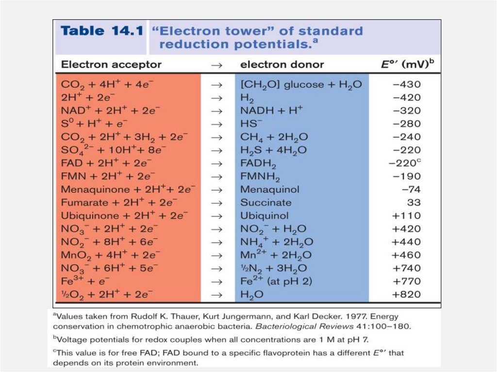
• To understand the energy gained in this system,
please practice using the “electron tower”
11.
12.
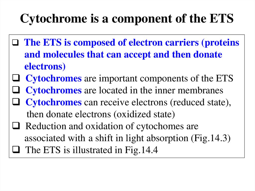
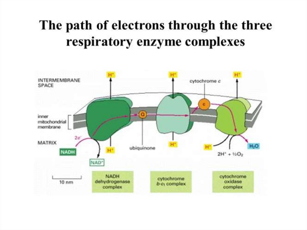
Cytochrome is a component of the ETSThe ETS is composed of electron carriers (proteins
and molecules that can accept and then donate
electrons)
Cytochromes are important components of the ETS
Cytochromes are located in the inner membranes
Cytochromes can receive electrons (reduced state),
then donate electrons (oxidized state)
Reduction and oxidation of cytochomes are
associated with a shift in light absorption (Fig.14.3)
The ETS is illustrated in Fig.14.4
13.
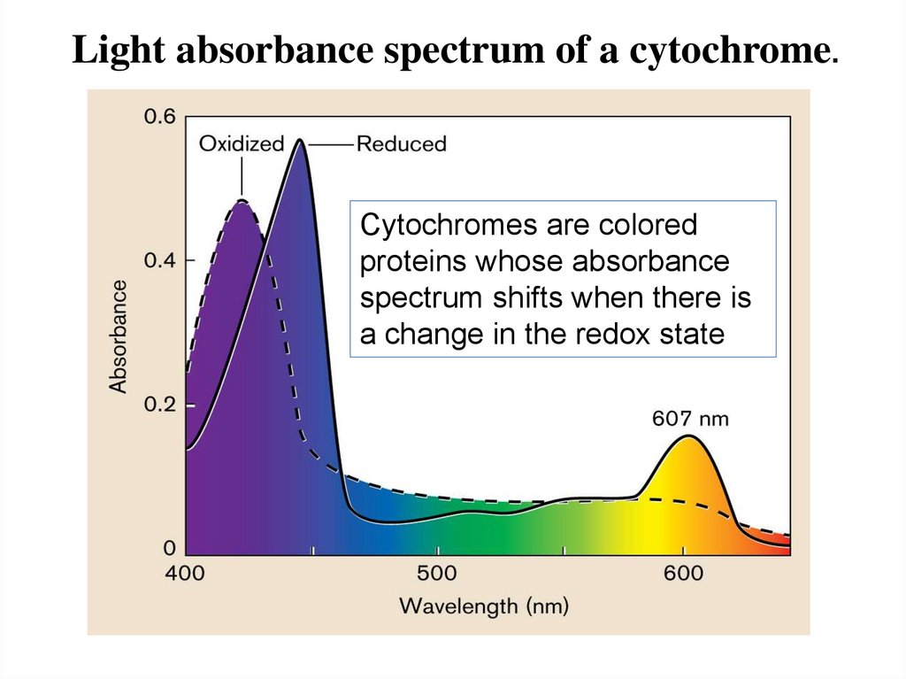
Light absorbance spectrum of a cytochrome.Cytochromes are colored
proteins whose absorbance
spectrum shifts when there is
a change in the redox state
14.
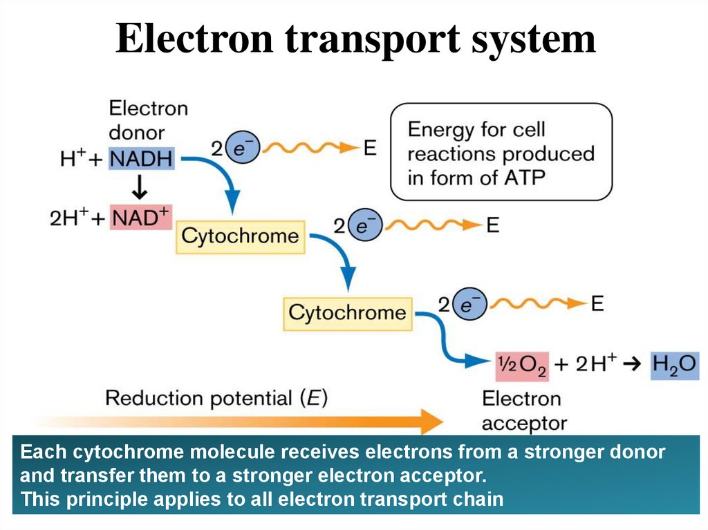
Electron transport systemEach cytochrome molecule receives electrons from a stronger donor
and transfer them to a stronger electron acceptor.
This principle applies to all electron transport chain
15.
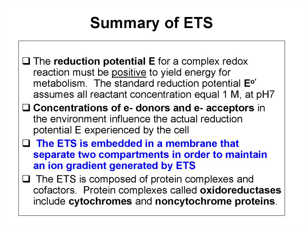
Summary of ETSThe reduction potential E for a complex redox
reaction must be positive to yield energy for
metabolism. The standard reduction potential Eo’
assumes all reactant concentration equal 1 M, at pH7
Concentrations of e- donors and e- acceptors in
the environment influence the actual reduction
potential E experienced by the cell
The ETS is embedded in a membrane that
separate two compartments in order to maintain
an ion gradient generated by ETS
The ETS is composed of protein complexes and
cofactors. Protein complexes called oxidoreductases
include cytochromes and noncytochrome proteins.
16.
Electron Transport Chain: Oxidativephosphorylation
https://www.youtube.com/watch?v=LsRQ5_EmxJA
• Excellent video on Electron Transport Chain:
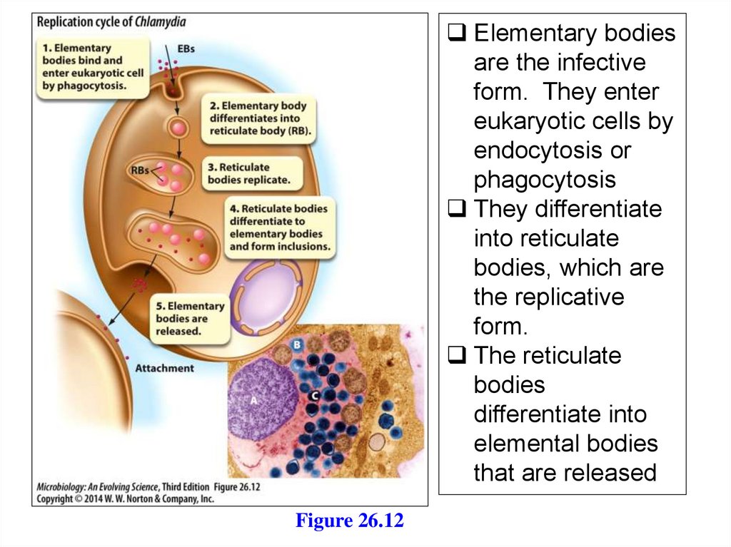
Chemiosmotic Theory.
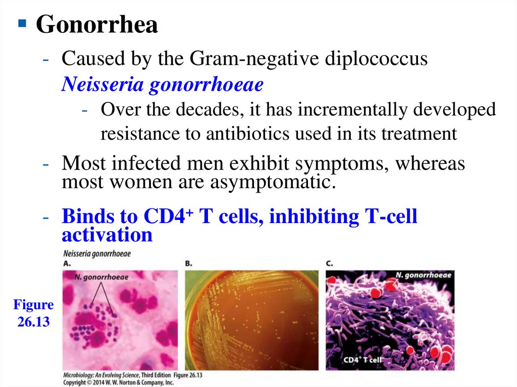
• Note that the author described the ETC in eukaryotes,
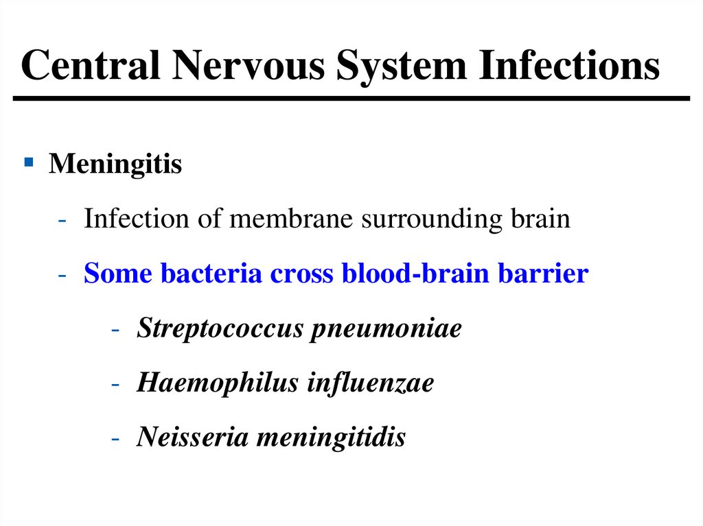
but the basic principles are similar in organotrophs.
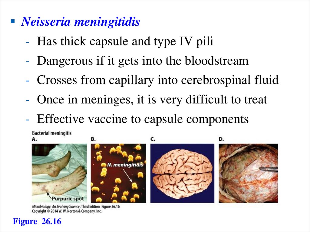
17.
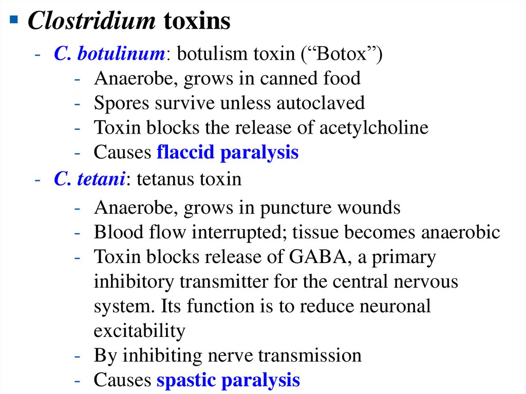
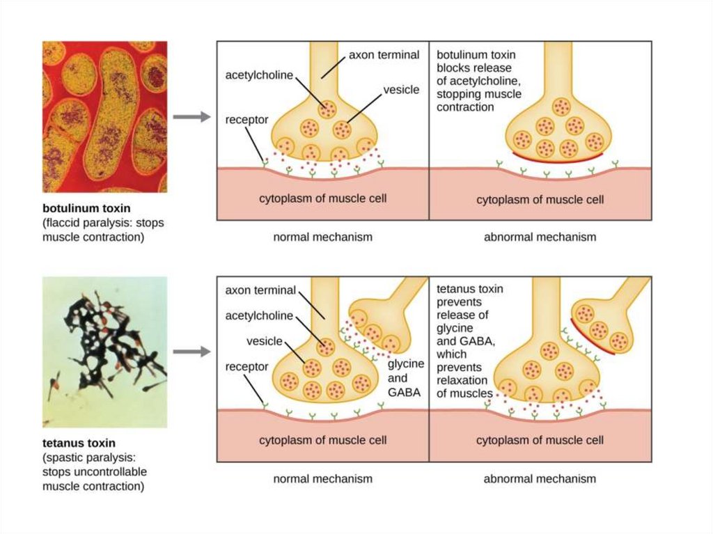
The proton motive forceThe sequential transfer of e- from one ETS protein to the
next yield energy to pump ions (in most cases H+) across the
membrane: Proton pump
Proton pumping generates a proton motive force , also called
proton potential, which is composed of the H+
concentration difference as well as the charge difference
across the membrane
The proton motive force (PMF) drives many different cell
processes:
ATP synthesis as discussed previously under nutrient
transports
Flagellar rotation (Bacteria swim using rotary motions
powered by a proton current
18.
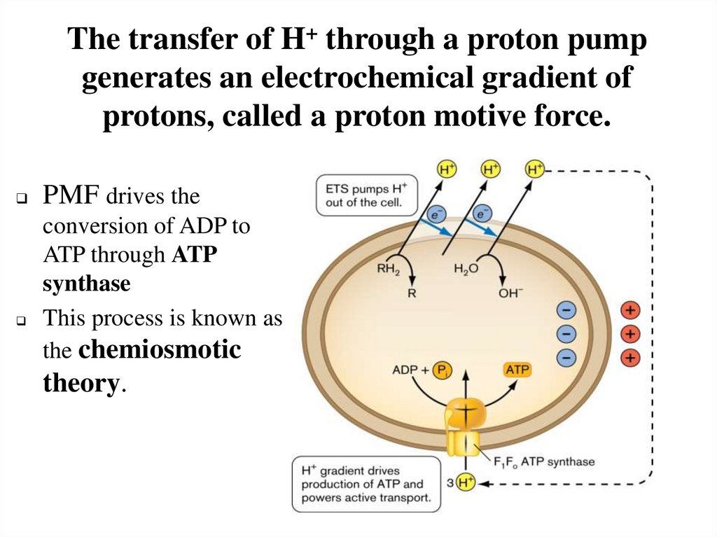
The transfer of H+ through a proton pumpgenerates an electrochemical gradient of
protons, called a proton motive force.
PMF drives the
conversion of ADP to
ATP through ATP
synthase
This process is known as
the chemiosmotic
theory.
19.
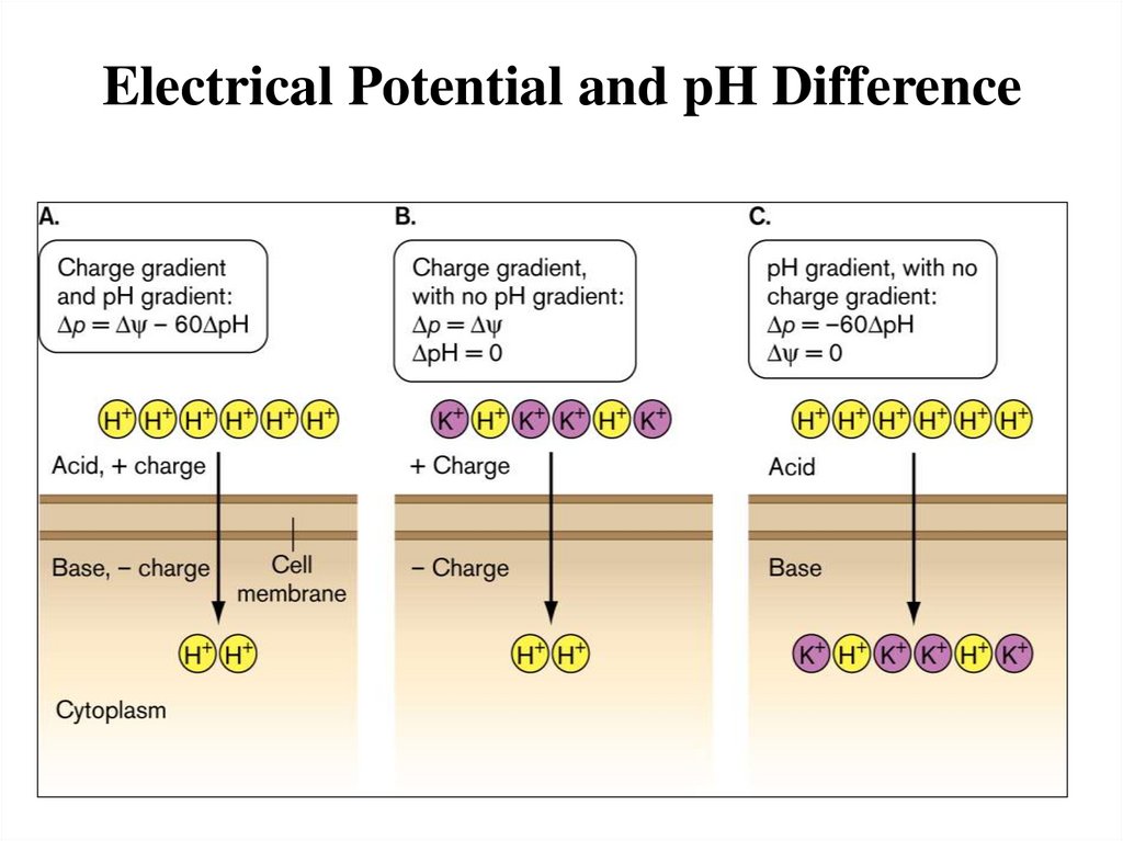
Electrical Potential and pH Difference20.
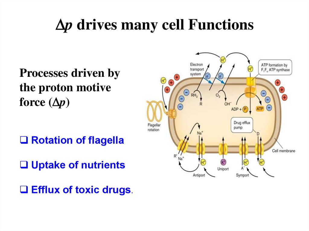
Dp drives many cell FunctionsProcesses driven by
the proton motive
force (Dp)
Rotation of flagella
Uptake of nutrients
Efflux of toxic drugs.
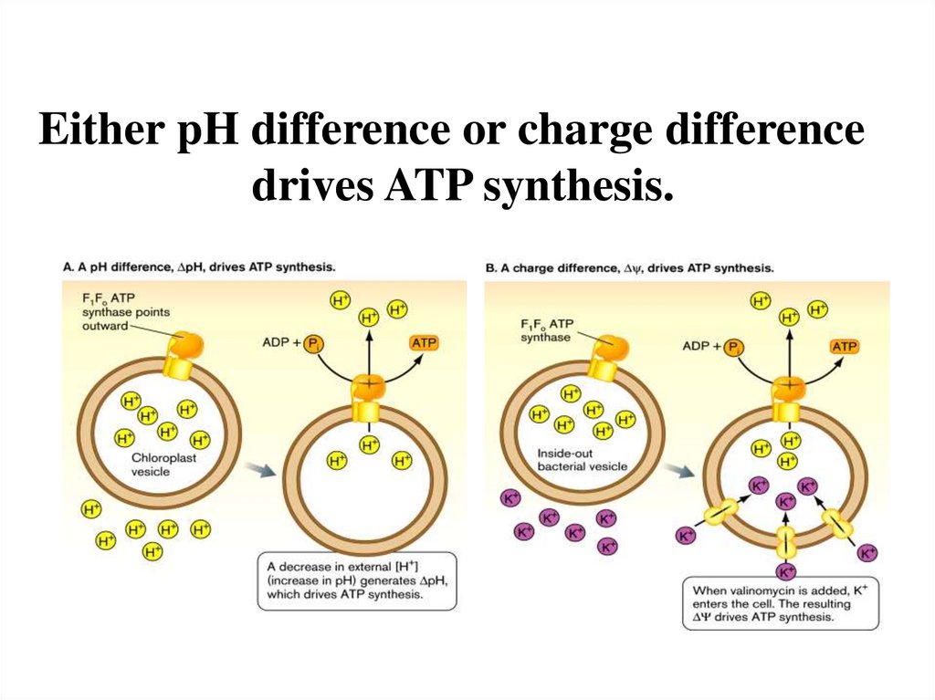
21.
Either pH difference or charge differencedrives ATP synthesis.
22.
The ETS componentsinclude enzymatic reactions
Oxidoreductases catalyzes the removal of remove e(oxidation of one substrate), and the donation of e(reduction of another substrate)
Dehydrogenases: Oxidoreductases that accept e- from
NADH or FADH2 are also called dehydrogenases
because their reaction releases H+
Oxidases catalyzes the removal of remove e-
23.
Oxidoreductase Protein ComplexesA respiratory electron transport system
includes at least 3 functional components
1. An initial substrate oxidoreductase (or
dehydrogenase)
2. A mobile electron carrier
3. A terminal oxidase
24.
25.
1. The substrate dehydrogenase receives a pair ofelectrons from an organic substrate, such as
NADH, or an inorganic substrate, such as H2.
2. It donates the electrons ultimately to a mobile
electron carrier, such as quinone.
Quinone picks up 2H+ from the solution and is
thus reduced to quinol. There are many
quinones, each with a different side chain; so for
simplicity they are collectively referred to as Q and
QH2.
3. The oxidation of NADH and reduction of Q is
coupled to pumping 4H+ across the membrane
26.
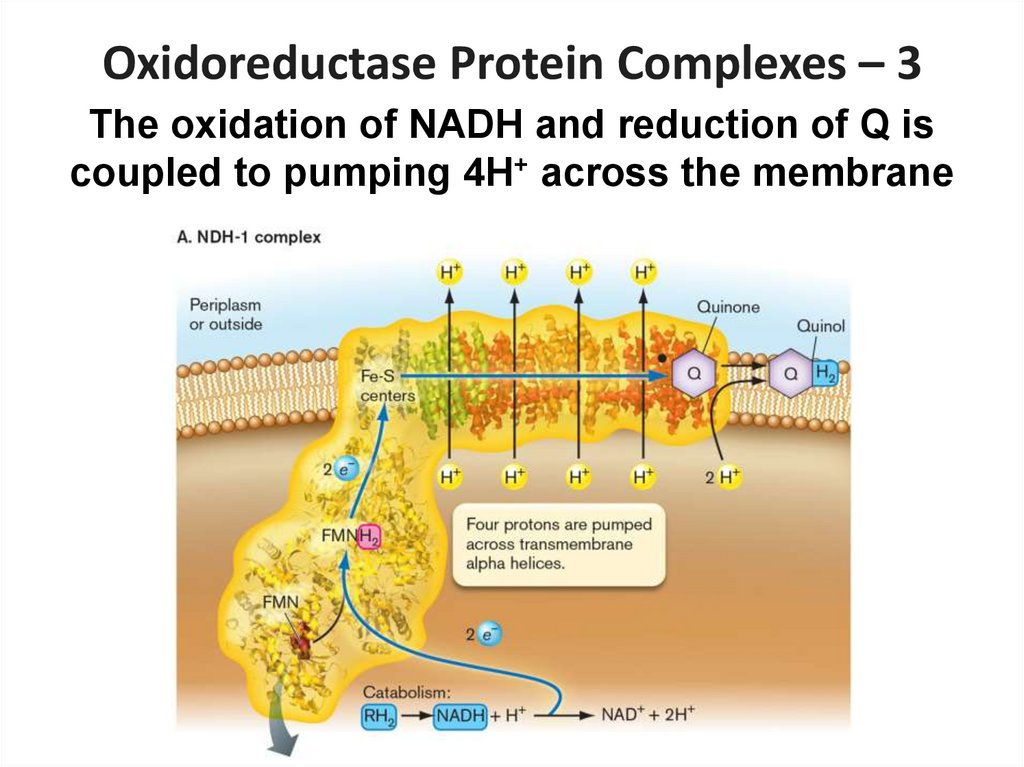
Oxidoreductase Protein Complexes – 3The oxidation of NADH and reduction of Q is
coupled to pumping 4H+ across the membrane
27.
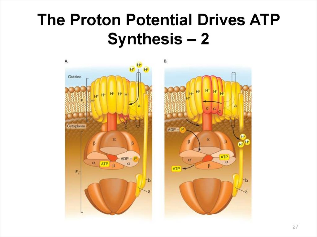
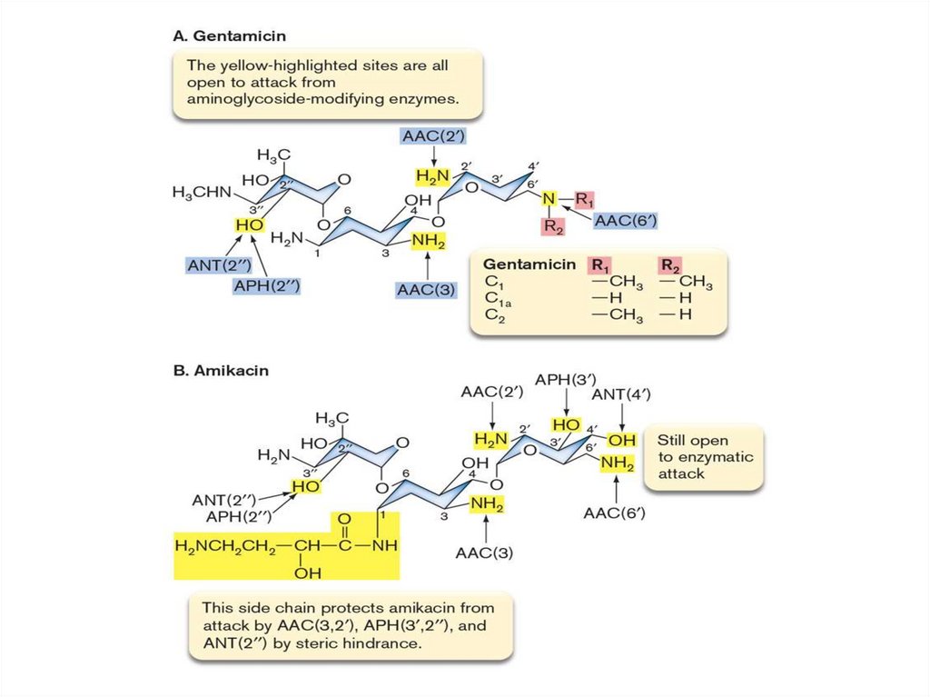
The Proton Potential Drives ATPSynthesis – 2
27
28.
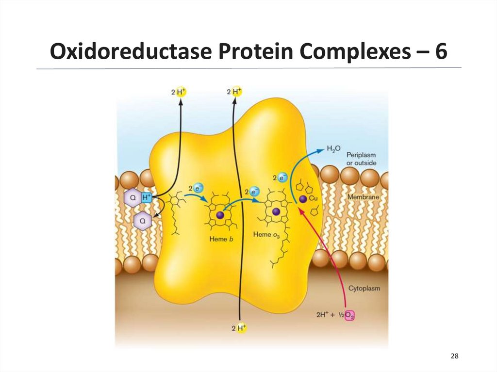
Oxidoreductase Protein Complexes – 628
29.
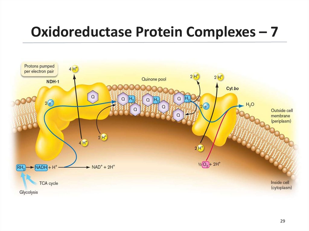
Oxidoreductase Protein Complexes – 729
30.
Mitochondrial Electron Transport30
31.
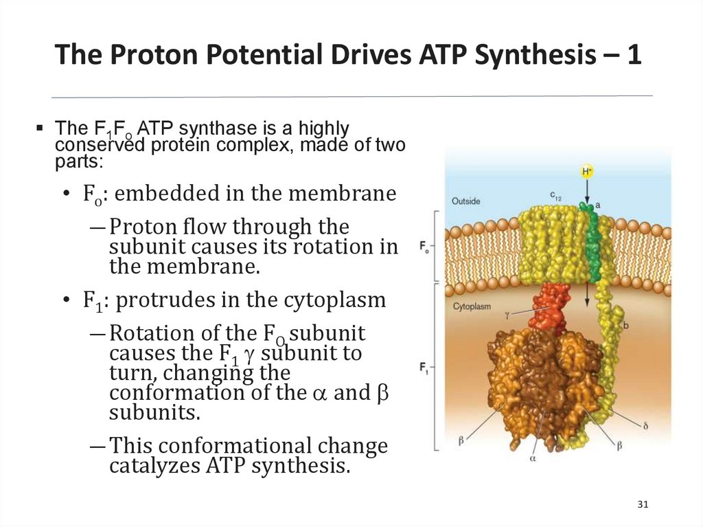
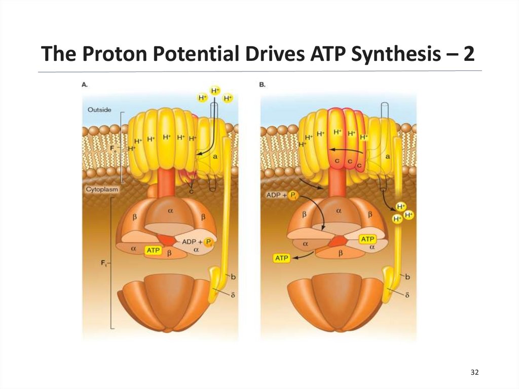
The Proton Potential Drives ATP Synthesis – 1The F1Fo ATP synthase is a highly
conserved protein complex, made of two
parts:
• Fo: embedded in the membrane
― Proton flow through the
subunit causes its rotation in
the membrane.
• F1: protrudes in the cytoplasm
― Rotation of the FO subunit
causes the F1 g subunit to
turn, changing the
conformation of the a and b
subunits.
― This conformational change
catalyzes ATP synthesis.
31
32.
The Proton Potential Drives ATP Synthesis – 232
33.
Electron Transport Chain• hhttp://www.youtube.com/watch?v=xbJ0nb
zt5Kw&feature=related
• NDSU Virtual Cell Animations Project animation 'Cellular Respiration
(Electron Transport Chain)'. For more information please see
http://vcell.ndsu.edu/animations
• https://www.youtube.com/watch?v=3y1dO
4nNaKY Gradients (ATP synthase)
• NDSU Virtual Cell Animations Project animation 'Gradients (ATP
Synthase)'. For more information please see
http://vcell.ndsu.edu/animations
34.
Terminal electron acceptors• In organotrophy or chemoorganotrophy: It is a
form of metabolism in which organic
molecules donate electrons, and the terminal
electron acceptor is O2.
• Alternative electron acceptor is Nitrate (NO3)
• Some electron acceptors are organic
molecules, such as fumarate (note that
fumaric acid is a key chemical intermediate in
the Krebs cycle)
35.
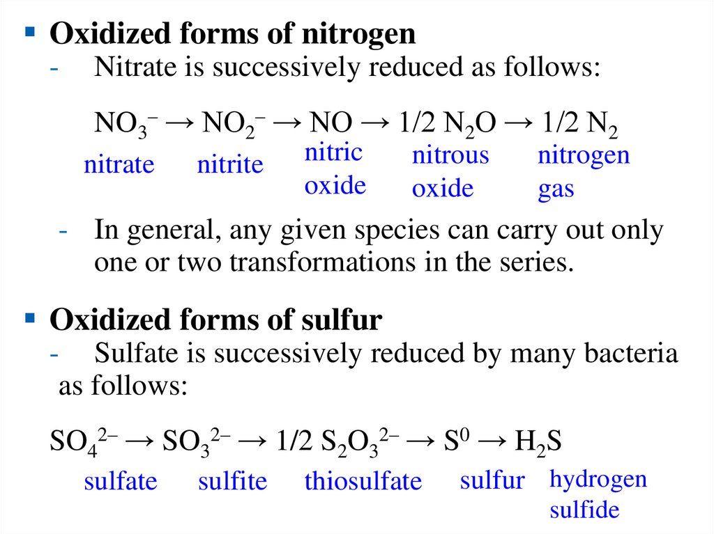
Oxidized forms of nitrogen-
Nitrate is successively reduced as follows:
NO3– → NO2– → NO → 1/2 N2O → 1/2 N2
nitrate
nitrite
nitric
oxide
nitrous
oxide
nitrogen
gas
- In general, any given species can carry out only
one or two transformations in the series.
Oxidized forms of sulfur
- Sulfate is successively reduced by many bacteria
as follows:
SO42– → SO32– → 1/2 S2O32– → S0 → H2S
sulfate
sulfite
thiosulfate
sulfur hydrogen
sulfide
36.
Anaerobic Respiration in OrganotrophsObligate aerobes are organisms that grow only
using O2 as a terminal electron acceptor.
- Include animals, plants, and many bacteria
Anaerobic respiration. Other prokaryotes use a
wide range of terminal electron acceptors, including
metals, oxidized ions of nitrogen, and sulfur.
- This anaerobic respiration generally occurs in
environments where oxygen is scarce
- Wetland soil and the human digestive tract
37.
Electron Acceptors and DonorsAnaerobic respiration is unique to prokaryotes.
- They usually possess alternative electron donors and
electron acceptors.
FYI
Figure 14.18
38.
Dissimilatory Metal ReductionAn important class of anaerobic respiration involves
the reduction of metal cations, or dissimilatory
metal reduction.
- In contrast to minerals reduced for the purpose of
incorporation into cell components (assimilatory
metal reduction)
The metals most commonly reduced through
anaerobic respiration are:
- Iron (Fe3+ → Fe2+)
- Manganese (Mn4+ → Mn2+)
39.
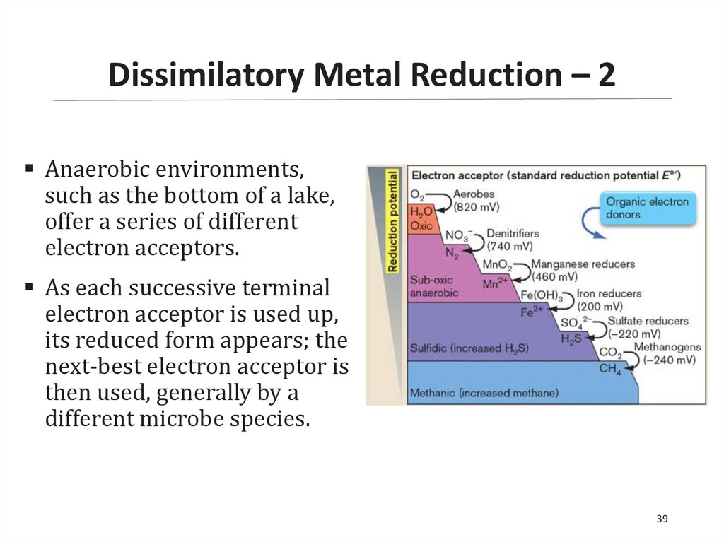
Dissimilatory Metal Reduction – 2Anaerobic environments,
such as the bottom of a lake,
offer a series of different
electron acceptors.
As each successive terminal
electron acceptor is used up,
its reduced form appears; the
next-best electron acceptor is
then used, generally by a
different microbe species.
39
40.
Chapter SummaryElectron transport systems (ETS) consist of membraneembedded proteins that transfer electrons from an initial
electron donor to a TEA that leaves the cell.
The ETS complexes generate a proton motive force that
can drive ATP synthesis and other cell functions.
Electron carriers contain metal ions and/or conjugated,
double-bonded ring structures.
An ETS includes at least three functional components:
- Substrate dehydrogenase, mobile electron carrier, and
terminal oxidase
The F1Fo ATP synthase is a membrane-embedded protein
complex.
- Three protons drive each F1Fo cycle, synthesizing one
molecule of ATP.
41.
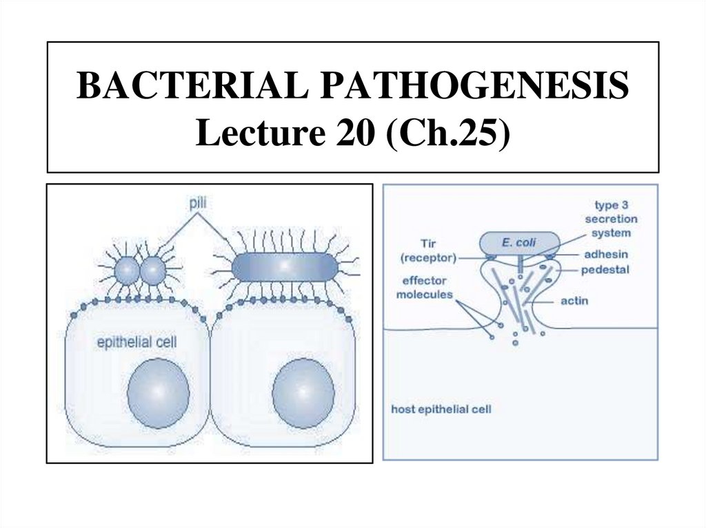
BACTERIAL PATHOGENESISLecture 20 (Ch.25)
42.
Chapter OverviewHost-pathogen interactions
How microbes attach to host cells
How toxins subvert host functions
How toxins and effectors are deployed
How pathogens survive within their hosts
Tools used to probe pathogenesis
42
43.
IntroductionMammals have elaborated physical, chemical, and
immunological defenses that protect against diseasecausing microbes.
- However, every fortress has its weakness.
- Pathogenic microbes exploit those weaknesses,
and the result is disease.
The fundamental question of microbial pathogenesis
is this:
- How an organism too small to be seen with the
naked eye can kill a human a million times larger?
44.
The Languageof Pathogenesis
45.
The Language of Pathogenesis – 1By definition, a parasite is an organism that
receives benefits at the expense of a host.
• In practice, the term “parasite” refers to
disease-causing protozoa and worms. Bacterial,
viral, and fungal parasitic agents of disease are
called pathogens.
• Ectoparasites live on the surface of the host;
• Endoparasites live inside the host’s body.
An infection occurs when a pathogen or parasite
enters or begins to grow on a host. However, the
term “infection” does not necessarily imply overt
disease.
46.
The Language of Pathogenesis – 1By definition, a parasite is an organism that
receives benefits at the expense of a host.
• In practice, the term “parasite” refers to diseasecausing protozoa and worms. Bacterial, viral, and
fungal parasitic agents of disease are called
pathogens.
• Ectoparasites live on the surface of the host;
• Endoparasites live inside the host’s body.
An infection occurs when a pathogen or parasite
enters or begins to grow on a host. However, the
term “infection” does not necessarily imply overt
disease.
47.
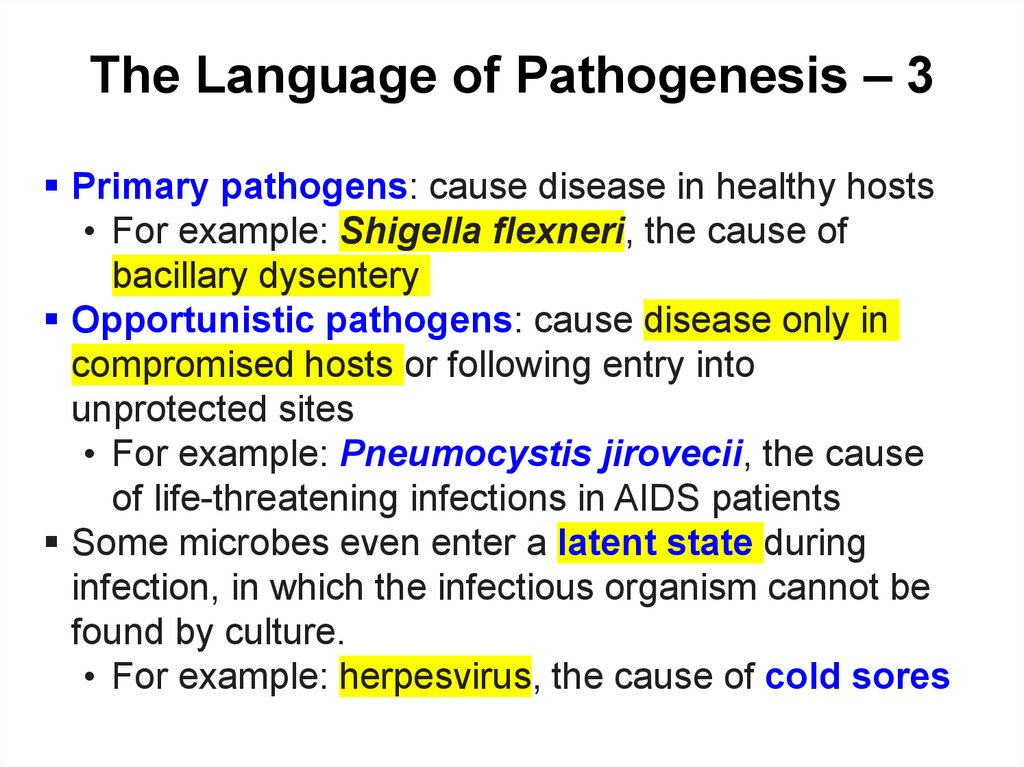
The Language of Pathogenesis – 3Primary pathogens: cause disease in healthy hosts
• For example: Shigella flexneri, the cause of
bacillary dysentery
Opportunistic pathogens: cause disease only in
compromised hosts or following entry into
unprotected sites
• For example: Pneumocystis jirovecii, the cause
of life-threatening infections in AIDS patients
Some microbes even enter a latent state during
infection, in which the infectious organism cannot be
found by culture.
• For example: herpesvirus, the cause of cold sores
48.
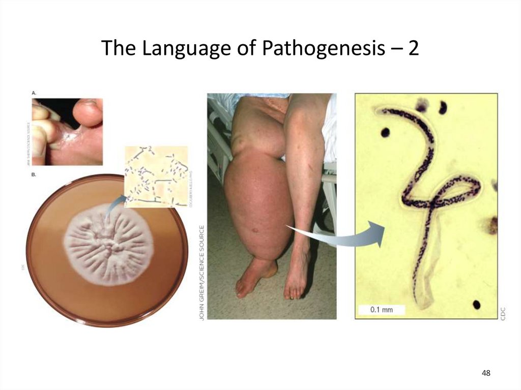
The Language of Pathogenesis – 248
49.
The Language of Pathogenesis – 4A. Pneumocytis jirovechi cysts in bronchoalveolar
materials
B. Cold sore produced by a reactivated
herpesvirus hiding latent in nerve cells
49
50.
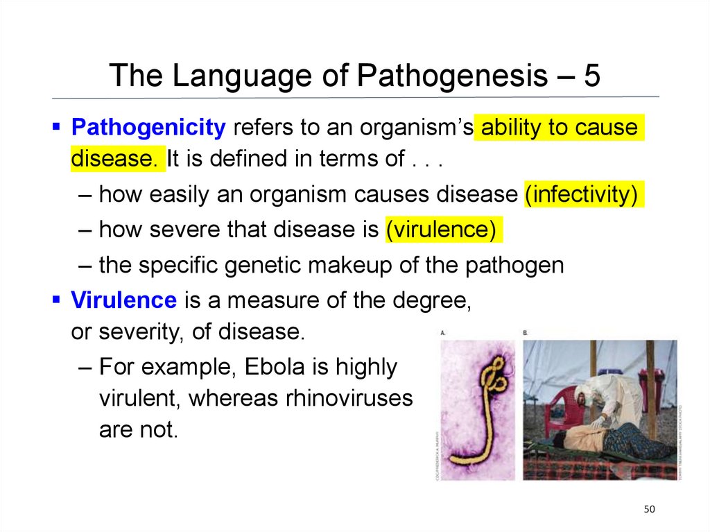
The Language of Pathogenesis – 5Pathogenicity refers to an organism’s ability to cause
disease. It is defined in terms of . . .
– how easily an organism causes disease (infectivity)
– how severe that disease is (virulence)
– the specific genetic makeup of the pathogen
Virulence is a measure of the degree,
or severity, of disease.
– For example, Ebola is highly
virulent, whereas rhinoviruses
are not.
50
51.
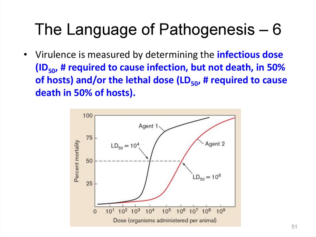
The Language of Pathogenesis – 6• Virulence is measured by determining the infectious dose
(ID50, # required to cause infection, but not death, in 50%
of hosts) and/or the lethal dose (LD50, # required to cause
death in 50% of hosts).
51
52.
ImmunopathogenesisIt is often “friendly fire” by our immune system reacting to a
pathogen that causes major tissue and organ damage.
The term immunopathogenesis applies when the immune
response to a pathogen is a contributing cause of pathology and
disease.
To fully understand any infectious disease, researchers must
study both the pathogenic mechanisms of the pathogen and the
disease symptoms caused by immunopathogenesis.
Ex. Lyme disease caused by the spirochete Borrelia burdorferi is
associated with stimulates the production of strong
inflammatory cytokines, including TNF alpha, IL-1
• beta and IL-6.
53.
Infection Cycles54.
Infection Cycles – 1• The infection cycle describes the route of transmission
of an infectious organism.
– Horizontal transmission: passage from one person
or animal to another within the same generation
• Can be direct (e.g., handshaking) or indirect (e.g.,
sharing contaminated objects)
• Fomites: inanimate objects (e.g., doorknobs, hand
towels, utensils)
• Vehicles: ingested or inhaled materials (e.g., food,
water, air)
– Vertical transmission: passage from a mother to her
fetus during pregnancy (transplacental) or birth
(parturition)
54
55.
56.
Infection Cycles – 3Complex infection cycles often involve
vectors as intermediaries (usually
arthropods like mosquitoes, ticks, mites, or
flies).
– For example, a mosquito vector transfers the
virus that causes yellow fever from infected to
uninfected individuals.
57.
Infection Cycles – 4A reservoir is an animal, bird, or arthropod that normally
harbors the pathogen, often without exhibiting disease.
In the case of yellow fever, the mosquito is not only the
vector but the reservoir as well, because the insect can
pass the virus to future generations of mosquitoes
through vertical transmission.
The virus causing eastern equine encephalitis (EEE),
however, uses birds as a reservoir.
Reservoirs are critically important for the survival of a
pathogen and as a source of infection.
58.
Portals of Entry• Infectious agents enter the body through one or
more portals of entry that are best suited to their
mechanism of pathogenesis.
– Mouth
– Respiratory tract
– Conjunctiva and mucous membranes
– Wounds, injuries, and skin lesions
– Parenteral route: direct injection into
bloodstream (e.g., tick and mosquito bites,
needle punctures)
58
59.
Effect of Infections on Microbiota• A pathogen’s growth and the host’s resulting immune
response also will affect the host’s normal microbiota.
• Numerous mechanisms affect microbial competition
and, ultimately, species diversity within the body.
– Diarrhea reduces the overall numbers of gut
microbiota.
– Intestinal pathogens occupy host binding sites and
alter available nutrients.
– Inflammation can benefit pathogens more than
normal microbiota.
59
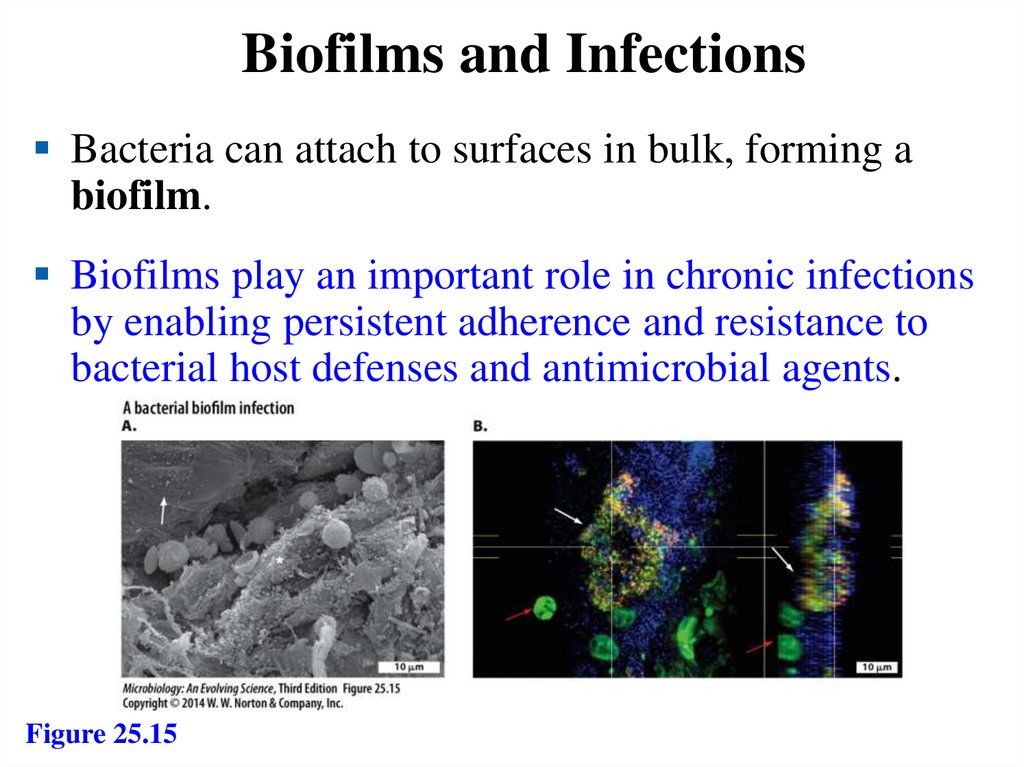
60.
Biofilms and InfectionsBacteria can attach to surfaces in bulk, forming a
biofilm.
Biofilms play an important role in chronic infections
by enabling persistent adherence and resistance to
bacterial host defenses and antimicrobial agents.
Figure 25.15
61.
Virulence Factors62.
Virulence Factors and How to FindThem – 1
• To cause disease, all pathogens must . . .
– Enter a host
– Find their unique niche
– Avoid, circumvent, or subvert normal host defenses
– Multiply
– Transmit to a new susceptible host
• Pathogens employ virulence factors, encoded by
virulence genes, to accomplish these goals. Virulence
factors include toxins, attachment proteins, capsules, and
other devices.
62
63.
Virulence Factorsand How to Find Them – 2
• A suspected virulence gene can be confirmed as having
a role in virulence or pathogenicity only if it fulfills a set of
molecular Koch’s postulates.
1. The phenotype under study should be associated with
pathogenic strains of a species.
2. Specific inactivation of the suspected virulence
gene(s) should lead to a measurable loss in virulence
or pathogenicity. The gene(s) should be isolated by
molecular methods.
3. Reversion or replacement of the mutated gene should
restore pathogenicity.
63
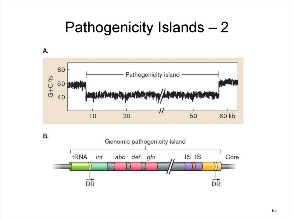
64.
Pathogenicity Islands – 1• Some virulence genes reside on plasmids or in
phage genomes.
• Virulence genes in bacterial pathogens often are
clustered into pathogenicity islands that encode
virulence functions.
• Most pathogenicity islands appear to have been
horizontally transmitted via conjugation or
transduction
– Unique GC/AT ratio
– Linkage to a tRNA gene
– Association with genes homologous to
phage/plasmid genes
64
65.
Pathogenicity Islands – 265
66.
Examples of Pathogen Evolution byHorizontal Gene Transfer – 1
• Dangerous pathogens caught in the act of evolving include:
– Escherichia coli O104:H4, which caused a major
European outbreak of hemolytic uremic syndrome
– Streptococcus agalactiae and Staphylococcus aureus.
– Streptococcus pyogenes (Group A Streptococcus)
• Large-scale genome sequencing data have recently
determined that epidemics are caused by clonal
replacement events rather than by reemergence of
preexisting clones.
66
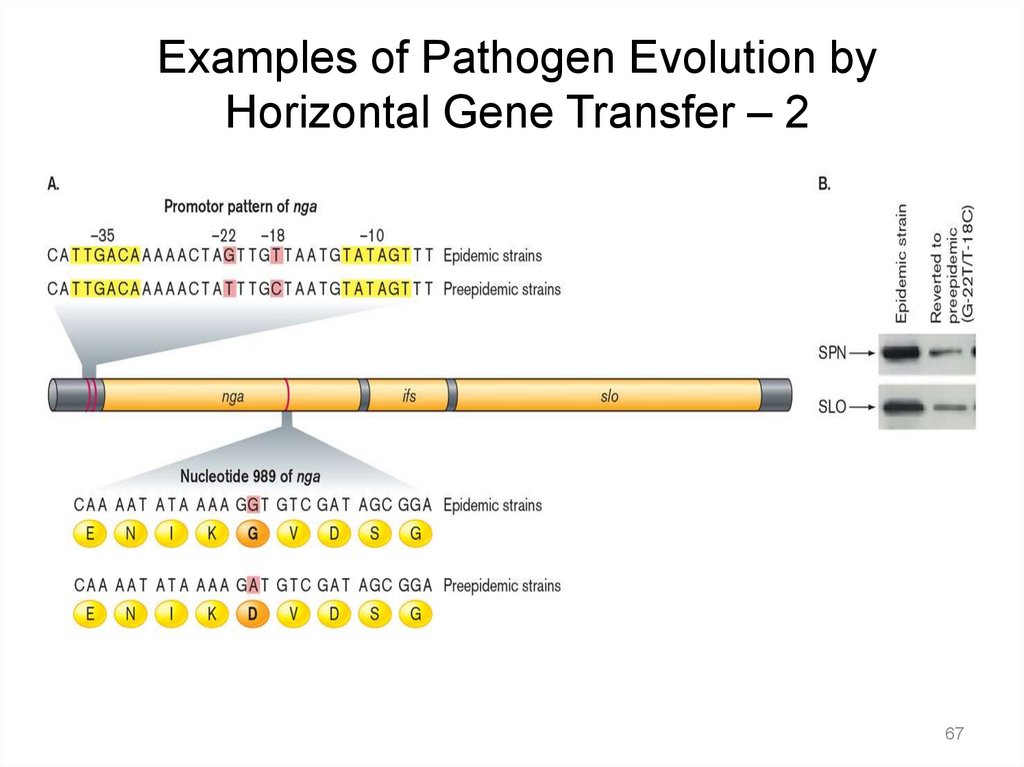
67.
Examples of Pathogen Evolution byHorizontal Gene Transfer – 2
67
68.
Microbial Attachment69.
25.2 Microbial Attachment: FirstContact – 1
• The human body has many ways to exclude
pathogens. How do bacteria manage to stick around
long enough to cause disease?
• The first step toward infection is attachment, or adhesion.
– Any microbial factor that promotes attachment is called
an adhesin.
– Viruses attach to host cells through their capsid or
envelope proteins.
– Bacteria use a variety of strategies, including pili
(fimbriae) and other nonpilus proteins, to bind to
specific host cell factors.
69
70.
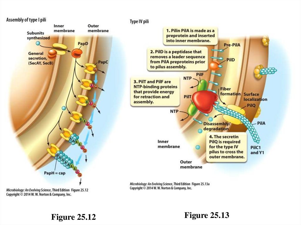
Pili – 1• Many bacteria typically attach to specific host cells using
hairlike appendages called pili (fimbriae). [Note that
fimbriae pili are not the same as conjugation, or sex pili
used for gene transfer.]
– Type I: adhere to carbohydrates on host membranes
• Produce a static attachment to host cell
• Grow from outer membrane of certain Gram-negative
bacteria
– Type IV: involved in “twitching motility”
• Produce a dynamic attachment via assembly and
disassembly
• Grow from inner membrane of many Gram-negative
bacteria
70
71.
Figure 25.12Figure 25.13
72.
Nonpilus Adhesins – 3• Why are some people susceptible to certain
infections, whereas others are not?
– Immunocompetence
– Receptor availability
• Pathogens rely on very specific surface structures
(receptors) to recognize and attach to appropriate
host cells.
– Person-to-person differences in receptor
structures are possible.
– Example: HIV binds C-C chemokine receptor
type 5 (CCR5); individuals with a CCR5
mutation are resistant to HIV infection!
72
73.
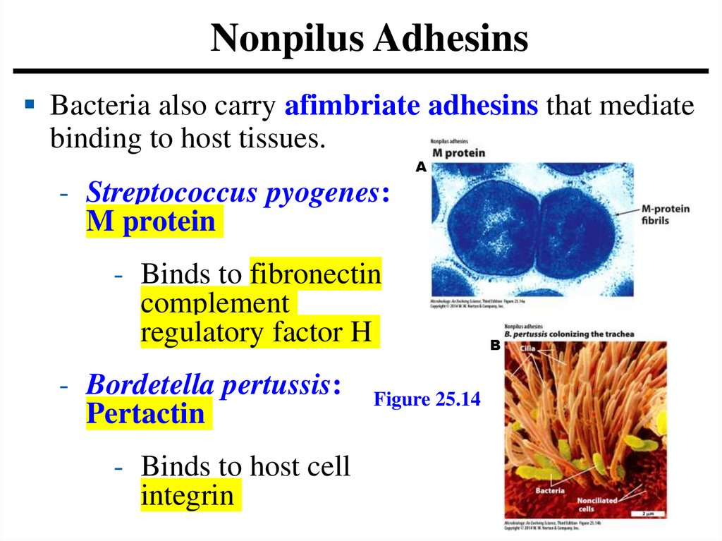
Nonpilus AdhesinsBacteria also carry afimbriate adhesins that mediate
binding to host tissues.
A
- Streptococcus pyogenes:
M protein
- Binds to fibronectin
complement
regulatory factor H
- Bordetella pertussis:
Pertactin
- Binds to host cell
integrin
Figure 25.14
B
74.
Microbial Toxins75.
25.3 Toxins Subvert Host Functions – 1• Bacterial toxins can be divided into two main types.
1. Exotoxins
• Proteins produced and secreted by various types
of bacteria
• Kill host cells and unlock their nutrients
2. Endotoxin
• A part of lipopolysaccharide (LPS) of Gramnegative bacteria
• Hyperactivate host immune systems to harmful
levels
75
76.
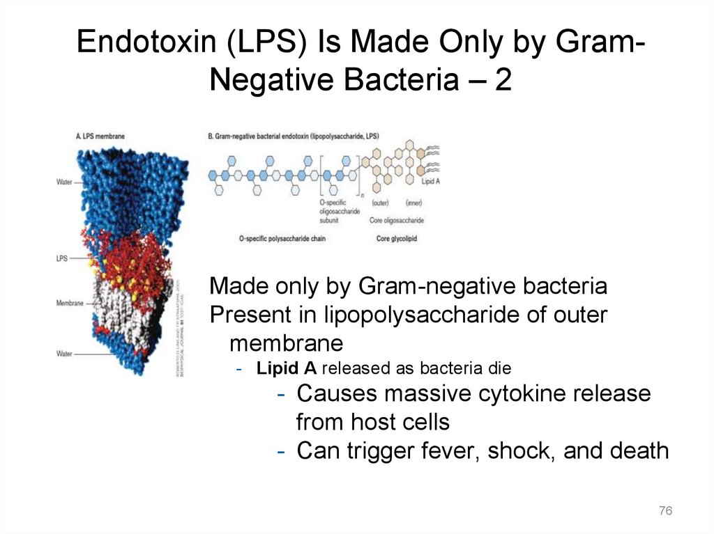
Endotoxin (LPS) Is Made Only by GramNegative Bacteria – 2Made only by Gram-negative bacteria
Present in lipopolysaccharide of outer
membrane
- Lipid A released as bacteria die
- Causes massive cytokine release
from host cells
- Can trigger fever, shock, and death
76
77.
Categories of Microbial Exotoxins – 1• Microbial exotoxins fall into several categories
based on their mechanisms of action.
• Plasma membrane
disruption
• Signal transduction
disruption
• Cytoskeleton
alterations
• Cell-cell adherence
• Protein synthesis
disruption
• Inhibit exocytosis
• Cell cycle disruption
• Vesicular traffic
• Superantigens
77
78.
Categories of Microbial Exotoxins – 2Pore-forming toxins
assemble in target
membranes and
cause leakage of
compounds into and
out of cells
Shiga toxin attaches to
ganglioside Gb3, enters the
cell, and cleaves 28SrRNA in
eukaryotic ribosomes to stop
translation
Enterotoxigenic E. coli heatstable toxin affects cGNP
production.The results is
altered electrolyte transport:
inhibition of Na+ uptake and
stimulation of Cl- transport in
response to the resulting
electrolyte imbalance, water
78
leaves the cell
79.
Membrane Disruption – 1• Some exotoxins disrupt host cell membranes by
forming pores that cause leakage of cell
constituents (host cell lysis).
– Hemolysins lyse red blood cells (and
sometimes other cells).
– Leukocidins lyse white blood cells (leukocytes).
– Some membrane-disrupting exotoxins function
as both hemolysins and leukocidins.
• Streptolysin S of Streptococcus pyogenes
79
80.
Membrane Disruption – 2• Two types of exotoxins disrupt host cell membranes.
– Pore-forming proteins insert themselves into
membranes by binding cholesterol and membrane
receptors
• Alpha toxin of Staphylococcus aureus
• Panton-Valentine toxin of MRSA (see Special
Topic 25.1)
• Listeriolysin O of Listeria monocytogenes
– Phospholipase enzymes hydrolyze phospholipids
into fatty acids
• Phospholipase C of Clostridium perfringens
80
81.
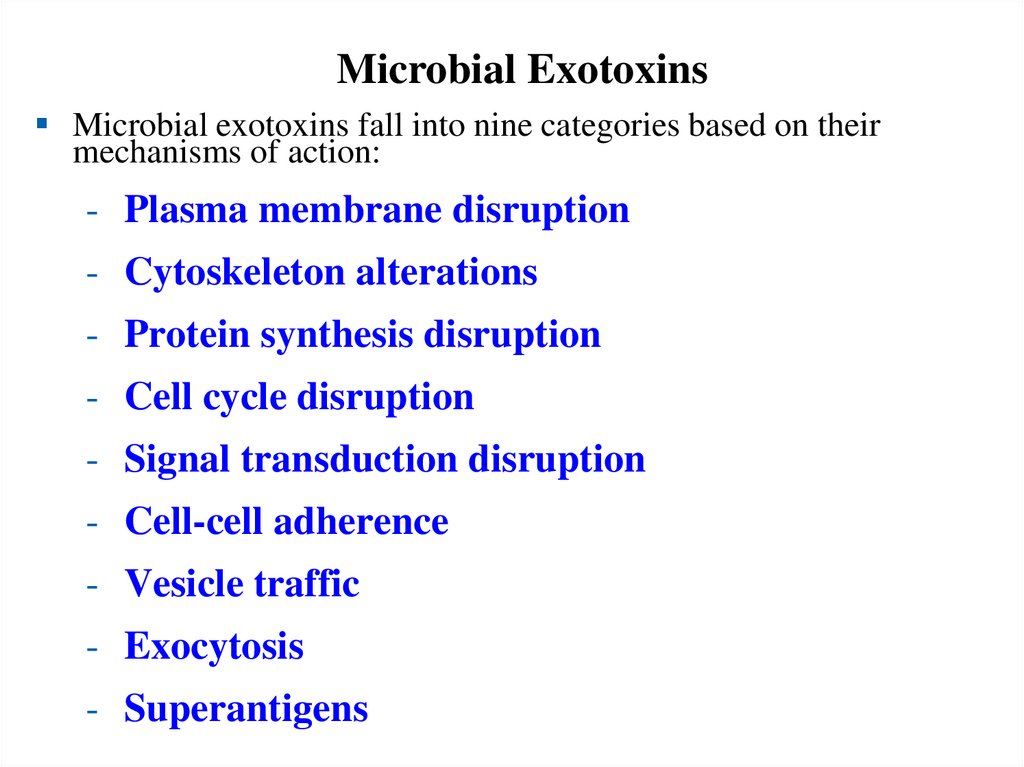
Microbial ExotoxinsMicrobial exotoxins fall into nine categories based on their
mechanisms of action:
- Plasma membrane disruption
- Cytoskeleton alterations
- Protein synthesis disruption
- Cell cycle disruption
- Signal transduction disruption
- Cell-cell adherence
- Vesicle traffic
- Exocytosis
- Superantigens
82.
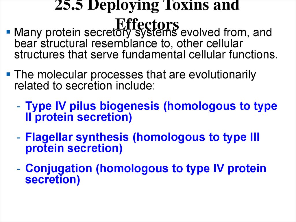
25.5 Deploying Toxins andEffectors
Many protein secretory systems evolved from, and
bear structural resemblance to, other cellular
structures that serve fundamental cellular functions.
The molecular processes that are evolutionarily
related to secretion include:
- Type IV pilus biogenesis (homologous to type
II protein secretion)
- Flagellar synthesis (homologous to type III
protein secretion)
- Conjugation (homologous to type IV protein
secretion)
83.
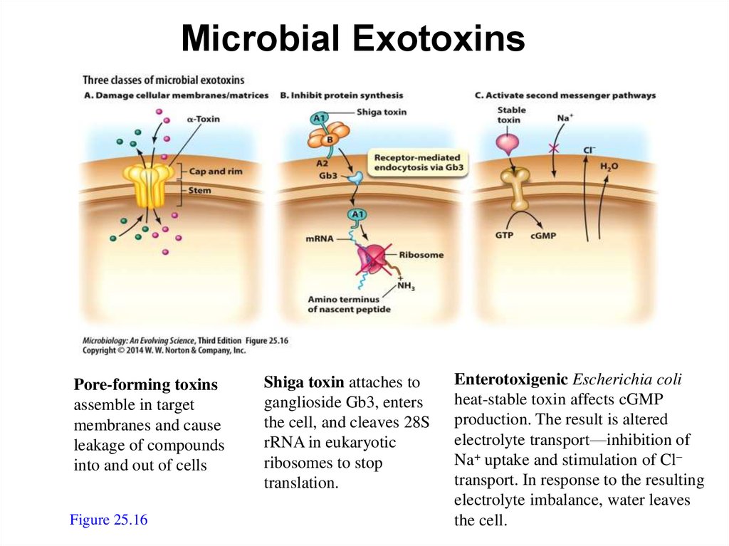
Microbial ExotoxinsPore-forming toxins
assemble in target
membranes and cause
leakage of compounds
into and out of cells
Figure 25.16
Shiga toxin attaches to
ganglioside Gb3, enters
the cell, and cleaves 28S
rRNA in eukaryotic
ribosomes to stop
translation.
Enterotoxigenic Escherichia coli
heat-stable toxin affects cGMP
production. The result is altered
electrolyte transport—inhibition of
Na+ uptake and stimulation of Cl–
transport. In response to the resulting
electrolyte imbalance, water leaves
the cell.
84.
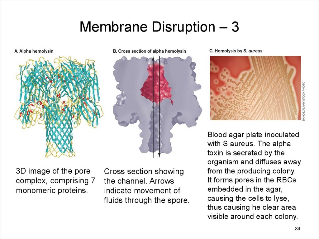
Membrane Disruption – 33D image of the pore
complex, comprising 7
monomeric proteins.
Cross section showing
the channel. Arrows
indicate movement of
fluids through the spore.
Blood agar plate inoculated
with S aureus. The alpha
toxin is secreted by the
organism and diffuses away
from the producing colony.
It forms pores in the RBCs
embedded in the agar,
causing the cells to lyse,
thus causing he clear area
visible around each colony.
84
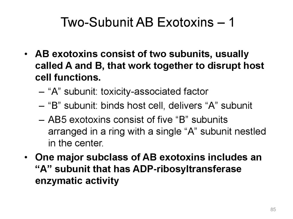
85.
Two-Subunit AB Exotoxins – 1• AB exotoxins consist of two subunits, usually
called A and B, that work together to disrupt host
cell functions.
– “A” subunit: toxicity-associated factor
– “B” subunit: binds host cell, delivers “A” subunit
– AB5 exotoxins consist of five “B” subunits
arranged in a ring with a single “A” subunit nestled
in the center.
• One major subclass of AB exotoxins includes an
“A” subunit that has ADP-ribosyltransferase
enzymatic activity
85
86.
Two-Subunit AB Exotoxins – 2Typical AB toxin consists
of an A subunit and a
pentameric B subunit
joined noncovalently
Many AB toxins are ADP-ribosyltransferase enzymes
that modify proteins structure and function
86
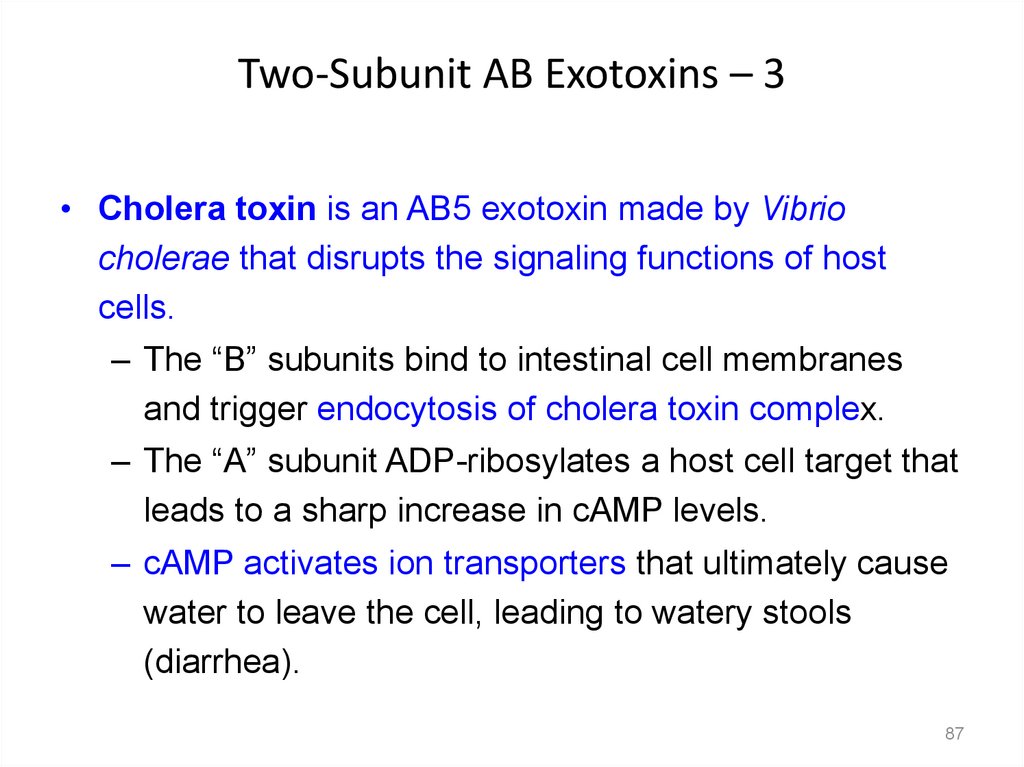
87.
Two-Subunit AB Exotoxins – 3• Cholera toxin is an AB5 exotoxin made by Vibrio
cholerae that disrupts the signaling functions of host
cells.
– The “B” subunits bind to intestinal cell membranes
and trigger endocytosis of cholera toxin complex.
– The “A” subunit ADP-ribosylates a host cell target that
leads to a sharp increase in cAMP levels.
– cAMP activates ion transporters that ultimately cause
water to leave the cell, leading to watery stools
(diarrhea).
87
88.
Two-Subunit AB Exotoxins – 4Vibrio cholerae
(SEM). Note
the slight curve
of the cell and
the presence of
a single polar
flagellum
Brush border of
intestine (TEM) V.
cholerae binds to
the fingerlike villi
on the apical
surface
V. cholerae,
binding to the
surface of a host
cell (SEM). Note
that V. cholerae
does not invade
the host cells
3D structure of
cholera toxin,
binding
ganglioside GM1
on the intestinal
cell surface
88
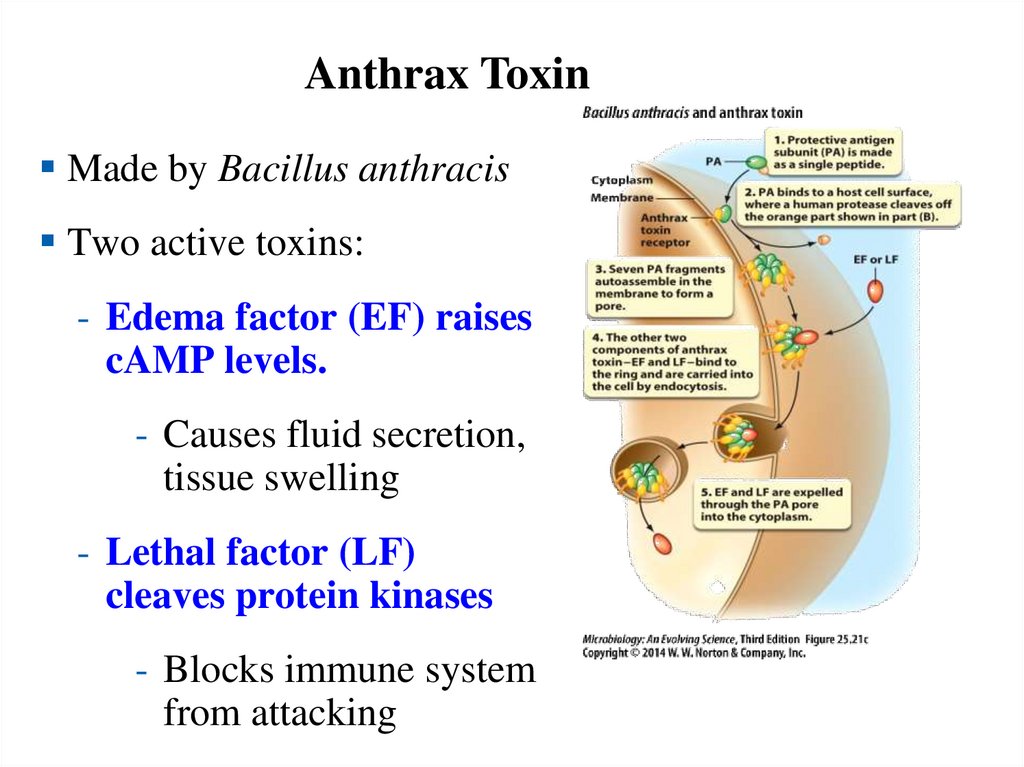
89.
Anthrax ToxinMade by Bacillus anthracis
Two active toxins:
- Edema factor (EF) raises
cAMP levels.
- Causes fluid secretion,
tissue swelling
- Lethal factor (LF)
cleaves protein kinases
- Blocks immune system
from attacking
90.
25.4 Deploying Toxins and Effectors – 1• Many protein secretory systems evolved from, and
bear structural resemblance to, other cell
structures that serve fundamental cell functions.
• We will look at several secretion systems and the
molecular processes with which they share an
evolutionary history.
– Type II secretion: homologous to type IV pilus
biogenesis
– Type III secretion: homologous to flagellar
synthesis
– Type IV secretion: homologous to conjugation
90
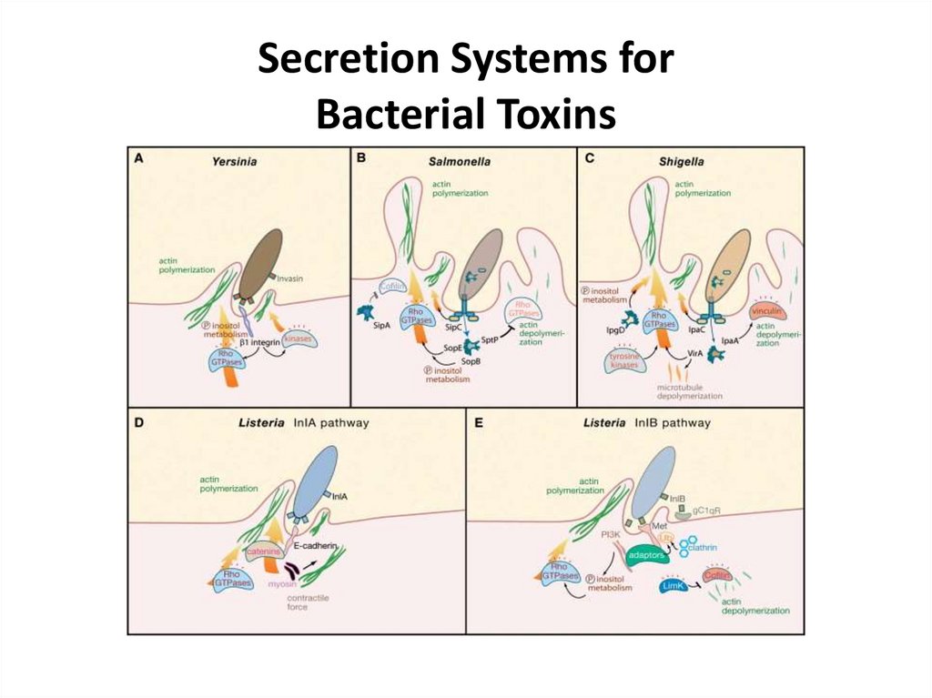
91.
Secretion Systems forBacterial Toxins
92.
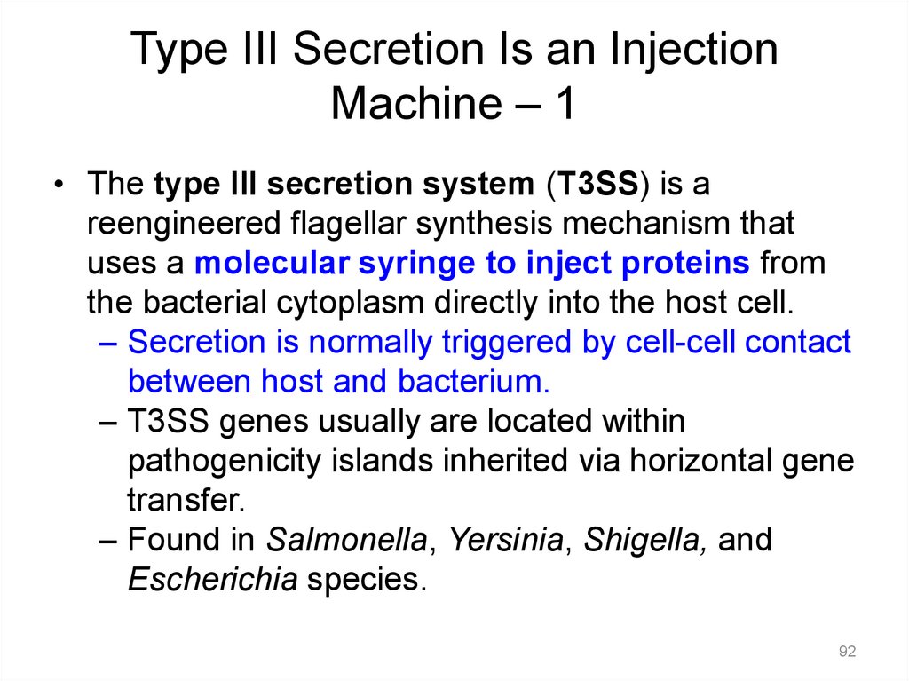
Type III Secretion Is an InjectionMachine – 1
• The type III secretion system (T3SS) is a
reengineered flagellar synthesis mechanism that
uses a molecular syringe to inject proteins from
the bacterial cytoplasm directly into the host cell.
– Secretion is normally triggered by cell-cell contact
between host and bacterium.
– T3SS genes usually are located within
pathogenicity islands inherited via horizontal gene
transfer.
– Found in Salmonella, Yersinia, Shigella, and
Escherichia species.
92
93.
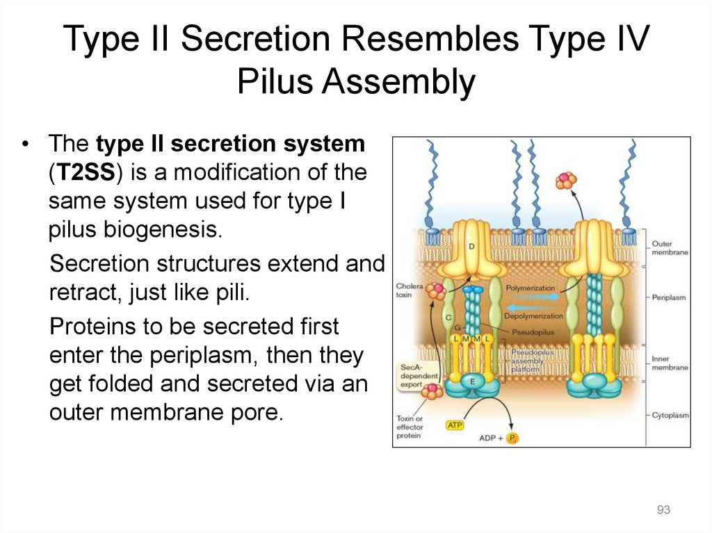
Type II Secretion Resembles Type IVPilus Assembly
• The type II secretion system
(T2SS) is a modification of the
same system used for type I
pilus biogenesis.
Secretion structures extend and
retract, just like pili.
Proteins to be secreted first
enter the periplasm, then they
get folded and secreted via an
outer membrane pore.
93
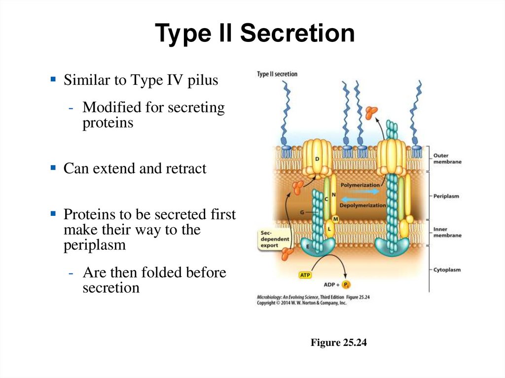
94.
Type II SecretionSimilar to Type IV pilus
- Modified for secreting
proteins
Can extend and retract
Proteins to be secreted first
make their way to the
periplasm
- Are then folded before
secretion
Figure 25.24
95.
Type III Secretion Is an Injection Machine–1
• The type III secretion system (T3SS) is a reengineered
flagellar synthesis mechanism that uses a molecular
syringe to inject proteins from the bacterial cytoplasm
directly into the host cell.
– Secretion is normally triggered by cell-cell contact
between host and bacterium.
– T3SS genes usually are located within pathogenicity
islands inherited via horizontal gene transfer.
– Found in Salmonella, Yersinia, Shigella, and
Escherichia species.
95
96.
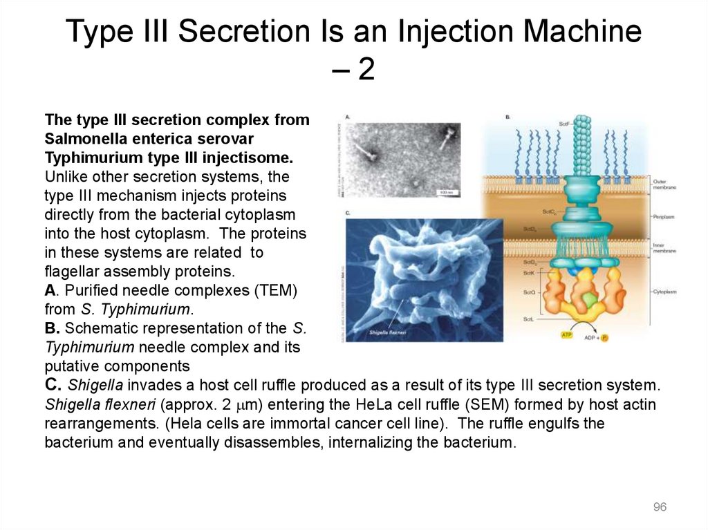
Type III Secretion Is an Injection Machine–2
The type III secretion complex from
Salmonella enterica serovar
Typhimurium type III injectisome.
Unlike other secretion systems, the
type III mechanism injects proteins
directly from the bacterial cytoplasm
into the host cytoplasm. The proteins
in these systems are related to
flagellar assembly proteins.
A. Purified needle complexes (TEM)
from S. Typhimurium.
B. Schematic representation of the S.
Typhimurium needle complex and its
putative components
C. Shigella invades a host cell ruffle produced as a result of its type III secretion system.
Shigella flexneri (approx. 2 mm) entering the HeLa cell ruffle (SEM) formed by host actin
rearrangements. (Hela cells are immortal cancer cell line). The ruffle engulfs the
bacterium and eventually disassembles, internalizing the bacterium.
96
97.
Type III Secretion Is an InjectionMachine – 4
• Some microbes do not rely solely on the natural array of
host receptors for attachment.
• Instead, these bacterial pathogens use a T3SS to insert
their own receptors into target cells.
– Bacteria inject Tir proteins into the host cell. These
proteins act as receptors for the outer membrane
protein intimin. Intimin binds to Tir to establish a
strong attachment.
– Used by enteropathogenic E. coli (EPEC) and
enterohemor-rhagic E. coli (EHEC).
97
98.
Type III Secretion Is an InjectionMachine – 5
A. Model of entropathogenic E. coli (EPEC)
attachment and pedestal formation on intestinal
cells. (1) EPEC attaches first, using type I pili. (2)
Bound EPEC uses a T3SS to inject Tir protein into the
host membrane and acts as a receptor for the EPEC
surface intimin. (3) Tir also communicates through
phosphorylation with other host factors that control actin
filamentation and cytoskeleton formation. Actin
polymerization raises the host membrane to produce a
pedestal upon which EPEC sits. B. Pedestal formation
(colorized SEM).
98
99.
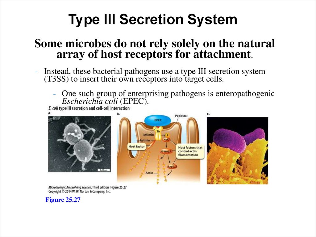
Type III Secretion SystemSome microbes do not rely solely on the natural
array of host receptors for attachment.
- Instead, these bacterial pathogens use a type III secretion system
(T3SS) to insert their own receptors into target cells.
- One such group of enterprising pathogens is enteropathogenic
Escherichia coli (EPEC).
Figure 25.27
100.
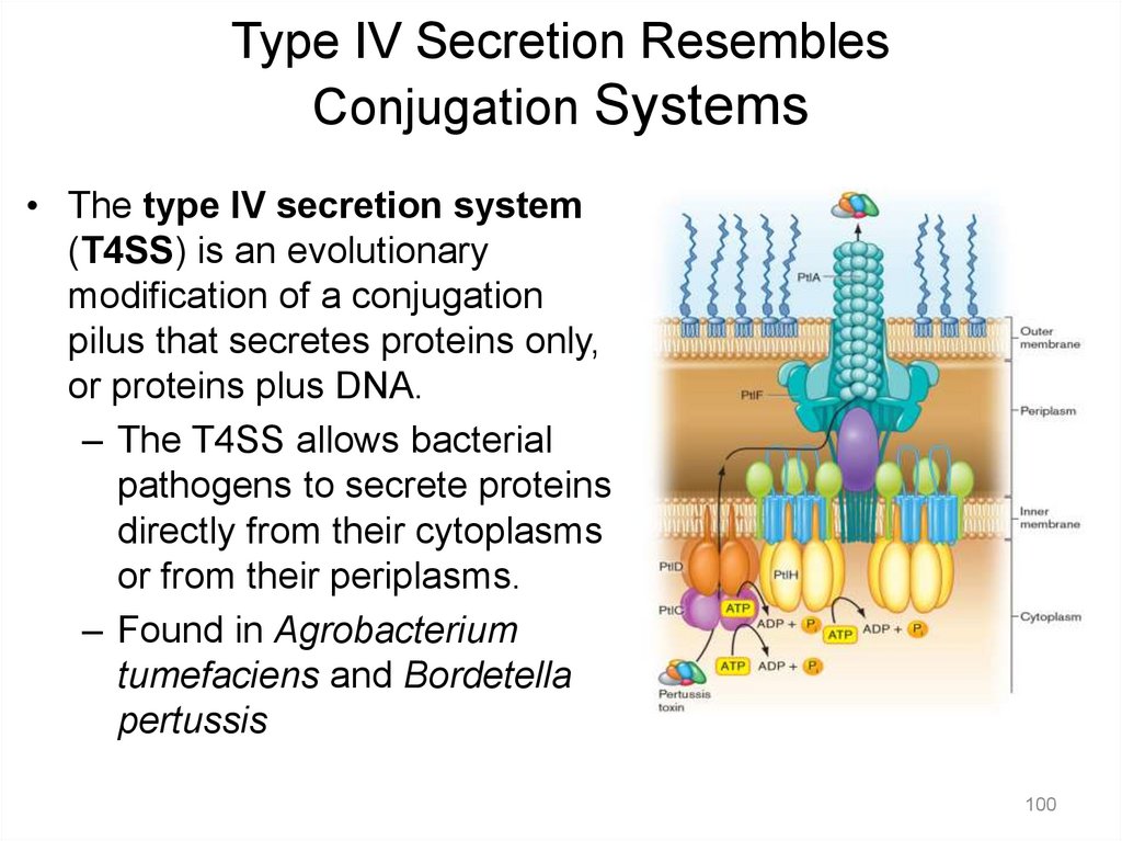
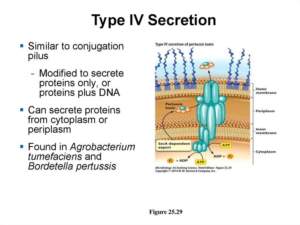
Type IV Secretion ResemblesConjugation Systems
• The type IV secretion system
(T4SS) is an evolutionary
modification of a conjugation
pilus that secretes proteins only,
or proteins plus DNA.
– The T4SS allows bacterial
pathogens to secrete proteins
directly from their cytoplasms
or from their periplasms.
– Found in Agrobacterium
tumefaciens and Bordetella
pertussis
100
101.
Type III SecretionUse a molecular syringe
to inject proteins from the
bacterial cytoplasm
directly into host cell
- Similar to flagellum
Genes usually located on
pathogenicity island
Found in Salmonella,
Yersinia, and Shigella
Figure 25.25
102.
Type IV SecretionSimilar to conjugation
pilus
- Modified to secrete
proteins only, or
proteins plus DNA
Can secrete proteins
from cytoplasm or
periplasm
Found in Agrobacterium
tumefaciens and
Bordetella pertussis
Figure 25.29
103.
Where Am I?Various sensing systems act in concert to recognize
any specific environmental niche.
- Two-component signal transduction systems
- Detect magnesium concentration, pH
- Both low in host cell vacuole
- Detects exotoxins made by other cells
- Delays toxin synthesis until many bacteria
present
- Possible pathway for preventing pathogen
growth?
104.
Intracellular PathogensCell ingests pathogens in phagosome.
- Some pathogens use hemolysin to break out.
- Shigella dysenteriae, Listeria monocytogenes
Phagosome fuses with acidic lysosome.
- Some pathogens secrete proteins to prevent fusion.
- Salmonella, Chlamydia, Mycobacterium,
Legionella
- Some pathogens mature in acidic environment.
- Coxiella burnetii: causes Q fever
105.
Chapter SummaryA pathogen is any microbial agent of disease.
- Primary pathogens cause disease in normal hosts.
- Opportunistic pathogens need immunocompromised
host.
Infection cycles can be direct or indirect.
Virulence factors may be encoded by gene clusters
on pathogenicity islands.
- Acquired by horizontal transmission
Adhesins mediate bacterial attachment to host
cells.
- Type I pili: static attachment
- Type IV pili: continually assembled and
disassembled
- Afrimbrial adhesins: pertactin and M protein
106.
MICROBIAL DISEASESCHAPTER 26
107.
Chapter OverviewSkin and soft-tissue infections
Respiratory tract infections
Gastrointestinal tract infections
Genitourinary tract infections
Cardiovascular system infections
Central nervous system infections
Systemic infections
108.
IntroductionMicrobial diseases are with us daily and are major
contributors to global mortality.
The need for investigations into microbial disease
mechanisms and the body’s ability to combat
infectious agents have been heightened by:
- The emergence of new pathogens, increasing
drug resistance, and threats of bioterrorism
In addition, effective diagnostic algorithms are
needed to quickly identify infectious diseases and
prevent their spread.
109.
Characterizing andDiagnosing Microbial Diseases
Microbial diseases may be classified based on several
criteria:
- By organism
- By organ system (used in this chapter)
- By portal of entry
Each approach has clear benefits and pitfalls.
Pathogens can be divided into four main groups based
on their route of infection:
- Food-borne, airborne, blood-borne, and sexually
transmitted
110.
Zoonotic DiseasesFrancisella tularensis
• Diarrheal disease of the travellers
• Tularemia
Coxiella burnetii
• Q fever
111.
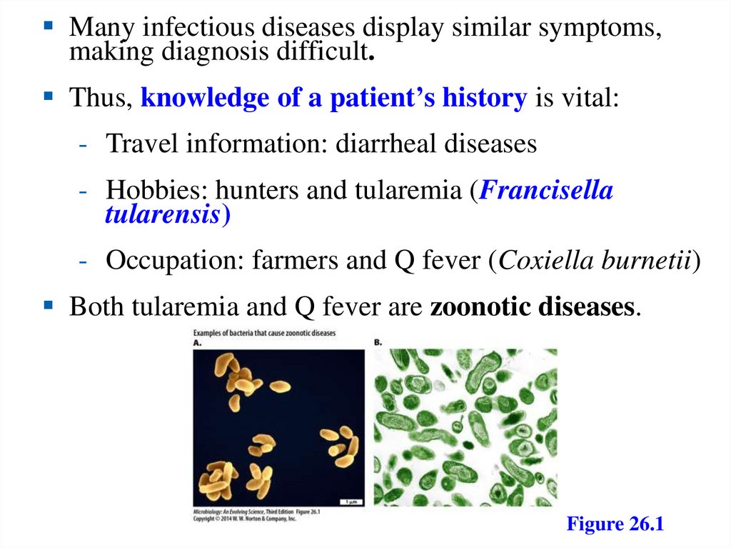
Many infectious diseases display similar symptoms,making diagnosis difficult.
Thus, knowledge of a patient’s history is vital:
- Travel information: diarrheal diseases
- Hobbies: hunters and tularemia (Francisella
tularensis)
- Occupation: farmers and Q fever (Coxiella burnetii)
Both tularemia and Q fever are zoonotic diseases.
Figure 26.1
112.
Skin and Soft-Tissue InfectionsStaphylococcus aureus
Streptococcus pyogenes, also called Group A
Streptococcus (GAS)
• Lancefield groups
• Lancefield grouping is
a serological method for
classifying streptococci into one of 20
groups (designated by a letter) based on
the presence of polysaccharide
and teichoic acid antigens in the bacterial
113.
Lancefield groupsLancefield grouping is a serological method for
classifying streptococci into one of 20 groups
(designated by a letter) based on the presence of
polysaccharide and teichoic acid antigens in the
bacterial cell wall (Lancefield 1933).
The technique is now performed using
commercial latex agglutination test kits, which allow
rapid detection of clinically important streptococcal
groups.
Some streptococci, for example S. pneumoniae,
have not been assigned to a group because their
antigen extracts fail to react with group antisera.
With the exception of S. pneumoniae all the equine
114.
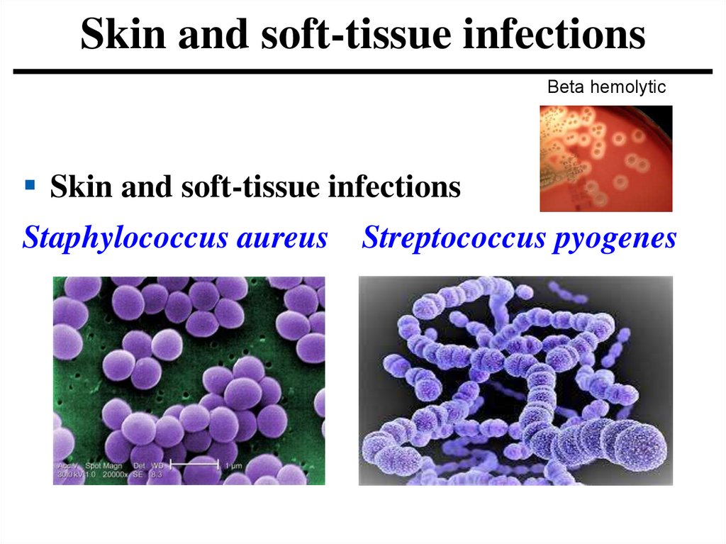
Skin and soft-tissue infectionsBeta hemolytic
Skin and soft-tissue infections
Staphylococcus aureus Streptococcus pyogenes
115.
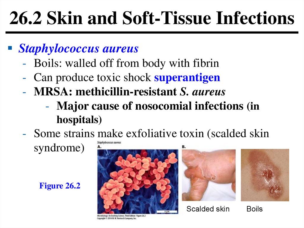
26.2 Skin and Soft-Tissue InfectionsStaphylococcus aureus
- Boils: walled off from body with fibrin
- Can produce toxic shock superantigen
- MRSA: methicillin-resistant S. aureus
- Major cause of nosocomial infections (in
hospitals)
- Some strains make exfoliative toxin (scalded skin
syndrome)
Figure 26.2
Scalded skin
Boils
116.
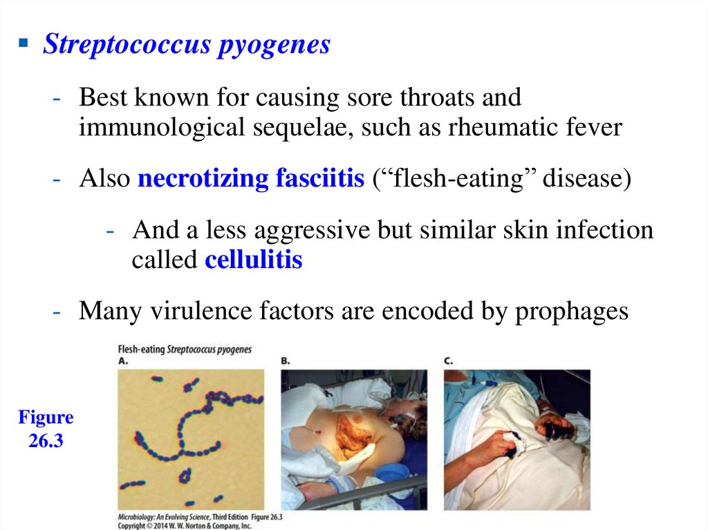
Streptococcus pyogenes- Best known for causing sore throats and
immunological sequelae, such as rheumatic fever
- Also necrotizing fasciitis (“flesh-eating” disease)
- And a less aggressive but similar skin infection
called cellulitis
- Many virulence factors are encoded by prophages
Figure
26.3
117.
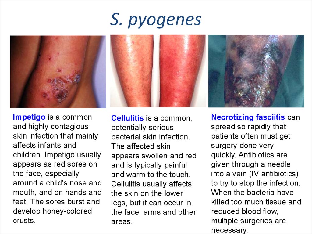
S. pyogenesImpetigo is a common
and highly contagious
skin infection that mainly
affects infants and
children. Impetigo usually
appears as red sores on
the face, especially
around a child's nose and
mouth, and on hands and
feet. The sores burst and
develop honey-colored
crusts.
Cellulitis is a common,
potentially serious
bacterial skin infection.
The affected skin
appears swollen and red
and is typically painful
and warm to the touch.
Cellulitis usually affects
the skin on the lower
legs, but it can occur in
the face, arms and other
areas.
Necrotizing fasciitis can
spread so rapidly that
patients often must get
surgery done very
quickly. Antibiotics are
given through a needle
into a vein (IV antibiotics)
to try to stop the infection.
When the bacteria have
killed too much tissue and
reduced blood flow,
multiple surgeries are
necessary.
118.
Respiratory Tract InfectionBordetella pertussis
Steptococcus pneumoniae
Mycobacterium tuberculosis
Pneumocystis Jirovecci
119.
26.3 Respiratory Tract InfectionsThe mucociliary escalator is primary respiratory defense.
- Bordetella pertussis (cause of whooping cough) inhibits
it by binding to lung cilia.
- https://www.youtube.com/watch?v=HMdrhwEnY6M
Introduction to Mucociliary Transport Video
Microscopy
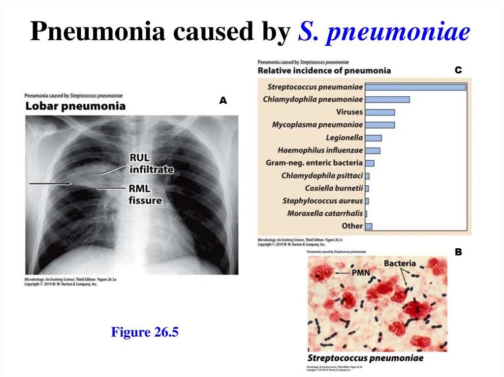
Pneumonia is a disease, not a specific infection.
- Caused by many different microbes
- Streptococcus pneumoniae is the main bacterium:
- Has capsule that prevents phagocytosis
- Can invade the bloodstream (bacteremia) and the
covering of the brain (meningitis)
120.
Pneumonia caused by S. pneumoniaeC
A
B
Figure 26.5
121.
Mycobacteriumtuberculosis
- An acid-fast bacillus
- An ancient and
reemerging pathogen
- Forms calcified tubercles
in the lung
- Can disseminate through
the bloodstream
Figure 26.7
- Has high mortality rate due to multidrug-resistant
strains and high susceptibility of HIV patients
122.
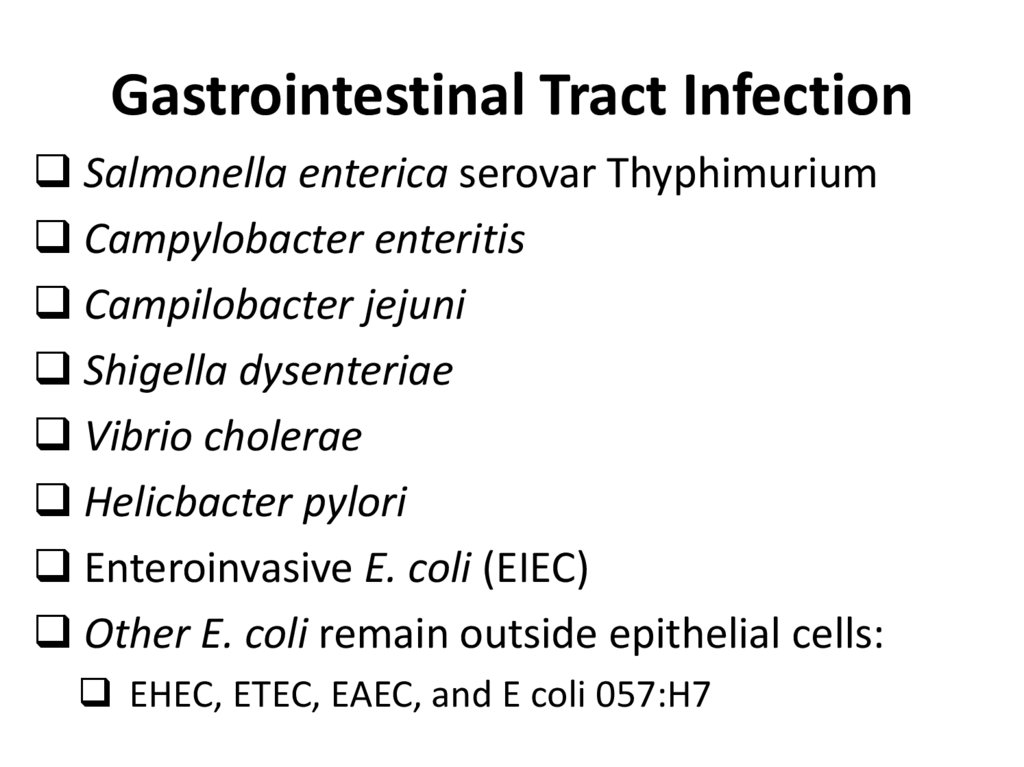
Gastrointestinal Tract InfectionSalmonella enterica serovar Thyphimurium
Campylobacter enteritis
Campilobacter jejuni
Shigella dysenteriae
Vibrio cholerae
Helicbacter pylori
Enteroinvasive E. coli (EIEC)
Other E. coli remain outside epithelial cells:
EHEC, ETEC, EAEC, and E coli 057:H7
123.
Gastrointestinal Tract InfectionsThe main symptoms of gastroenteritis are watery
diarrhea and vomiting. The most frequent causes of
self-limiting diarrheal disease include
Salmonella enterica serovar Typhimurium and
Bacteria of the genus Campylobacter, such as C.
enteritis is common cause of intestinal infection
A more severe form of gastroenteritis is called
dysentery (diarrhea with passage of blood or mucus)
- Bacterial dysentery: Shigella species, including
S. dysenteriae
124.
Remarkably, Staphylococcus aureus causesgastrointestinal disease without ever producing
infection.
Some strains can secrete enterotoxins into tainted foods
such as pies, turkey dressing, or potato salad, causing food
poisoning
The most important treatment for diarrhea is rehydration
therapy
Antibiotics are often inappropriate when treating
diarrhea
- Ineffective against viruses; bacterial gastroenteritis
resolves spontaneously
- In some cases, antibiotic treatment can actually
trigger gastrointestinal disease.
- Example: clindamycin can kill competing bacteria,
thus allowing Clostridium difficile to thrive
- Causes pseudomembranous enterocolitis
125.
Pseudomembranous colitis refers to
swelling or inflammation of the large
intestine (colon) due to an overgrowth of
Clostridium difficile (C. difficile) bacteria.
This infection is a common cause of
diarrhea after antibiotic use.
126.
Enterobacterial toxin-producing strains- Inject toxin via type III secretion
- Bacteria invade epithelial mucosa.
- Salmonella
- Shigella, enteroinvasive Escherichia coli (EIEC)
- Produce Shiga toxin
- Blocks host protein synthesis, damages
endothelia
- Capillary damage, loss of blood, clots
- Bacteria remaining outside epithelial cells
- E. coli: EHEC (O157:H7), ETEC, EAEC
- Entero-hemorrhagic, -toxigenic, -aggregative
- O157 = serotype of LPS; H7 = serotype of flagella
127.
Other bacterial agents of gastrointestinal disease- Campylobacter jejuni
- Most frequent bacterial cause of diarrhea
- Vibrio cholerae: cholera
- Helicobacter pylori: gastric ulcers
- Secretes urease:
urea → NH4+
- Neutralizes stomach acid
- Burrows into protective
mucous layer
- Associated with gastric
cancer
Figure 26.8
128.
Genitourinary tract infectionsUropathogenic E. coli (UPEC)
129.
26.5 Genitourinary Tract InfectionsThe urinary tract includes the kidneys, ureters,
urinary bladder, and urethra.
Active infection of the urinary tract occurs in one of
three basic ways:
- Infection from the urethra to the bladder
- Descending infection from the kidneys
- Ascending infection to the kidney
Most UTIs are caused by Gram-negative rods
from the GI tract.
- Only 5% are caused by Gram-positive
bacteria and fungi.
130.
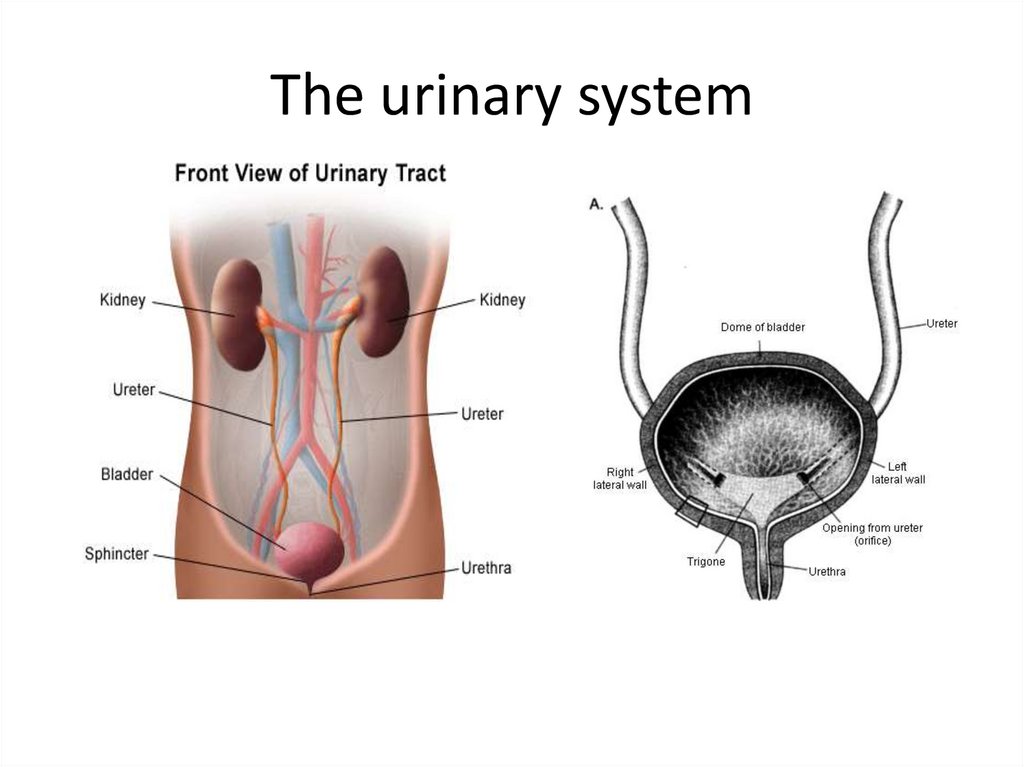
The urinary system131.
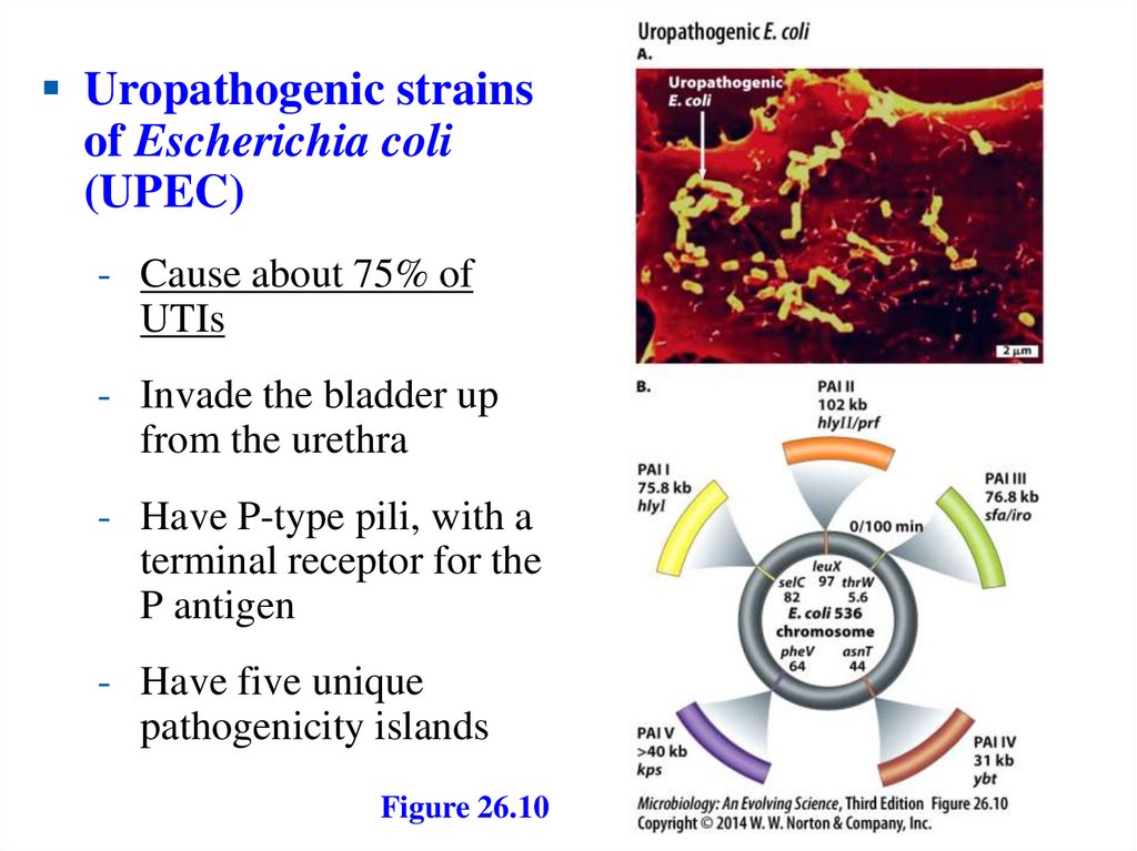
Uropathogenic strainsof Escherichia coli
(UPEC)
- Cause about 75% of
UTIs
- Invade the bladder up
from the urethra
- Have P-type pili, with a
terminal receptor for the
P antigen
- Have five unique
pathogenicity islands
Figure 26.10
132.
Sexually Transmitted DiseasesSyphilis by Treponema pallidum
Chlamydia trachomatis
Chlamydia pneumoniae
133.
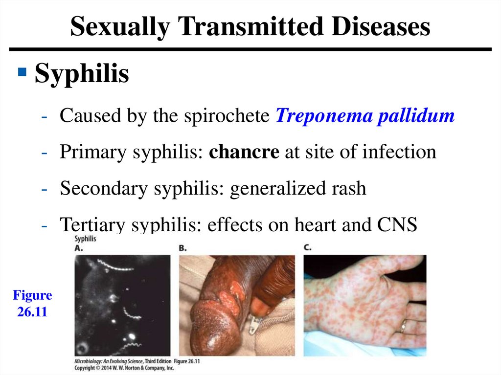
Sexually Transmitted DiseasesSyphilis
- Caused by the spirochete Treponema pallidum
- Primary syphilis: chancre at site of infection
- Secondary syphilis: generalized rash
- Tertiary syphilis: effects on heart and CNS
Figure
26.11
134.
Chlamydia- Most frequently reported STD in the United
States
- Caused by unusual Gram-negative bacteria
- Chlamydia trachomatis
- Chlamydia pneumoniae
- Obligate intracellular pathogens
- Both cause STDs, as well as
pneumonia and trachoma of the eye
- Left untreated, infection can cause serious
health problems in both females and males
135.
TrachomaTrachoma is a bacterial infection that affects the eyes.
It is caused by the bacterium Chlamydia trachomatis.
Trachoma is contagious, spreading through contact
with the eyes, eyelids, and nose or throat secretions of
infected people. It can also be passed on by handling
infected items, such as handkerchiefs.
At first, trachoma may cause mild itching and irritation
of your eyes and eyelids. Then you may notice swollen
eyelids and pus draining from the eyes. Untreated
trachoma can lead to blindness.
136.
Elementary bodiesare the infective
form. They enter
eukaryotic cells by
endocytosis or
phagocytosis
They differentiate
into reticulate
bodies, which are
the replicative
form.
The reticulate
bodies
differentiate into
elemental bodies
that are released
Figure 26.12
137.
Gonorrhea- Caused by the Gram-negative diplococcus
Neisseria gonorrhoeae
- Over the decades, it has incrementally developed
resistance to antibiotics used in its treatment
- Most infected men exhibit symptoms, whereas
most women are asymptomatic.
- Binds to CD4+ T cells, inhibiting T-cell
activation
Figure
26.13
138.
Central Nervous System InfectionsMeningitis
- Infection of membrane surrounding brain
- Some bacteria cross blood-brain barrier
- Streptococcus pneumoniae
- Haemophilus influenzae
- Neisseria meningitidis
139.
Neisseria meningitidis- Has thick capsule and type IV pili
- Dangerous if it gets into the bloodstream
- Crosses from capillary into cerebrospinal fluid
- Once in meninges, it is very difficult to treat
- Effective vaccine to capsule components
Figure 26.16
140.
Clostridium toxins- C. botulinum: botulism toxin (“Botox”)
- Anaerobe, grows in canned food
- Spores survive unless autoclaved
- Toxin blocks the release of acetylcholine
- Causes flaccid paralysis
- C. tetani: tetanus toxin
- Anaerobe, grows in puncture wounds
- Blood flow interrupted; tissue becomes anaerobic
- Toxin blocks release of GABA, a primary
inhibitory transmitter for the central nervous
system. Its function is to reduce neuronal
excitability
- By inhibiting nerve transmission
- Causes spastic paralysis
141.
142.
Cardiovascular DiseasesInfections of the cardiovascular system include:
- Endocarditis: inflammation of the heart’s
inner lining
- Septicemia: presence of microbes in the
blood
- Bacteremia: presence of bacteria in the blood
- Can develop from a local infection situated
anywhere in the body
- Caused by Gram-positives, Gramnegatives, aerobes, and anaerobes
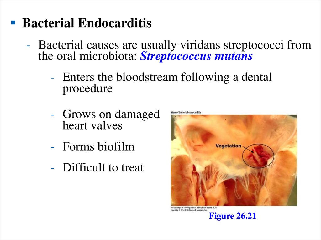
143.
Bacterial Endocarditis- Bacterial causes are usually viridans streptococci from
the oral microbiota: Streptococcus mutans
- Enters the bloodstream following a dental
procedure
- Grows on damaged
heart valves
- Forms biofilm
- Difficult to treat
Figure 26.21
144.
Systemic InfectionsSepticemia disseminating throughout body
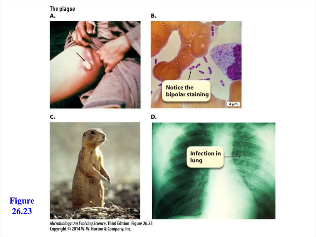
Plague
- Caused by the bacterium Yersinia pestis
- Bite of flea introduces organism
- Moves to lymph nodes: bubonic plague
- Moves to bloodstream: septicemic plague
- Inhaled: pneumonic plague
- Highly infectious
- Virulence factors inhibit phagocytosis
- Type III secretion system injects virulence proteins
145.
Zoonotic diseaseA zoonosis is another name for zoonotic disease. This
type of disease passes from an animal or insect to a
human. Some don’t make the animal sick but will
sicken a human.
Zoonotic diseases range from minor short-term
• illness to a major life-changing illness. Certain
• ones can even cause death.
Zoonosis include those caused by a virus, bacteria,
fungus, and parasites.
Zoonotic diseases spread by mosquitos and ticks are
some of the most serious of these diseases
(Healthline)
146.
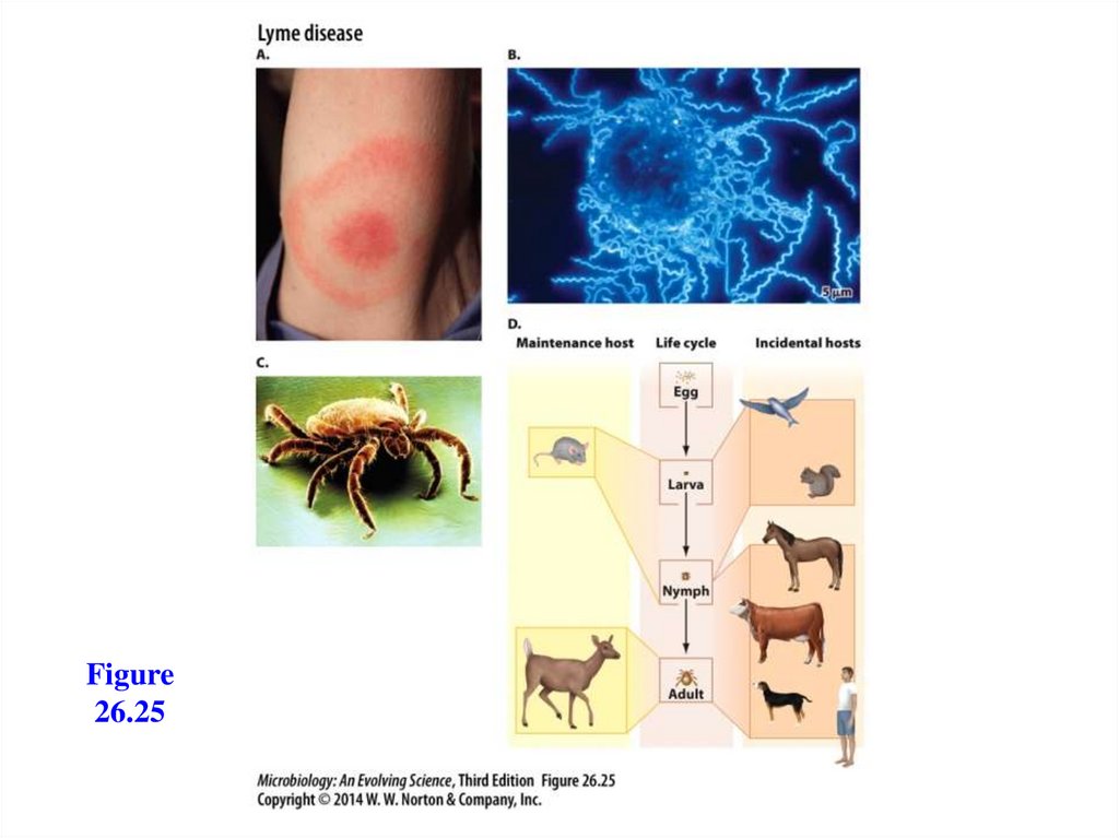
Lyme disease- Caused by Borrelia burgdorferi, a spirochete
- Transmitted by ticks
- Bacterium can travel to any part of the body
- Has three stages:
- Stage 1: a bull’s-eye rash (erythema migrans)
- Stage 2: joint, muscle, and nerve pain
- Stage 3: arthritis, with WBCs in the joint
fluid
- Treatment with antibiotics is recommended for
all stages
147.
Figure26.25
148.
ImmunizationVaccines are typically given in childhood
- Most are administered as multiple booster doses
- Except influenza: new vaccine every year
- Serious side effects are very rare
Herd immunity
- Vaccinating a large percentage of a community
effectively conveys herd immunity by interrupting
transmission of contagious diseases
- Example: gardasil—human papillomavirus vaccine
- Only works for diseases spread person to person
149.
Chapter SummaryPathogens can be classified as food-borne, airborne, bloodborne, or sexually-transmitted.
Patients histories are vital in diagnosing diseases.
Skin and soft-tissue infections include:
- Boils; scalded skin syndrome (Staphylococcus aureus)
- Necrotizing fasciitis (Streptococcus pyogenes)
- Measles: rubella and rubeola (viral infections)
Respiratory tract infections include pneumonia (caused by a
variety of organisms) and tuberculosis.
The main causes of GI tract infections include:
- Bacteria: EHEC, Salmonella, Shigella, H. pylori
- Protozoa: Entamoeba, Cryptosporidium, Giardia
- Viruses: rotavirus (single greatest cause of
gastroenteritis)
150.
Chapter SummaryThe main cause of UTIs is uropathogenic Escherichia coli (UPEC)
Sexually transmitted diseases include:
Syphilis, gonorrhea; chlamydia (bacterial diseases)
Trichomoniasis (protozoan disease), AIDS (viral disease)
Pathogens that cause CNS infections include:
Neisseria meningitidis (meningitis), Clostridium botulinum
toxin (flaccid paralysis), C. tetanus toxin (spastic paralysis)
Cardiovascular system infections include:
-
Septicemia, endocarditis, malaria
Pathogens that cause systemic infections include:
Yersinia pestis (plague), Borrelia burgdorferi (Lyme disease)
Herd immunity can protect unimmunized people.
151.
FYIViral diseases
Paramyxovirus
Herpes virus
Togavirus
Influenza virus and rhinovirus
Rotavirus
Hepatitis virus
Human immunodeficiency virus
152.
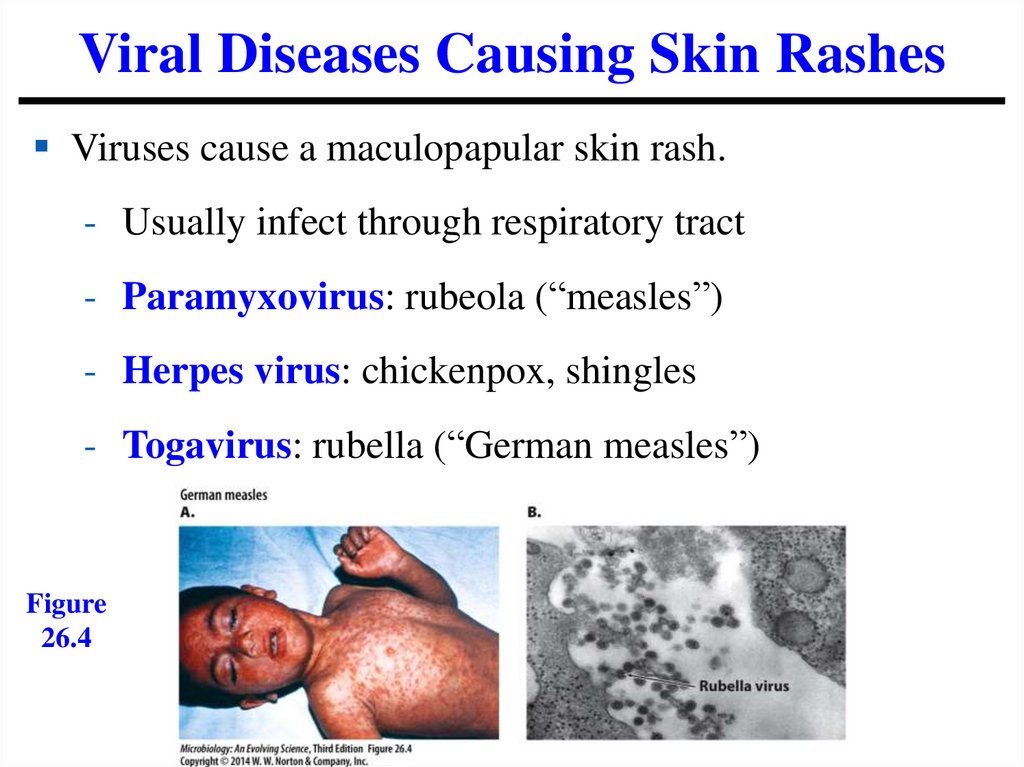
Viral Diseases Causing Skin RashesViruses cause a maculopapular skin rash.
- Usually infect through respiratory tract
- Paramyxovirus: rubeola (“measles”)
- Herpes virus: chickenpox, shingles
- Togavirus: rubella (“German measles”)
Figure
26.4
153.
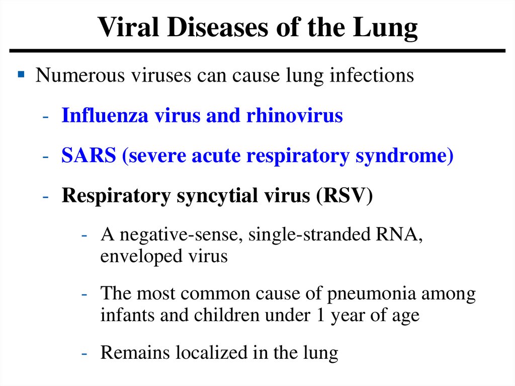
Viral Diseases of the LungNumerous viruses can cause lung infections
- Influenza virus and rhinovirus
- SARS (severe acute respiratory syndrome)
- Respiratory syncytial virus (RSV)
- A negative-sense, single-stranded RNA,
enveloped virus
- The most common cause of pneumonia among
infants and children under 1 year of age
- Remains localized in the lung
154.
Gastroenteritis caused by viruses- Rotavirus is the single greatest cause of
gastroenteritis
- Double-stranded RNA viruses
- Highly infectious, spreading by the fecal-oral route
- Endemic around the globe; affects all age groups
155.
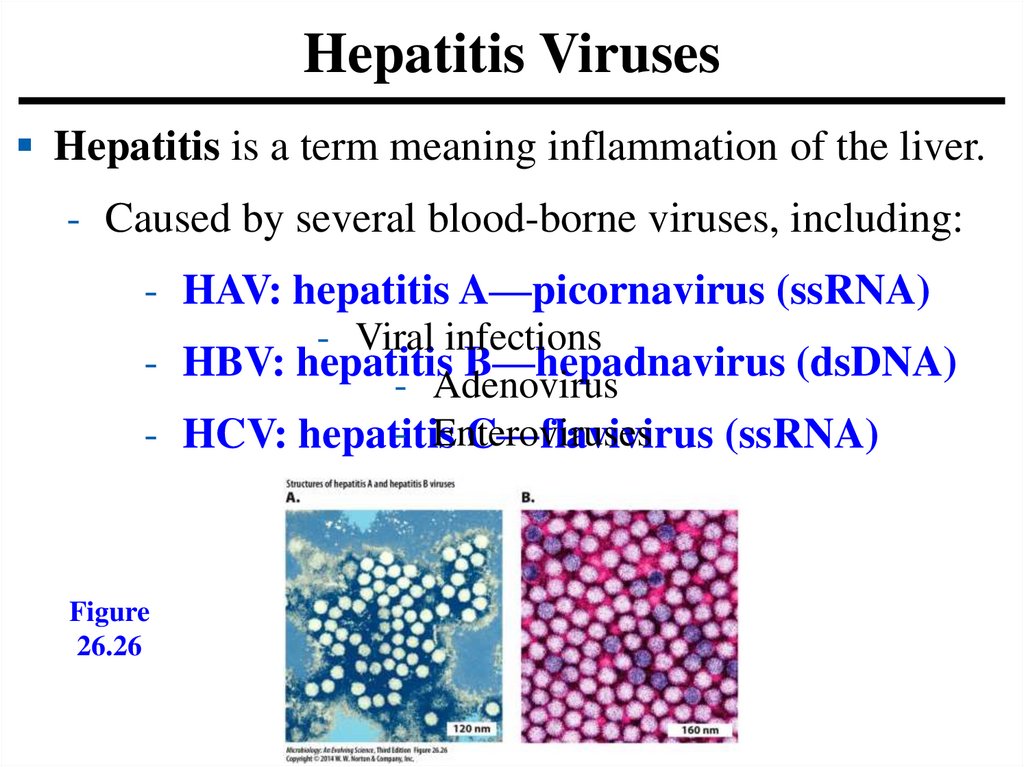
Hepatitis VirusesHepatitis is a term meaning inflammation of the liver.
- Caused by several blood-borne viruses, including:
- HAV: hepatitis A—picornavirus (ssRNA)
- Viral infections
- HBV: hepatitis B—hepadnavirus (dsDNA)
- Adenovirus
- Enteroviruses
- HCV: hepatitis
C—flavivirus (ssRNA)
Figure
26.26
156.
Viral endocarditisAssociation between enterovirus
endomyocardial infection and late severe
cardiac events in some adult patients
receiving heart transplants.
157.
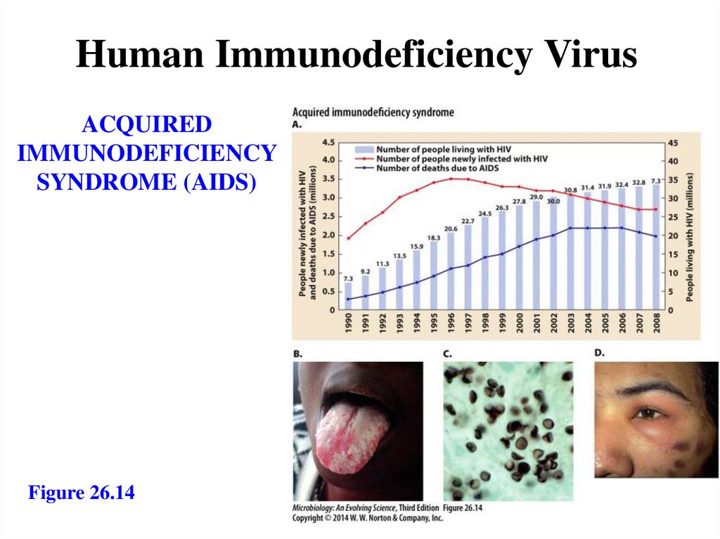
Human Immunodeficiency VirusACQUIRED
IMMUNODEFICIENCY
SYNDROME (AIDS)
Figure 26.14
158.
Acquired immunodeficiency syndrome- HIV: a lentiviral retrovirus
- Attacks CD4+ T cells, glial cells
- First stage: AIDS-related complex
- Fever, headache, rash
- Second stage: AIDS
- Depletion of T cells
- Opportunistic infections
- Oral candidiasis
- Pneumocystosis
- Third stage: AIDS-related dementia
- Fourth stage: rare cancers
- Kaposi’s sarcoma via herpes virus type 8
infection
159.
Fungal DiseasesMost fungi are not dangerous: Mild fungal skin diseases can
look like a rash and are very common. Ex. Trichophyton rubrum
(ectopathogen) causes Athtete’s foot.
Fungal diseases in the lungs are often similar to other illnesses
such as the flu or tuberculosis.
Blastomyces dermatitidis can cause skin, and bone lesions, and
metastatic or disseminating lesions in the lung, causing acute
pneumonia
Some fungal diseases like fungal meningitis and bloodstream
infections are less common than skin and lung infections but
can be deadly. It can be caused by Candida albicans,
Cryptococcus neoformans and Histoplasma.
160.
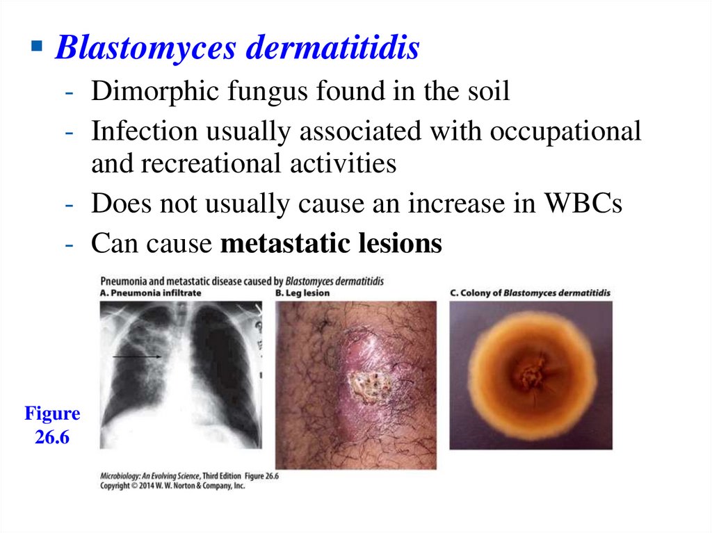
Blastomyces dermatitidis- Dimorphic fungus found in the soil
- Infection usually associated with occupational
and recreational activities
- Does not usually cause an increase in WBCs
- Can cause metastatic lesions
Figure
26.6
161.
Pathogenic Monocellular EukaryotesAmoeba
Entamoeba
Cryptosporidium
Naegleria
Acanthamoba
Giarda lamblia
Trichomonas
162.
Pathogenic Ameba• Unicellular eukaryotic organism
• Entamoeba histolytica causes amebic
dysentery
• Which is a more severe form of gastroenteritis,
• e.g. diarrhea with passage of blood or mucus.
163.
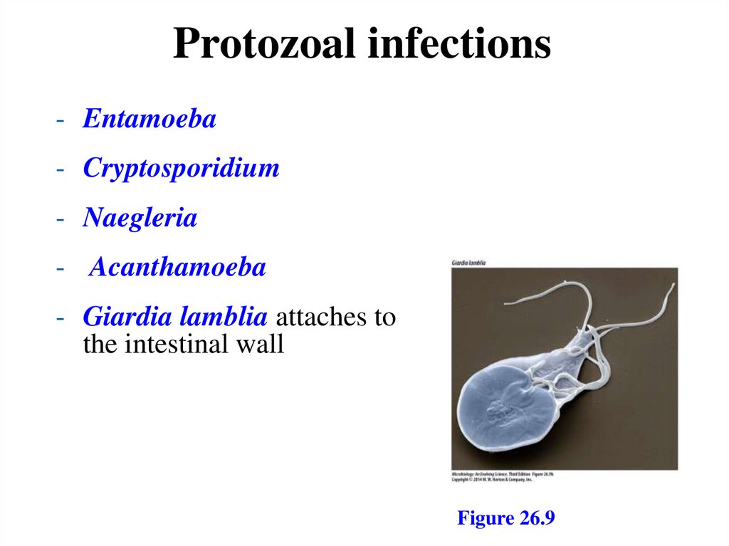
Protozoal infections- Entamoeba
- Cryptosporidium
- Naegleria
- Acanthamoeba
- Giardia lamblia attaches to
the intestinal wall
Figure 26.9
164.
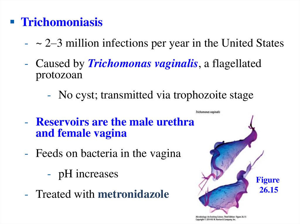
Trichomoniasis- ~ 2–3 million infections per year in the United States
- Caused by Trichomonas vaginalis, a flagellated
protozoan
- No cyst; transmitted via trophozoite stage
- Reservoirs are the male urethra
and female vagina
- Feeds on bacteria in the vagina
- pH increases
- Treated with metronidazole
Figure
26.15
165.
Figure26.23
166.
PrionsProteinaceous infectious particles
Cause spongiform encephalopathies
- Improperly folded proteins form aggregates that
damage the brain
- Most mammals suffer from these diseases
A
B
Figure 26.20
C
167.
MalariaCauses 1–3 million deaths per year.
Four protozoan Plasmodium species: P. falciparum,
P. malariae, P. vivax, and P. ovale.
- P. falciparum is the most deadly of all.
- Infects liver, red blood cells (RBCs).
- New merozoites are released every 48–72 hours.
- Many parasites are killed in each generation.
- Others switch protein placed on RBC surface.
- 60 var genes encode different surface proteins;
thus parasite constantly eludes immune system.
- Chloroquine resistance is a problem.
168.
Antimicrobial Chemotherapyand Discovery
Lecture 22 (Ch. 27)
169.
Chapter OverviewThe golden age of antibiotic discovery
Basic concepts of antimicrobial therapy
Measuring drug susceptibility
Mechanisms of action
Challenges of drug resistance
The future of drug discovery
Antiviral agents
Antifungal agents
170.
IntroductionThe discovery of antibiotics about 80 years ago
has played a major role in increasing life
expectancy throughout the world.
- From 45 to 50 years (prior to 1918) to nearly
79 years now
But antibiotics may soon become useless.
- Their overuse and misuse have led to the
development of antibiotic-resistant strains
171.
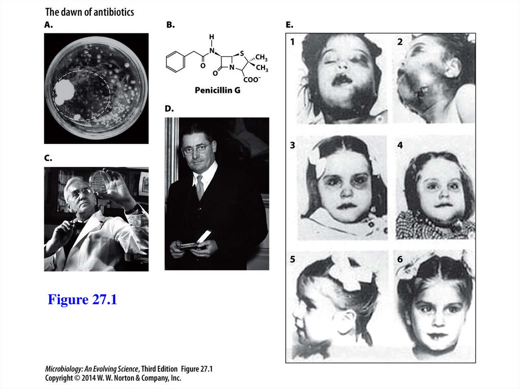
The Golden Age of Antibiotic DiscoveryAntibiotics are compounds produced by one microbe
that adversely affect other microbes.
The modern antibiotic revolution began in 1928 with the
discovery of penicillin by Alexander Fleming.
- A contaminating mold had inhibited the growth of
Staphylococcus aureus colonies on a plate.
- The mold was identified as Penicillium notatum.
- Penicillin was purified in the early 1940s by Howard
Florey and Ernst Chain.
- Has saved millions of lives since!
172.
Figure 27.1173.
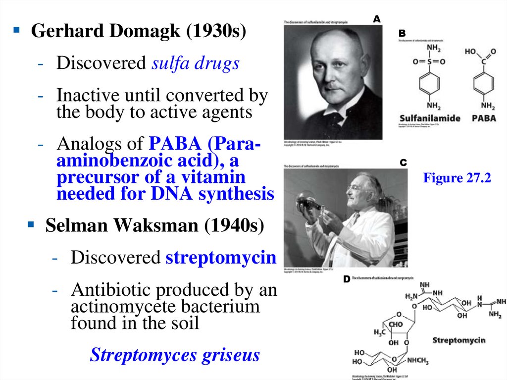
AGerhard Domagk (1930s)
B
- Discovered sulfa drugs
- Inactive until converted by
the body to active agents
- Analogs of PABA (Paraaminobenzoic acid), a
precursor of a vitamin
needed for DNA synthesis
C
Figure 27.2
Selman Waksman (1940s)
- Discovered streptomycin
- Antibiotic produced by an
actinomycete bacterium
found in the soil
Streptomyces griseus
D
174.
Fundamentals of Antimicrobial TherapyAntibiotics comprise mostly of chemotherapeutic agents
used to treat microbial diseases.
The term “antibiotic” originally referred to any
compound produced by one microbial species that
could kill or inhibit the growth of other microbes.
- Today the term “antibiotic” is also used for synthetic
chemotherapeutic agents, such as sulfonamides,
that are clinically useful but chemically synthesized.
Many natural and synthetic compounds affect microbial
growth, but their utility in a clinical setting is dictated by
certain key characteristics.
175.
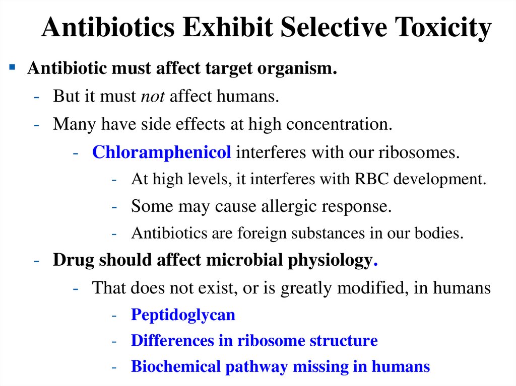
Antibiotics Exhibit Selective ToxicityAntibiotic must affect target organism.
- But it must not affect humans.
- Many have side effects at high concentration.
- Chloramphenicol interferes with our ribosomes.
- At high levels, it interferes with RBC development.
- Some may cause allergic response.
- Antibiotics are foreign substances in our bodies.
- Drug should affect microbial physiology.
- That does not exist, or is greatly modified, in humans
- Peptidoglycan
- Differences in ribosome structure
- Biochemical pathway missing in humans
176.
Antimicrobials Have a LimitedSpectrum of Activity
Broad spectrum
- Effective against many species
Narrow spectrum
- Effective against few or a single species
Source of antibiotics
- Most discovered as natural products
- Often modified by artificial means to:
- Increase efficacy
- Decrease toxicity to humans
177.
Antibiotics Are Classifiedas Bacteriostatic or Bactericidal
Bactericidal antibiotics kill target organisms.
- Many drugs only affect growing cells.
- Inhibitors of cell wall synthesis
- Only effective if organism is building new cell wall
- Example: penicillin
Bacteriostatic antibiotics prevent growth of organisms.
- Cannot kill organism
- Immune system removes intruding microbe
178.
Measuring Drug SusceptibilityOne critical decision a clinician must make when
treating an infection is which antibiotic to prescribe
for the patient.
There are several factors to consider, including:
- The relative effectiveness of different
antibiotics on the organism causing the
infection
- The average attainable tissue levels of each
drug
179.
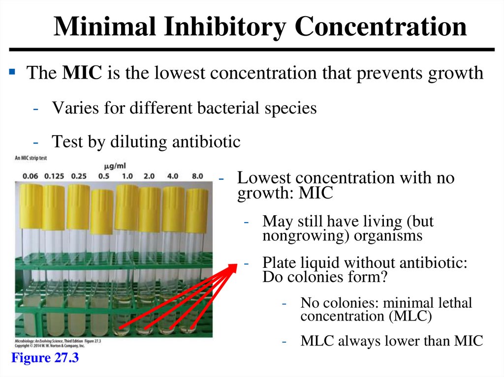
Minimal Inhibitory ConcentrationThe MIC is the lowest concentration that prevents growth
- Varies for different bacterial species
- Test by diluting antibiotic
- Lowest concentration with no
growth: MIC
- May still have living (but
nongrowing) organisms
- Plate liquid without antibiotic:
Do colonies form?
- No colonies: minimal lethal
concentration (MLC)
- MLC always lower than MIC
Figure 27.3
180.
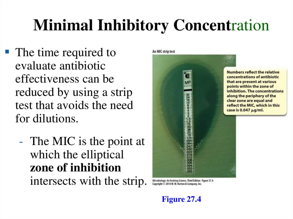
Minimal Inhibitory ConcentrationThe time required to
evaluate antibiotic
effectiveness can be
reduced by using a strip
test that avoids the need
for dilutions.
- The MIC is the point at
which the elliptical
zone of inhibition
intersects with the strip.
Figure 27.4
181.
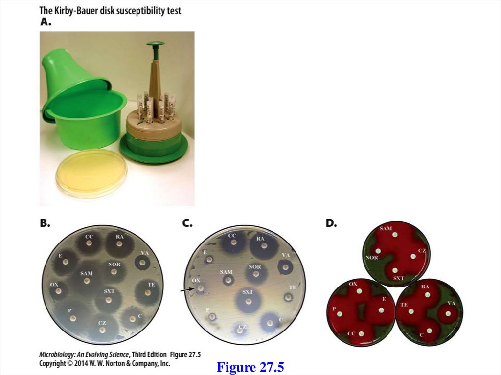
Kirby-Bauer Disk Susceptibility TestClinical labs can receive up to 100 or more isolates in one
day, so individual MIC determinations are impractical.
The Kirby-Bauer assay tests strain sensitivity to multiple
antibiotics.
- Uses a series of round filter paper disks impregnated
with different antibiotics.
- A dispenser delivers up to 12 disks simultaneously to
the surface of an agar plate covered by a bacterial lawn.
- During incubation, the drugs diffuse away from the
disks into the surrounding agar and inhibit growth of
the lawn.
- Size of cleared zones reflects relative sensitivity
182.
Kirby-Bauer Disk Susceptibility TestThe following are standardizations used to make
the test reproducible and easier:
- Size of the agar plate: 150 mm
- Depth of the media
- Media composition: Mueller-Hinton agar
- The number of organisms spread on the agar
plate
- Size of the disks: 6 mm
- Concentrations of antibiotics in the disks
- Incubation temperature: 37oC
183.
Figure 27.5184.
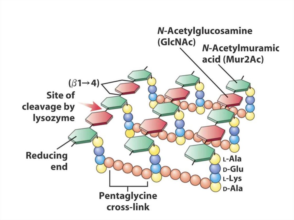
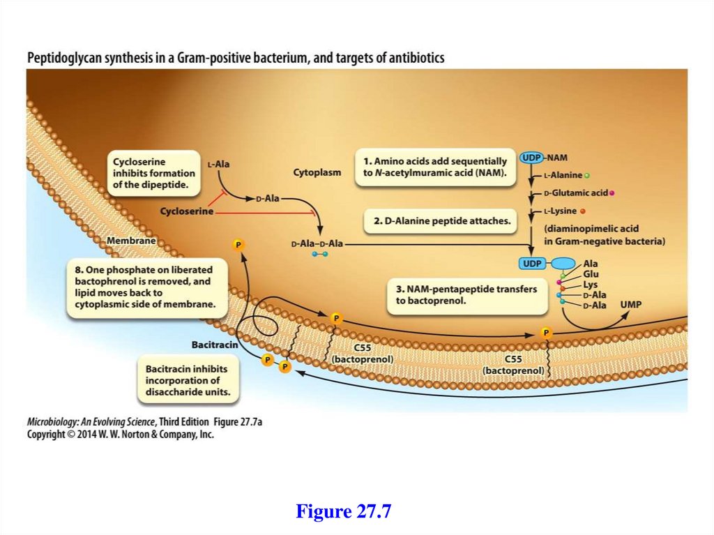
Cell Wall AntibioticsPeptidoglycan synthesis is rather complex.
- However, it may be summarized in these four steps:
1. Precursors are made in the cytoplasm.
- UDP-NAG and UDP-NAM-peptide
2. They are carried across the cell membrane by a
lipid carrier: bactoprenol.
- The carrier is then recycled.
3. The precursors are polymerized to the existing
cell wall structure by transglycosylases.
4. The peptide side chains are cross-linked by
transpeptidases.
185.
186.
Figure 27.7187.
Figure 27.7188.
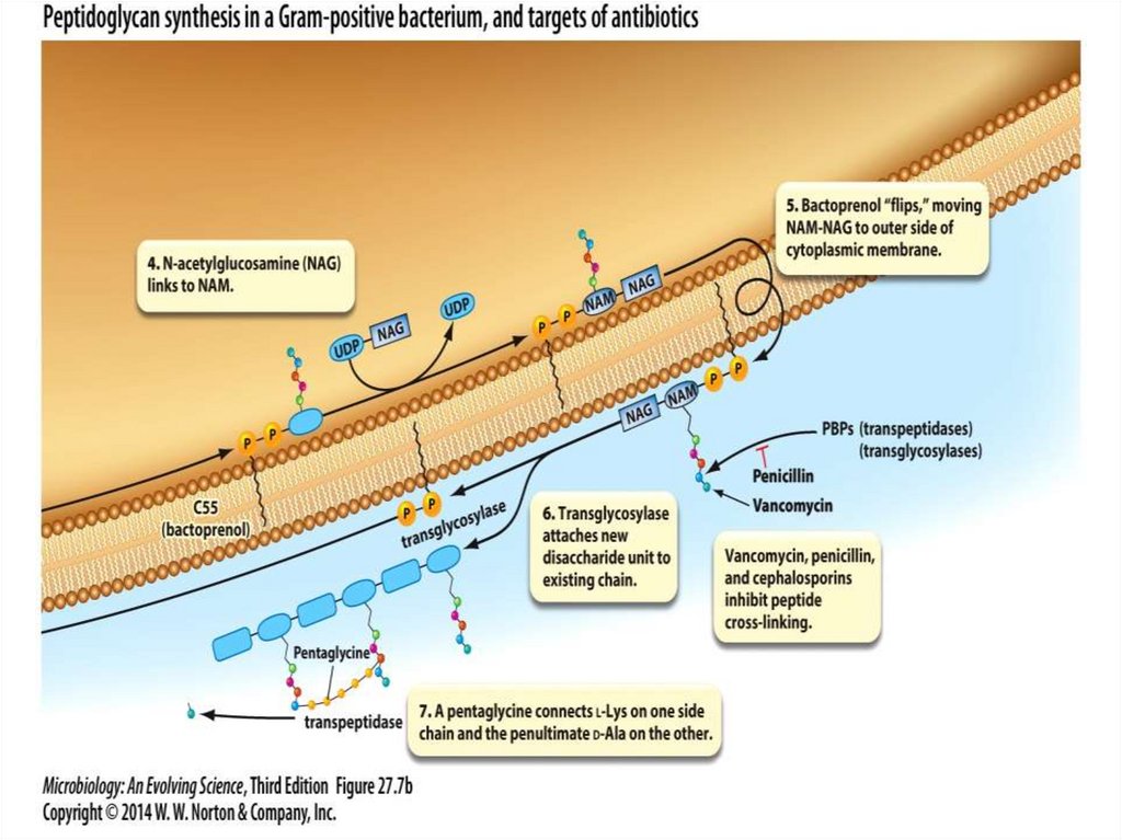
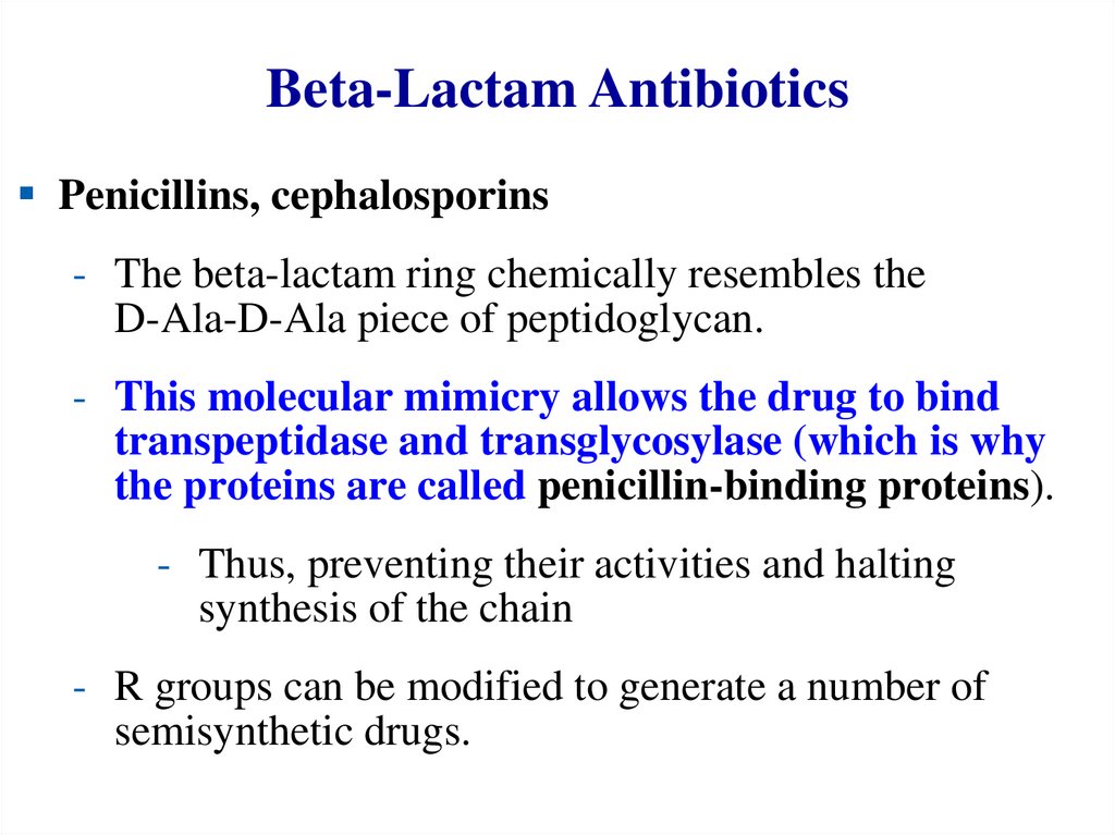
Beta-Lactam AntibioticsPenicillins, cephalosporins
- The beta-lactam ring chemically resembles the
D-Ala-D-Ala piece of peptidoglycan.
- This molecular mimicry allows the drug to bind
transpeptidase and transglycosylase (which is why
the proteins are called penicillin-binding proteins).
- Thus, preventing their activities and halting
synthesis of the chain
- R groups can be modified to generate a number of
semisynthetic drugs.
189.
Other Antibiotics That Inhibit Synthesis ofthe Cell Wall
Vancomycin: binds ends of peptides
- Prevents action of transglycosylases and
transpeptidases
- Same step as penicillin, but different activity
Cycloserine: inhibits formation of the D-ala-D-ala
dipeptide precursor
Bacitracin: blocks the lipid carrier
- Disaccharide subunits do not reach periplasm
190.
Drugs That Disrupt Cell MembranesGramicidin
- Cyclic peptide produced
by Bacillus brevis
- Forms a cation channel,
through which ions leak
Polymyxin
- Produced by Bacillus polymyxa
- Destroys cell membrane, just like a detergent
- Used only topically
Figure
27.11
191.
Drugs That Affect DNA Synthesis andIntegrity
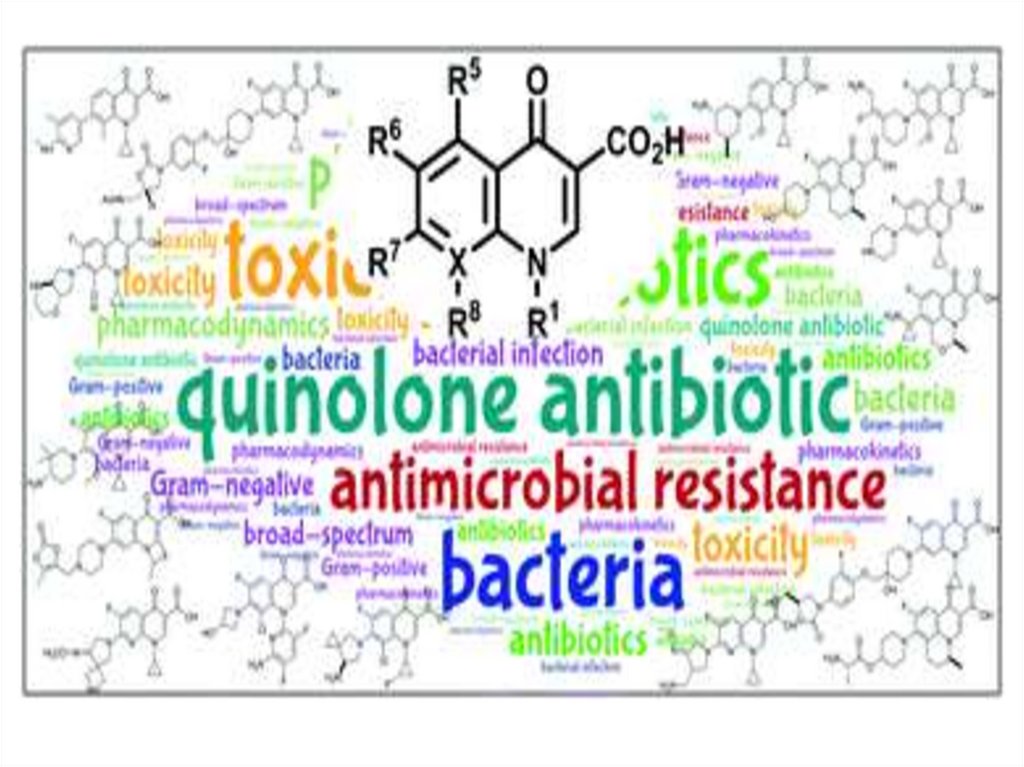
Quinolones: nalidixic acid, ciprofloxacin
- Block bacterial DNA gyrase, and so prevent
DNA replication
Metronizadole
- Nontoxic, unless metabolized by anaerobe
ferredoxin
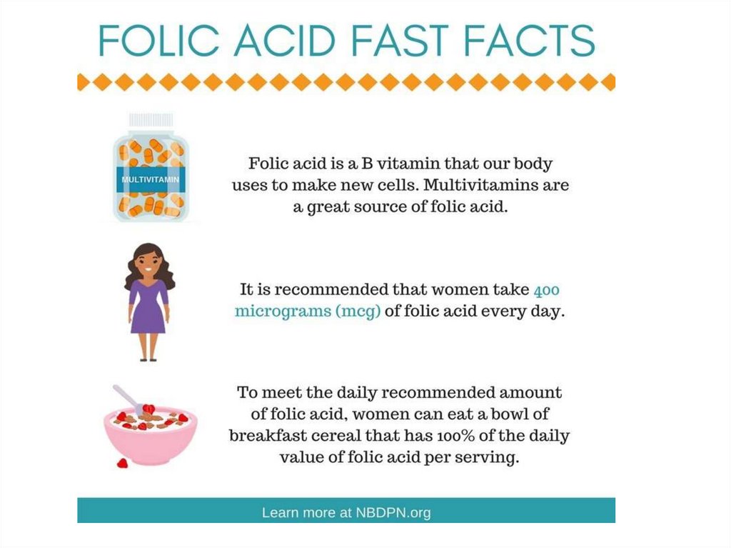
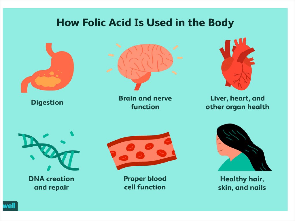
Sulfa drugs
- Analogs of PABA, a precursor of folic acid
- Needed for DNA synthesis
- Supplied in our diet, thus no folic acid
synthesis to inhibit
192.
193.
Figure27.12
194.
195.
196.
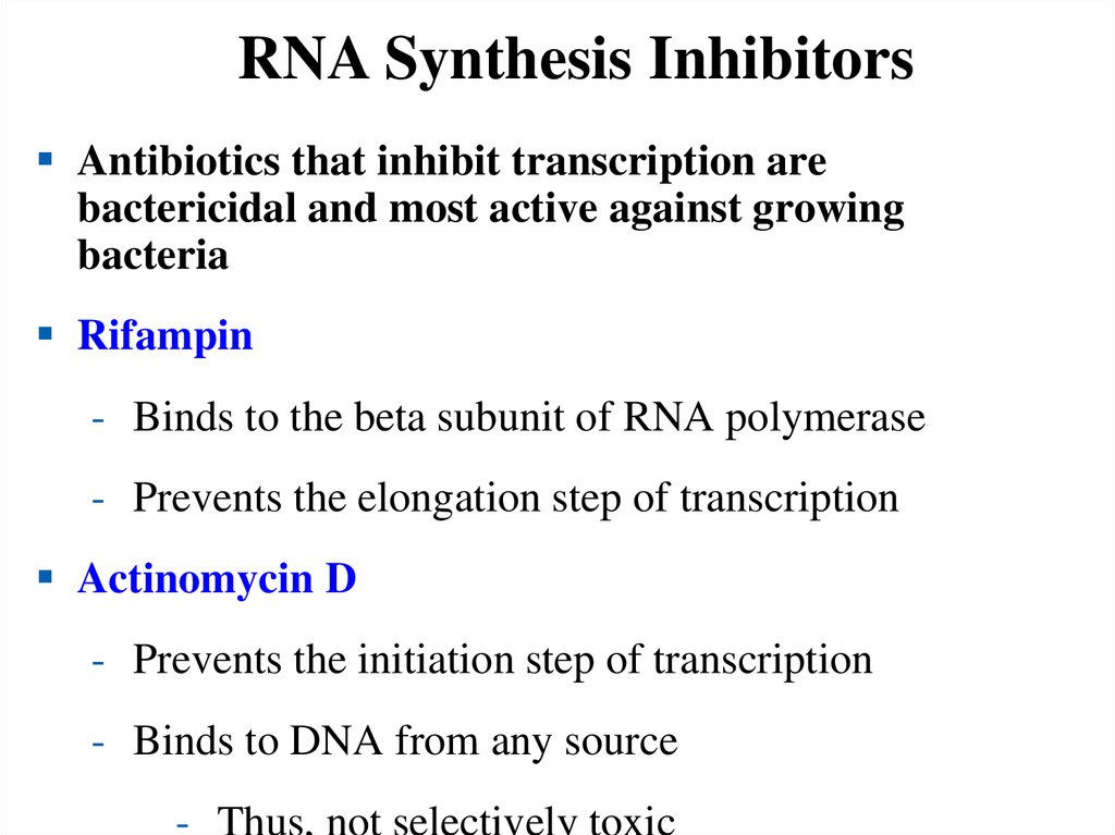
RNA Synthesis InhibitorsAntibiotics that inhibit transcription are
bactericidal and most active against growing
bacteria
Rifampin
- Binds to the beta subunit of RNA polymerase
- Prevents the elongation step of transcription
Actinomycin D
- Prevents the initiation step of transcription
- Binds to DNA from any source
- Thus, not selectively toxic
197.
Figure 27.14198.
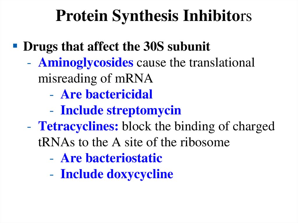
Protein Synthesis InhibitorsDrugs that affect the 30S subunit
- Aminoglycosides cause the translational
misreading of mRNA
- Are bactericidal
- Include streptomycin
- Tetracyclines: block the binding of charged
tRNAs to the A site of the ribosome
- Are bacteriostatic
- Include doxycycline
199.
Protein Synthesis InhibitorsDrugs that affect the 50S subunit
- Macrolides: inhibit translocation
- Lincosamides: inhibit translocation
- Chloramphenicol: inhibits peptidyl
transferase activity
- Oxazolidinones: prevent formation of the
70S ribosome initiation complex
- Streptogramins
- Streptogramin A: blocks tRNA binding
- Streptogramin B: blocks translocation
200.
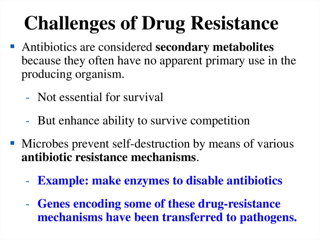
Challenges of Drug ResistanceAntibiotics are considered secondary metabolites
because they often have no apparent primary use in the
producing organism.
- Not essential for survival
- But enhance ability to survive competition
Microbes prevent self-destruction by means of various
antibiotic resistance mechanisms.
- Example: make enzymes to disable antibiotics
- Genes encoding some of these drug-resistance
mechanisms have been transferred to pathogens.
201.
Antibiotic resistance is agrowing problem worldwide
- Antibiotics are overused
- Overprescribed; used in farm
animal feed
This exerts selective pressure for
drug-resistant strains
- Streptococcus pneumoniae
- Acinetobacter baumanii
- Resistant to multiple drugs
202.
There arefour basic
forms of
antibiotic
resistance
Figure 27.18
203.
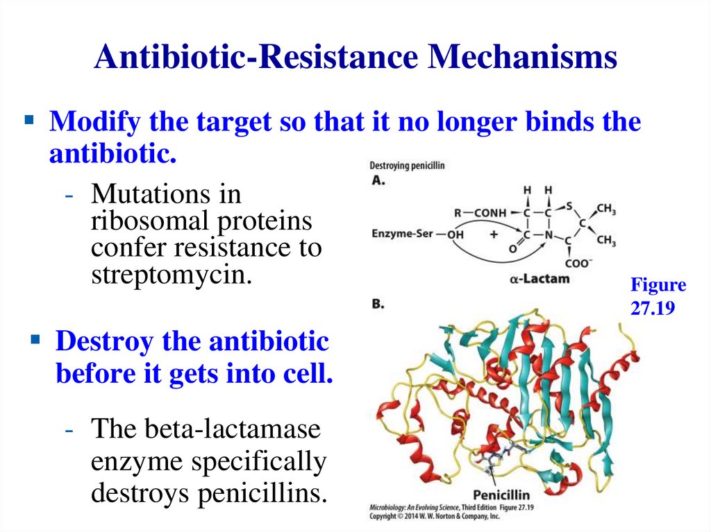
Antibiotic-Resistance MechanismsModify the target so that it no longer binds the
antibiotic.
- Mutations in
ribosomal proteins
confer resistance to
streptomycin.
Figure
27.19
Destroy the antibiotic
before it gets into cell.
- The beta-lactamase
enzyme specifically
destroys penicillins.
204.
Antibiotic Resistance MechanismsAdd modifying groups that inactivate the
antibiotic.
- Three classes of enzymes are used to modify
and inactivate the aminoglycoside antibiotics.
Pump the antibiotic out
of the cell.
- Specific and nonspecific
transport proteins
- Similar strategy is used
in cancer cells.
Figure 27.21
205.
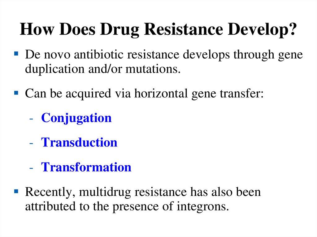
How Does Drug Resistance Develop?De novo antibiotic resistance develops through gene
duplication and/or mutations.
Can be acquired via horizontal gene transfer:
- Conjugation
- Transduction
- Transformation
Recently, multidrug resistance has also been
attributed to the presence of integrons.
206.
IntegronsIntegrons are genetic mechanisms that
allow bacteria to adapt and evolve rapidly
through the stockpiling and expression of
new genes. These genes are embedded in
a specific genetic structure called gene
cassette that generally carries one
promoterless open reading frame together
with a recombination site.
207.
The Future of Drug DiscoveryEvolutionary pressure is constant.
- Requires constant search for new antibiotics
The modern drug discovery process is outlined as such:
- Identify new targets using genomics.
- Design compounds to inhibit targets.
- Alter compound structure to optimize MIC.
- Determine spectrum of compound.
- Narrow or broad?
- Determine pharmaceutical properties.
- Not toxic to animals; persistence in body
208.
Antibiotics from the seaThe hero marine bacteria, Planctomycetes,
naturally produce antibiotic compounds to
fight against other bacteria. Thanks to
Jogler's lab work, a whopping 79 new
cultures of Planctomycetes could pave the
way for a new source of antibiotics and help
those who suffer from antibiotic-resistant
infections.Dec 13, 2019
209.
Potential targets for rational antimicrobial drug designinclude proteins expressed only in vivo or proteins
expressed both in vivo and in vitro.
Candidate antimicrobial compounds can be designed to
bind and inhibit the active site of a known enzyme
Combinatorial chemistry is used to make random
combinations of compounds that can be tested for enzyme
inhibitory activity and antimicrobial activity.
Intriguing ideas that may lead to novel antimicrobial
therapies include:
- Nanotubes to poke holes in bacterial cell membranes
- Molecules that “cork” the type III secretion apparatus
- Interfering with the quorum-sensing mechanisms
210.
Methods to Identify Drug-ResistantPathogens
The proportion of antibiotic-resistant infections has
doubled since 2002, rising from 5.2% to 11% of all
infections.
The faster a clinical lab can identify a pathogen’s antibiotic
susceptibility, the more quickly a clinician can prescribe an
appropriate narrow-spectrum antibiotic.
• Traditional MIC tests take up to 3 days to complete;
using automated methods cuts this down to 2 days.
• Multiplex PCR platforms can detect pathogenspecific or drug resistance gene DNA sequences
within an hour.
211.
Evolving, and Sharing, DrugResistance Genes
• De novo antibiotic resistance develops through gene
duplication and/or mutations.
• Antibiotic resistance also can be acquired via horizontal
gene transfer (conjugation, transduction, and
transformation ).
– A study in 2015 found that antibiotic-resistant
microbes occur naturally in uncontacted Amazon
communities.
– Recently, multidrug resistance has been
attributed to the presence of highly-mobile gene
expression elements called integrons.
211
212.
How Did We Get into This Mess? – 2Another proposed source of antibiotic resistance is the
widespread practice of adding antibiotics to animal feed.
• Giving animals subtherapeutic doses of antibiotics
in their food makes for larger, and therefore more
profitable, animals.
• Some estimates suggest that 80% of all antibiotics
used in the United States (up until 2017) were fed
to healthy livestock.
• Feeding growth-promoting antibiotics to cattle can
stimulate the spread of pathogenicity genes
between bacteria.
213.
How Did We Get into This Mess? – 3Many of these situations have conspired to produce
incredibly dangerous bacteria resistant to almost every
antibiotic known.
ESKAPE pathogens
• Term coined by the Infectious Diseases Society of
America
• Six highly resistant bacterial species that collectively
cause about two-thirds of all U.S. nosocomial infections
Enterococcus faecium
Staphylococcus aureus
Klebsiella pneumoniae
Acinetobacter baumannii
Pseudomonas aeruginosa
Enterobacter sp.
214.
How Did We Get into This Mess? – 4215.
How Did We Get into This Mess? – 5When should antimicrobials be used? Antibiotic
stewardships are coordinated interventions that improve
and measure antibiotic use.
• Do not use antibiotics to treat viral infections.
• Do not use an antibiotic if a patient’s microbiome
includes a strain that is resistant to the drug.
• Know which antibiotic resistant strains are prevalent in
the community or hospital before prescribing.
• Consider how long the patient needs to take the
antibiotic.
• De-escalate antibiotic usage whenever possible.
216.
Biofilms, Persisters, and the Mystery ofAntibiotic Tolerance
Why do some infections return after bactericidal antibiotic
treatment is discontinued?
A subpopulation of dormant organisms, called persister
cells, often arises within a population of antibioticsusceptible bacteria.
• The stalled metabolism of persisters renders them
tolerant to bactericidal antibiotics during treatment.
• Persister cells can be found in any biofilm or
population of late-exponential-phase cells.
• Tolerance provides antibiotic resistance at the price
of not growing.
217.
218.
Fighting Resistance and Finding NewDrugs – 1
The prudent use of current antibiotics and innovative
strategies for finding new ones hopefully will enable
us to continue to control evolving pathogens.
1. Directly countering drug resistance
―Dummy target compounds inactivate resistance
enzymes (e.g., clavulanic acid)
―Alter antibiotic’s structure so that it sterically
hinders access of bacterial modifying enzymes
219.
Fighting Resistance and Finding NewDrugs – 2
2. Finding new antibiotics
―Brute-force screening of microbes, plants, and
animals
―Combinatorial chemistry
―Genome sequence analysis to identify potential
bacterial molecular targets
―Photosensitive chemicals
―Interfering with quorum-sensing mechanisms
―CRISPR-based strategies for reversing antibiotic
resistance
220.
Fighting Resistance and Finding New Drugs –3
3. Antipersister and antibiofilm approaches
• Kill persisters directly
• Prevent persister formation
• Interfere with biofilm formation
• Induce biofilm dispersal
The progression of these strategies has been slow
because the road to FDA approval, though
necessary for safety reasons, is long (8 to 10 years)
and expensive, and it can discourage some
companies from investing in antibiotic discovery.
221.
Introduction to theImmune System
The Innate and
Adaptive Immunity
222.
What is the immune system?pollen
viruse
ss
Foreign
invaders
The immune system is a
system of defense to
protect the body against
foreign invaders (foreign
antigens)
Exterior defense:
Physiological barriers,
chemical barriers,
mechanical barriers,
223.
Exterior DefenseBarrier types
Example of
external barriers
Example of external barriers
Skin covering the body, cilia in the lining of the lungs, nose and
throat.
Chemical barriers Lysozyme in tears, pH, and mucin at mucosal surface
Mechanical
barriers
Microbiological
barriers
Sneezing, coughing, tear flow, and flushing of urinary tract
Normal microflora, commensal microorganisms in the gut and
vagina
224.
Interior or Systemic defenseIn the body there are cells and soluble factors that deal
with foreign antigens, which have come into the body.
In humans and higher vertebrates there are two lines of
internal defense, and distinct cells and soluble factors are
involved in each type of immunity, and hence they are
divided into:
1. Cells and factors of the innate immunity
2. Cells and soluble factors of the adaptive immunity
225.
Components of the immune systemThe immune system is composed of specialized organs, tissues,
cells and soluble factors that work in concert in order to mount an
adequate immunological response.
Organs
Tissues
Thymus
Spleen
Lymph nodes
Bone marrow
Lymphoid tissues
Cells
Soluble Factors
White blood cells
Red blood cells and platelets
are also involved in the
inflammation
Complement system
Antimicrobial peptides
Antibodies
Cytokines
Chemotactic factors
226.
Components of the ImmuneSystem
The immune system is composed
of lymphoid organs,
tissues, cells and soluble factors.
Organs of the immune system and their functions
• Primary lymphoid organ: The thymus is a bilobed
organ where immature T cells (also called progenitor T
cells) undergo maturation
• Secondary lymphoid organs include the spleen and
lymph nodes. These organs contain B cells, T cells and
macrophages.
– The spleen is the largest secondary lymphoid organ where antigens from the blood
are trapped
– Lymph nodes are encapsulated organs that are located at the junction of lymphatic
vessels
227.
The Bone MarrowThe bone marrow consists of connective tissue that is
contained in the cavity of most bones in the body.
There are two types of bone marrow:
Yellow marrow rich in fat cells
Red marrow containing developing blood cells.
Red bone marrow is primarily a site of
development of all blood cells from a common
precursor, the hematopoietic stem cell by a
process called hematopoiesis.
All blood cell types mature in the bone marrow
except for the progenitor T cells, which undergo
maturation in the thymus.
228.
The ThymusThe thymus is an encapsulated bilobed organ that is located in the
thoracic cavity, between the chest bone and the heart. The thymus
contains lobules, which are organized into 2 regions: a cortex and a
medulla. The cortex contains immature T cells, whereas mature T
cells can be found in the medulla. T cell maturation occurs in the
cortical medullary region. It is noteworthy that only around 10 % of the
progenitor T cells could reach maturation, indicating that around 90% of
progenitor T cells are eliminated during the maturation process. The
thymus increases in size after birth, reaching its largest size at puberty,
after which time it progressively decreases in size with age. Thymic
involution is associated with reduced thymopoiesis and
immunosenescence.
229.
The thymusThe thymus is a bilobed organ that is divided into lobules. Immature thymocytes
are localized in the vortex, and T cell maturation occurs in the cortical
medullary region
230.
The lymph nodesThe lymph nodes are small encapsulated organs that are located at
major junctions of lymphatic vessels. The lymph node is organized
into 3 regions, including the cortex or B cell zone that contains resting
B cells in primary follicles and proliferating B cells in germinal
centers, the paracortex or T cell zone, and the medulla, where T, B
cells, and macrophages can be found.
Lymph nodes are irrigated by both the blood circulatory system and the
lymphatic system. Blood enters the lymph node via the artery to the
arterioles and comes out through the venules and vein.
The lymph comes into the lymph node via the afferent vessels or
ducts, and out via the efferent duct. Therefore, lymph nodes trap
antigens from both the blood circulatory and lymphatic systems.
231.
Lymphnodes
Proliferatin
g B cells in
Afferent duct
High
endothelial
venule
(HEV)
T cell
zone
B cell zone
Efferent
duct
Lymph nodes are encapsulated organs where antigens in both the blood and
the lymph are trapped and where immune cells (B and T cells, macrophages,
dendritic cells and follicular dendritic cells) are located and ready to interact
with each other in order to mount an adequate immunological response
232.
The SpleenThe spleen is the largest encapsulated secondary lymphoid organ. In humans,
the spleen is located below the rib cage and to the left of the abdomen. The
spleen contains two regions that are designated as the red pulp and white pulp.
The red pulp contains worn out red blood cells that are destined for
destruction, hence it is referred to as a cemetery for RBCs. The process of
removing worn out and dead RBCs is called hemocatharsis, meaning
cleansing of the blood. The white pulp is rich in white blood cells and it
forms a sheath around the central arteriole. The sheath is designated as
periarteriolar lymphoid sheath (PALS), which is a tissue rich in T cells
(mostly CD4+ T cells and CD8+ T cells, and in smaller numbers B cells that
are located in follicles and germinal centers, and macrophages that are located
in the marginal zone. .
233.
Thespleen
The spleen is the largest secondary lymphoid organ, in which antigens in the blood are
trapped Blood circulation gets into the spleen via the artery and arterioles and come out
via venules and the vein, and hence the spleen serves as a trap of foreign antigens that
are coming from the blood circulation
234.
HematopoiesisAll blood cells are
derived from a
common precursor,
the hematopoietic
stem cell via three
lineages of
differentiation: The
lymphoid lineage,
the myeloid
lineage, and the
erythrocyte/megak
aryocyte lineage
Blood circulation
Lymphoid lineage
Erythrocyte and
Megakaryocyte lineage
Myeloid lineage
Tissues
235.
Non-Immune cells: Erythrocytes & Megakaryocytes.Erythrocytes, also referred to as red blood cells
(RBCs) are the most abundant cells in peripheral blood.
RBCs do not have a nucleus. These cells are not
immune cells, but they play a role in the binding and
removal of immune complexes from the blood
circulation.
Megakaryocytes are large cells (at least 20 X larger
than the RBCs). These cells are the precursors to
platelets, and around 4000 platelets can be generated
by the fragmentation of one megakaryocyte. Platelets
are necessary for blood clotting, and they also play an
important role in the inflammatory response.
Megakaryocyte
Erythrocytes or
Red blood cells
236.
Cells of the myeloid lineageMonocytes are the largest white blood cells,
which constitutes 3–7% of the leukocytes in the
circulating blood. They are characterized by a
kidney-shaped nucleus, grey cytoplasm and
small red-blue granules upon staining by the
Giemsa stain. Pictures of monocytes and
macrophages are available at. Monocytes can
leave the blood circulation and differentiate
into tissue macrophages and dendritic cells.
237.
MacrophagesMacrophages are larger than the monocytes, with larger
cytoplasm t hat is heavily vacuolated. Macrophages
assume different shapes and are given different names
based on their location. Ex., Alveolar macrophages in the
lung, peritoneal macrophages, Kupffer cells that are
resident macrophages in the liver, brain microglia
(resident macrophages in the brain), spleen sinus
macrophages, lymph node sinus macrophages, Kidney
intraglomerular mesangial cells…etc. Macrophages
display abundant dendrites when exposed to bacteria
(Fig.2.7A). Macrophages have the ability to perform
phagocytosis of foreign antigens, and hence like the
neutrophils, they are designated as
“professional”phagocytes.
238.
Monocyte-Derived Dendriticcells
Monocyte-Derived Dendritic
cells belong to a heterogeneous
population of professional
antigen-presenting cells
(APCs).
Monocyte-derived DCs
(mDCs), which are also called
myeloid DCs are the best
understood types of DCs.
The mDCs are characterized by a
Dendritic cell
exposed to virus
239.
Polymorphonuclear granulocytesNeutrophils (50-70% of leukocytes) have a multilobed
nucleus and cytoplasmic granules that are small and
stained in pale blue with the neutral Giemsa stain.
The primary function of neutrophils is to perform
phagocytosis of foreign antigens in the blood
circulation.
These cells will also respond to chemotactic factors
and chemokines generated at the site of injury and/or
inflammation. The chemokine CXCL8 produced by
activated macrophages facilitate neutrophil
extravasation, and the chemotactic factor
complement C5a direct them to the site of the
infection
240.
• Eosinophils (>0-5% of leukocytes) havea bilobed nucleus and cytoplasmic
granules that are stained in red with the
acidic dye eosin. They are proinflammatory white blood cells, which
play a role in the immune defense
against small parasites, such as helminth
worms.
Eosinophil
Basophils (>0-2% of leukocytes) have a bilobed nucleus
and cytoplasmic granules stained in blue with the basic dye
Alcian blue. Basophils are also involved in the immune
response to parasitic infection.
Basophil
241.
Mast CellsMast cells are only found in tissues, in both
connective tissue (Connective tissue mast cells or
CTMCs) and mucosa (mucosa-associated mast
cells or MMCs).
Mast cells show some similar morphological
characteristics to basophils, such as the presence
of a blue cytoplasm with deep purple granules by
histochemical staining method using combined
Alcian Blue-Safranin O.
A direct precursor to mast cells has not yet been
identified.
Inactive mast
cell
Degranulated
mast cell
242.
Cells of the innate immunityNeutrophil
Eosinophil
Mast cell
Basophil
Dendritic cell
Monocyte
Macrophage
243.
RecapCells of the innate immunity
• Polymorphonuclear granulocytes (Neutrophils, Eosinophils and
Basophils)
• Monocytes that differentiate into tissue Macrophages and Denditic cells
• Mast cells in connective tissue and mucosa-associated tissues
• Natural Killer cells (NK cells).
• Note: Megakaryocytes and Erythrocytes are not immune cells, but they
do contribute to the immune system, Megakaryocytes being precursors
to platelets, and Erythrocytes help in the removal of immune complexes
from the blood circulatory system.
244.
Cells of theAdaptive Immunity
245.
Cells of theadaptive
immunity
246.
Clonal selection of B cell by antigen anddifferentiation into plasma cells producing antibodies
247.
T-helper cell and B-cell Interaction248.
The Killing Action of Cytotoxic TCells
249.
RecapThe cells of the adaptive immunity include B
and T cells
• B cells which differentiate into plasma cells
producing antibodies
• T cells which include
TCR-1 T cells
TCR-2 T cells, which are divided into
• CD4+ T helper cells by producing cytokines
• CD8+ Cytotoxic T cells which kill infected cells by induction of
250.
The Immune Responses251.
Lecture contentsThe systemic Immunological response to foreign antigens
involves two lines of defense
First line of defense
• Involves cells and soluble factors of the innate immunity
• Immediate response
Second line of defense
• Delayed response
• Specific response
• Interaction between innate and adaptive components
252.
Innate Immunity or First Line ofDefense
The importance of the innate immunity is the readiness of
innate immune cells and soluble factors in the elimination of
foreign antigens and infected cells.
Our focus is on the following:
1. Phagocytosis of foreign antigens by neutrophils and
macrophages.
2. The inflammatory response to an infection and/or injury.
3. Lysis of Gram-negative bacteria ny the complement system.
4. Detection and killing of infected or transformed cells by NK
cells.
5. Antimicrobial factors.
253.
PhagocytosisNeutrophils and macrophages are professional
phagocytic cells
• Macrophages are the main phagocytes in
tissues.
• Neutrophils are the main phagocytes in the
blood circulatory system and, when called by
macrophages, they extravasate and migrate to
the site of an infection and/or inflammation.
The next slide will show the main four steps in
phagocytosis
254.
PhagocytosisMacrophages and
neutrophils are referred to
as professional phagocytic
cells or phagocytes.
Phagocytosis is facilitated
by the coating of the antigen
with opsonins, such as
antibodies or complement
component C3b
255.
Steps inStep 1 phagocytosis
• Binding of bacterium by surface receptors on
phagocytes. Ex. CD14 binding LPS on gramnegative bacteria, and receptors for opsonins
which are proteins that can bind both to bacteria
and the corresponding receptors on phagocytes,
thus facilitating phagocytosis. Examples of
opsonins include antibodies and the
complement component C3b.
Step 2
• Internalization of the bacterium is facilitated by
the cross-linking of the bacterium and the
phagocyte.
256.
Step 3• Internalization of the bacterium in a phagosome
Step 4
• Fusion of the lysosome with the phagosome, thus forming a
phagolysosome, where the bacterium is destroyed by up to 40 acidic
lysosomal enzymes, such as lysozyme digesting peptidoglycan cell wall,
hydrolytic enzymes or acid hydrolases such as nucleases, proteases,
lipases, phosphatase, etc., and protein such as lactoferrin that binds iron
and deprive iron-dependent bacteria.
• Oxygen-dependent killing of bacteria. The bacterium is also destroyed
by reactive oxygen species (ROS), such as superoxide anion (O2−),
hydrogen peroxide (H2O2), and hydroxyl radical (HO•), consist of
radical and non-radical oxygen species formed by the partial reduction
of oxygen. Cellular ROS are generated endogenously as in the process
of mitochondrial oxidative phosphorylation,
257.
Killing of bacteria in thephagolysosome
258.
The beneficialacute inflammatory response
259.
InflammationThe five cardinal signs of
inflammation
Redness
Heat
Pain
Swelling
Loss of function
Question: What can cause these
signs?
260.
The Inflammatory ResponseBeneficial acute
inflammatory response to
injury and infection
Macrophages performing
phagocytosis of bacteria
Activated macrophages
producing cytokines to
activate other immune
cells in tissue, to mobilize
circulatory neutrophils
and platelets, to induce
fever by pyrogens, etc.
261.
The 5 signs of inflammation1. Redness resulted from vasodilation caused by histamine and
prostaglandin produced by mast cells.
2. Swelling resulted from exudates caused by vasodilation and smooth
muscle contraction.
3. Heat resulted from the effect of pyrogens (produced by activated
macrophages: IL-1 beta and IL-6 on the hypothalamus.
4. Pain resulted from the stimulation of nerve ending by bradykinin and
prostaglandin.
5. Loss of function caused by disruption of tissue structure.
262.
Neutrophil undergoing NETosisNeutrophils can extravasate (get out of
blood vessel) in response to the
chemokine CXCL8 produced by
macrophage and the chemotactic factor
C5a from complement activation.
CXCL8 induces the expression of
adhesion molecules on neutrophils (LFAI) and on endothelial cells (ICAM-I).
C5a is a chemotactic factor that attracts
neutrophils to the site of the
infection/inflammation.
Neutrophils do not recirculate, and they
die by apoptosis or NETosis at the site
of the infection/inflammation.
263.
Functions of the complementsystem
Lysis of foreign cells for the insertion of on of membrane
attack complexes (MAC) into the membrane of Gramthem more attractive to phagocytic
bacteria The
cells such as macrophages (a process
Productionknown
of reactive
proteins:
as opsonization).
Some
complement components also promote
C3b: Opsonin
inflammation by stimulating cells to release
C3a and C5a
at low concentration: chemotactic factors
histamine and by attracting phagocytic
C3a and C5a
are anaphylatoxins
cellsattohigher
the siteconcentration,
of infection.
that bind to their respective receptors on mast cells,
triggering the release of factors of anaphylaxis
264.
Complement activation• The complement system is composed of over 30 heat-labile
proteins that are produced mainly by the liver. These proteins
circulate in the blood and extracellular fluid in an inactivated
state until a complement component encounters a foreign cell,
leasing to a sequence of enzymatic reaction on the surface of
the foreign cell, such as E. coli (Gram- bacteria).
• Complement activation occurs by three pathways:
– The Alternative pathway which is also called the properdin pathway
– The Lectin pathway
– The Classical pathway
265.
Initiation or Activation of the
Complement
The Alternative pathway is initiated by the binding of a fluid phase C3 convertase that
consists of iC3 associated with Factor Bb (iC3Bb), which cleaves complement C3 into 2
fragments C3a and C3b that binds to the bacterial surface, followed by the formation of
the C3 convertase C3bBb on bacterial surface. Binding of the protein properdin to
C3bBb extends the half life of this C3 convertase .
The Lectin pathway is initiated by the binding of a Mannan-binding lectin (MBL) complex
to mannose residues on the bacterial surface. Mannan-binding lectin complex consists of
one molecule of Mannan-binding lectin associated with two molecule of MBL associated
serine protease 1 (MBL-SP1) and two molecule of MBL-associated serine protease 2
(MBL-SP2), which results in the activation of a serine protease. Cleavage of complement
C4 and C2 by this enzyme results in the formation of C4bC2b (C4b2b), which is a C3
convertase.
The Classical pathway is initiated by the binding of antibodies of the IgG and IgM classes
to the bacterial surface, which is followed by the binding of C1 complex (C1q associated
with two C1r and two C1s to the antibodies), which results in the activation of a serine
protease. Like in the Lectin pathway, cleavage of complement C4 and C2 by this enzyme
results in the formation of C4bC2b (C4b2b), which is a C3 convertase.
266.
Formation of C5 convertase andMAC
Formation of C5 convertase. This step is similar in the three
pathways: C3 convertase cleaves C3 into C3a and C3b fragments.
Binding of C3b to C3 convertase results in the formation of C5
convertase: C3bBb3b in the Alternative pathway, and C4b2b3b
in the Lectin and Classical pathways.
Formation of the membrane attack complex (MAC). This step is
identical in all three pathways. C5 convertase cleaves C5 into two
fragments C5a and C5b, which gets associated sequentially with C6,
C7 and C8, forming the complex C5b678 which inserts into the
membrane of the bacteria. Finally, C9 binds to C8 of C5b678,
followed by C9 polymerization (C5b678(C9)n in the membrane of
the bacteria, thus creating a channel or pore, leading to bacterial
lysis.
267.
Membrane attack complexMAC
https://www.youtube.com/watch?v=
vbWYz9XDtLw
The Classical Pathway of the
complement system
268.
Antimicrobial soluble factorsAlpha and beta defensins
Alpha-2 macroglobulins
Lysozyme
Polyamines
Kinins
Pentrexins
269.
Antimicrobial FactorsAntimicrobial factors include:
• Antimicrobial peptides: Alpha and beta defensins that disrupt
microbial membranes.
• Alpha-2 macroglobulin inhibits potentially damaging proteases
• Pentraxins: Pentameric plasma proteins of the innate immunity that
bind microorganisms and host phagocytes, thus facilitating
phagocytosis.
• Lysozyme: Enzyme digesting bacterial cell wall.
• Polyamines: Molecules causing the sequestration of lipoteichoic acid
of Gram+ bacteria.
• Kinins: plasma proteins of the blood coagulation with antifungal and
anti bacterial properties.
270.
Natural Killer CellsNK cells have the natural (or innate) ability to detect and kill infected, stressed out and
abnormal cells, such as virus- or bacteria-infected cells, or cancer cells. NK cells are
derived from the lymphoid lineage, but they do not have the characteristics of the cells of
the adaptive immunity, hence they are considered as members of the innate immunity.
A distinctive feature in NK cells is the expression of inhibitory receptors and activating
receptors.
The killer inhibitory receptors that recognize MHC-I expressed on cells in the body contain
a region designated as Immunoreceptor Tyrosine-based Inhibition Motif (ITIM) in their
cytoplasmic tail.
In contrast NK cells have the activating receptors, such as NKG2D that binds to MICA
and MICB that are expressed by cells that are under stress, such as tumor cells or infected
cells. The activating receptors contain a region in their cytoplasmic tail designated as
Immunoreceptor Tyrosine-based Activation Motif (ITAM)
271.
Cells of the Lymphoid lineage• B lymphocytes or B cells. These cells express B cell receptors
for antigens (BCRs), which are membrane-associated surface
immunoglobulins (Igs) or antibodies (Abs).
• The surface Abs expressed by naïve mature B cells (mature B
cells that have not yet encountered the corresponding antigens)
are of the IgM and IgD classes. There are around 105 surface
immunoglobulins on naïve B cells.
• Upon binding antigen B cell get activated and proliferate, and the
cells generated undergo differentiation into effector cells, which
are in this case antibody forming cells or plasma cells, and
memory B cells as described previously.
272.
Cytokines Produced by Macrophages in theInflammatory Response
Killing of cancer cells, virus or
bacteria-infected cells by
causing these cells to undergo
apoptosis, similarly to
cytotoxic T cells (described in
later slides)
273.
Function of NK cellsA. NK cell has inhibitory receptors
that bind to body cell expressing
MHC-I. This is a mechanism to
prevent the killing of body cell by
NK cells.
B. Some cancer cells that fail at
expressing MHC-I are killed by
NK cells.
C. Cancer cells are also killed by NK
cells if they are coated by
antibodies to cancer antigens.
This process is called ADCC
(Antibody-Dependent Cellular
Cytotoxicity).
274.
275.
The adaptive immune response276.
The Adaptive ImmunityThe Adaptive Immunity is also called Acquired Immunity. This form of
immunity is characterized by specificity in binding antigen and memory in
response upon subsequent encounter with the same antigen.
The cells of the acquired immunity are the B cells and T cells. Common
characteristics between these cells are their high specificity in the recognition of
antigens. Only a few cells can recognize a given antigen, and these cells get
activated and then proliferate and differentiate into effector cells and memory
cells
Our focus is on B cells, T helper cells (Th), and cytotoxic T (Tc) cells. The
effector B cells are plasma cells that produce specific antibodies against the
antigen, whereas the effector Th cells produce cytokines that activate other
immune cells, and effector Tc specifically kill altered or infected cells in the
body. While B cell receptor (BCR) can bind antigen directly, TCR-2 of Th can
only bind antigen peptide in association with MHC class II, and TCR-2 of
Tc can only bind antigen peptide in association with MHC-I.
277.
B lymphocytesAs illustrated, there is a repertoire of B
cells that differ from each other in
antigen binding specificity. BCRs
consist of cell surface antibody
molecules that are represented in
different colors.
A. Clonal selection by antigen
B. Clonal activation
C. Clonal proliferation and
differentiation into plasma cells
and memory cells
D. Secondary immune response is
mediated by activation of memory
cell which responds much faster
and at much higher levels
compared to the initial B cell
A
B
C
D
278.
T Lymphocytes or T cells.These cells are cells of the adaptive cell-mediated immunity.
There are two types of T cells, which differ in T cell receptors for
antigen (abbreviated as TCRs): These cells include the TCR-1 T
cells and TCR-2 T cells based on the type of TCRs.
TCR-1 is a dimer composed of a g and a d chains and, like BCR, it
can bind directly foreign antigen. These cells do not have the
characteristics of cells of the adaptive immunity, and hence will not be
addressed in this lecture.
TCR-2 is a dimer composed of a a and a b chains. TCR-2 T cells
cannot bind directly antigens and they are divided into two groups of
cells based on the presence of CD4 (example Th cells) or CD8 (example
Tc cells) surface molecules.
279.
CD4+ T cells, also called CD4 T cells include T helper cells (Th),follicular T helper cells (Tfh), and regulatory T cells or Tregs. We will
focus first on Th cells. As mentioned above, these cells cannot bind directly
protein antigens, but only foreign antigen peptide that is associated with
MHC class II molecule (abbreviated as Agp:MHC-II complex) that are
expressed on antigen-presenting cells (APCs : Macrophages, B cells and
Dendritic cells). TCR-2 T cells undergo clonal selection upon binding the
corresponding Agp:MHC complexes, they get activated and undergo clonal
proliferation and differentiation into effector cells that can carry out their
specific functions, and memory cells.
CD8+ T cells that are also referred to as cytotoxic T lymphocytes
(CTLs) bind to Agp:MHC-I complex that are expressed on infected or
altered cells in the body and kill these cells by the induction of apoptosis.
280.
Functions of T-helper (Th) cells vs cytotoxic T (Tc) cellsElimination of infected body cells
Elimination of extracellular antigens
281.
Killing of Target cells by CytotoxicT cells
Tc recognizes target cell by TCR-2 of Tc binding to
Agp:MHC-I complex that are expressed on the surface
of the host infected cells
• Activated Tc expresses perforin molecules, which
polymerize in the target cell membrane and create a
polyperforin channel through which serine protease
granzymes are transferred from Tc to the target cells
• Granzymes induce the apoptosis of the target cell.
282.
283.
Antigen Processing &Presentation
There are two pathways of antigen processing and presentation.
1. The exogenous or endocytic pathway
– The protein antigen is extracellular.
– Endocytosis of extracellular antigen.
– Processing in an endosome by proteases into antigen peptides.
– Association of peptide antigen (Agp) in association with MHC-II.
– Presentation of Agp: MHC-II complex on the antigen processing .
cell (APC: dendritic cells, B cells & macrophages) to cognate
CD4+ helper T cell.
284.
2. The endogenous or cytosolic pathway ofantigen processing.
– The foreign or worn-out protein antigen is
inside of a host cell.
– Processing of the protein antigen occurs in the
proteasome in the cytoplasm
– Antigen peptides enter the RER through the
membrane-associated TAP1 and TAP2.
– The antigen peptide is loaded onto the antigen
loading complex (which is composed of MHC-I
associated with calreticulin, ERP57, and Tapasin
285.
Endocytic orexogenous
pathway of
antigen and
presentation
to Th cells
Only Antigen
Presenting cells
(APCs) can
present antigen
peptide:MHCII comlex to Th
cells. APCs
include
Macrophages, B
cells and
dendritic cells
Endogenous
pathway of
antigen and
presentatio
n to Th cells
All cells in the
body can
process
intracellular
antigen for
presentation
to Tc
286.
Principles of Innateand Adaptive Immune Response
• https://www.ncbi.nlm.nih.gov/books/NBK27
090/
• https://www.birmingham.ac.uk/Documents/c
ollege-mds/facilities/cis/.../Chapter1.pdf
• https://www.youtube.com/watch?v=skPtWoc
TKdU
• https://www.youtube.com/watch?v=sYjtMP