


 biology
Similar presentations:


")


True Pathogens of the Enterobacteriaceae
1.
2.
True Pathogensof the
Enterobacteriaceae:
~~~~~~~~~~~~~~~~~~~~~~~~~~~~~~~~~~~~~
Salmonella,
Shigella & Yersinia
3.
Anatomy of Digestive TractDigestive tract is a “tube” (from mouth to anus);
technically “outside” of the body
Lumen = space within tubular or hollow organ such as an
artery, vein, or intestine
Intestinal lumen = the inside of the intestine
Mesentery = membrane attaching organ (e.g.,
intestine) to body wall; often has lymphoid tissue
Food is moved down tract via peristalsis
Entire length of digestive tract epithelium is covered
by mucosal membrane (mucosa) with mucus that
is secreted from specialized glands
Surface area of intestine increased by presence of
villi (finger-like projections) and microvilli that
absorb nutrients and other products of digestion
4.
Anatomy of Digestive Tract (cont.)Mouth, pharynx, esophagus & esophageal sphincter
Stomach and pyloric valve (sphincter)
Small intestine (about 23 feet in length)
Duodenum (~10” in length) (bile & pancreatic ducts carry
digestive juices secreted by gall bladder, liver & pancreas)
Jejunum (~8 feet in length)
Ileum (final 3/5 of length) and ileocecal valve
Absorbs bile salts & nutrients, including vitamin B12
Large intestine
Cecum(caecum) (blind pouch where appendix also enters)
Colon (ascending, transverse, descending, sigmoid)
Rectum and anus (with internal and external sphincters)
5.
General Characteristics of SalmonellaColiform bacilli (enteric rods)
Motile gram-negative facultative anaerobes
Non-lactose fermenting
Resistant to bile salts
H2S producing
6.
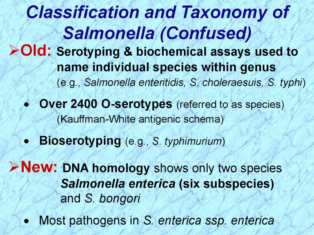
Classification and Taxonomy ofSalmonella (Confused)
Old: Serotyping & biochemical assays used to
name individual species within genus
(e.g., Salmonella enteritidis, S. choleraesuis, S. typhi)
Over 2400 O-serotypes (referred to as species)
(Kauffman-White antigenic schema)
Bioserotyping (e.g., S. typhimurium)
New: DNA homology shows only two species
Salmonella enterica (six subspecies)
and S. bongori
Most pathogens in S. enterica ssp. enterica
7.
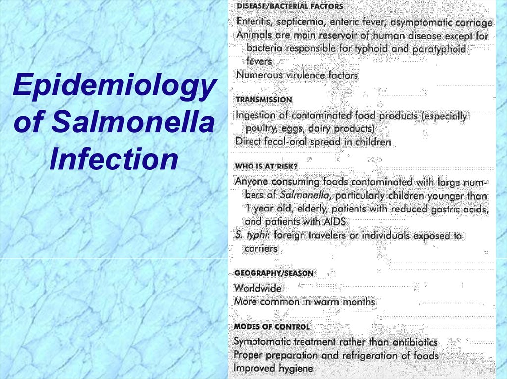
Epidemiologyof Salmonella
Infection
8.
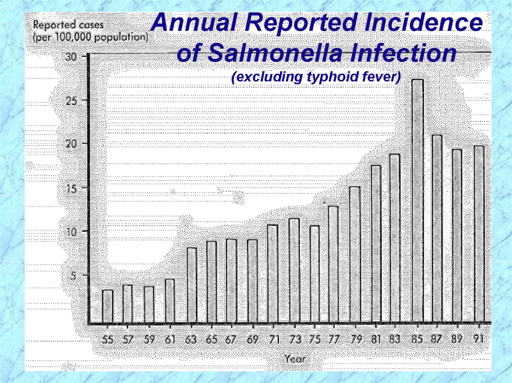
Annual Reported Incidenceof Salmonella Infection
(excluding typhoid fever)
9.
Clinical Syndromes of SalmonellaSalmonellosis = Generic term for disease
Clinical Syndromes
Enteritis (acute gastroenteritis)
Enteric fever (prototype is typhoid fever and
less severe paratyphoid fever)
Septicemia (particularly S. choleraesuis, S. typhi,
and S. paratyphi)
Asymptomatic carriage (gall bladder is the
reservoir for Salmonella typhi)
10.
Epidemiology and Clinical Syndromesof Salmonella (cont.)
Enteritis
Most common form of salmonellosis with major
foodborne outbreaks and sporadic disease
High infectious dose (108 CFU)
Poultry, eggs, etc. are sources of infection
6-48h incubation period
Nausea, vomiting, nonbloody diarrhea, fever,
cramps, myalgia and headache common
S. enteritidis bioserotypes (e.g., S. typhimurium)
11.
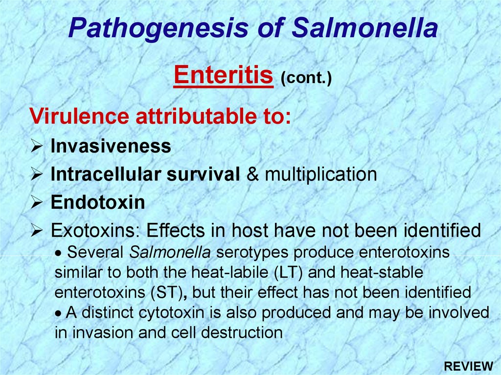
Pathogenesis of SalmonellaEnteritis (cont.)
Virulence attributable to:
Invasiveness
Intracellular survival & multiplication
Endotoxin
Exotoxins: Effects in host have not been identified
Several Salmonella serotypes produce enterotoxins
similar to both the heat-labile (LT) and heat-stable
enterotoxins (ST), but their effect has not been identified
A distinct cytotoxin is also produced and may be involved
in invasion and cell destruction
12.
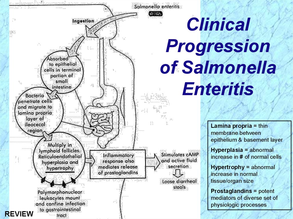
Pathogenesis of Salmonella (cont.)Invasiveness in Enteritis (cont.)
Penetrate mucus, adhere to and invade into
epithelial layer (enterocytes) of terminal small
intestine and further into subepithelial tissue
Bacterial cells are internalized in endocytic
vacuoles (intracellular) and the organisms multiply
PMN’s confine infection to gastrointestinal (GI) tract,
but organisms may spread hematogenously
(through blood, i.e., septicemia) to other body sites
Inflammatory response mediates release of
prostaglandins, stimulating cAMP and active fluid
secretion with loose diarrheal stools; epithelial
destruction occurs during late stage of disease
13.
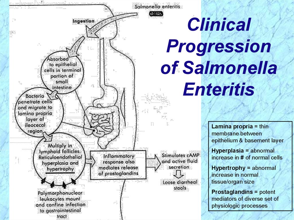
ClinicalProgression
of Salmonella
Enteritis
Lamina propria = thin
membrane between
epithelium & basement layer
Hyperplasia = abnormal
increase in # of normal cells
Hypertrophy = abnormal
increase in normal
tissue/organ size
Prostaglandins = potent
mediators of diverse set of
physiologic processes
14.
Epidemiology & Clinical Syndromes (cont.)Enteric Fevers
S. typhi causes typhoid fever
S. paratyphi A, B (S. schottmuelleri) and C
(S. hirschfeldii) cause milder form of enteric fever
Infectious dose = 106 CFU
Fecal-oral route of transmission
Person-to-person spread by chronic carrier
Fecally-contaminated food or water
10-14 day incubation period
Initially signs of sepsis/bacteremia with sustained
fever (delirium) for > one week before abdominal
pain and gastrointestinal symptoms
15.
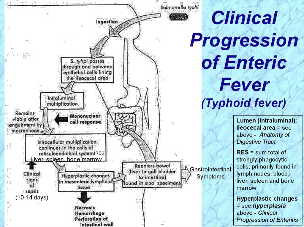
Pathogenesis of Salmonella (cont.)Enteric Fevers (cont.)
Virulence attributable to:
Invasiveness
Pass through intestinal epithelial cells in ileocecal region,
infect the regional lymphatic system, invade the bloodstream,
and infect other parts of the reticuloendothelial system
Organisms are phagocytosed by macrophages and
monocytes, but survive, multiply and are transported to the
liver, spleen, and bone marrow where they continue to replicate
Second week: organisms reenter bloodstream and cause
prolonged bacteremia; biliary tree and other organs are
infected; gradually increasing sustained fever likely from
endotoxemia
Second to third week: bacteria colonize gallbladder, reinfect
intestinal tract with diarrheal symptoms and possible necrosis
of the Peyer’s patches
16.
ClinicalProgression
of Enteric
Fever
(Typhoid fever)
Lumen (intraluminal);
ileocecal area = see
above - Anatomy of
Digestive Tract
(RES)
Liver, spleen, bone marrow
(10-14 days)
RES = sum total of
strongly phagocytic
Gastrointestinal cells; primarily found in
lymph nodes, blood,
Symptoms
liver, spleen and bone
marrow
Hyperplastic changes
= see hyperplasia
above - Clinical
Progression of Enteritis
17.
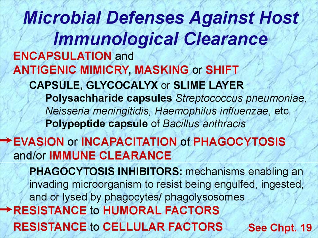
Microbial Defenses Against HostImmunological Clearance
ENCAPSULATION and
ANTIGENIC MIMICRY, MASKING or SHIFT
CAPSULE, GLYCOCALYX or SLIME LAYER
Polysachharide capsules Streptococcus pneumoniae,
Neisseria meningitidis, Haemophilus influenzae, etc.
Polypeptide capsule of Bacillus anthracis
EVASION or INCAPACITATION of PHAGOCYTOSIS
and/or IMMUNE CLEARANCE
PHAGOCYTOSIS INHIBITORS: mechanisms enabling an
invading microorganism to resist being engulfed, ingested,
and or lysed by phagocytes/ phagolysosomes
RESISTANCE to HUMORAL FACTORS
RESISTANCE to CELLULAR FACTORS
See Chpt. 19
18.
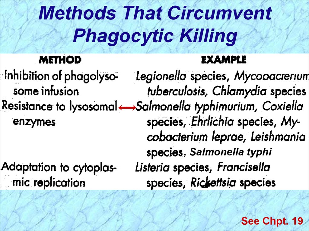
Methods That CircumventPhagocytic Killing
, Salmonella typhi
See Chpt. 19
19.
Epidemiology & Clinical Syndromes (cont.)Septicemia
Can be caused by all species, but more
commonly associated with S. choleraesuis, S.
paratyphi, S. typhi, and S. dublin
Old, young and immunocompromised (e.g.,
AIDS patients) at increased risk
20.
Epidemiology & Clinical Syndromes (cont.)Asymptomatic Carriage
Chronic carriage in 1-5% of cases following S.
typhi or S. paratyphi infection
Gall bladder usually the reservoir
Chronic carriage with other Salmonella spp.
occurs in <<1% of cases and does not play a
role in human disease transmission
21.
Treatment, Prevention and Controlof Salmonella Infections
Enteritis:
Antibiotics not recommended for enteritis
because prolong duration
Control by proper preparation of poultry & eggs
Enteric fever:
Antibiotics to avoid carrier state
Identify & treat carriers of S. typhi & S. paratyphi
Vaccination can reduce risk of disease for
travellers in endemic areas
22.
23.
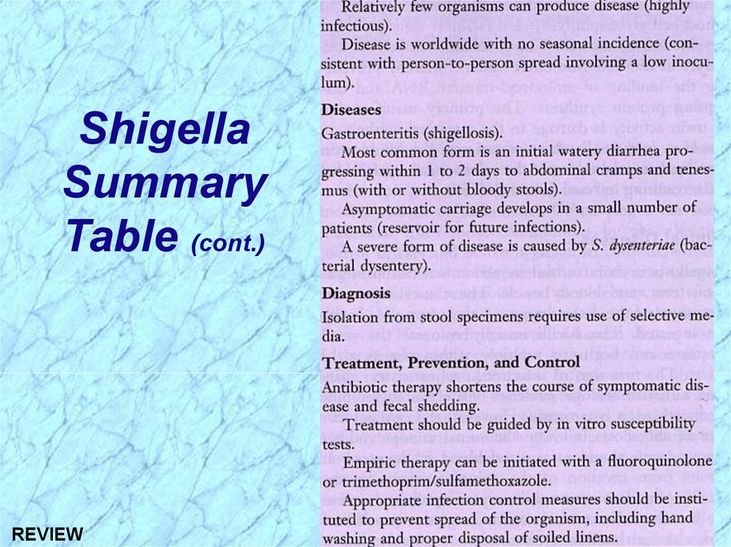
General Characteristics of ShigellaColiform bacilli (enteric rods)
Nonmotile gram-negative facultative anaerobes
Four species
Shigella sonnei (most common in industrial world)
Shigella flexneri (most common in developing countries)
Shigella boydii
Shigella dysenteriae
Non-lactose fermenting
Resistant to bile salts
24.
Epidemiology and Clinical Syndromesof Shigella
Shigellosis = Generic term for disease
Low infectious dose (102-104 CFU)
Humans are only reservoir
Transmission by fecal-oral route
Incubation period = 1-3 days
Watery diarrhea with fever; changing to dysentery
Major cause of bacillary dysentery (severe 2nd stage)
in pediatric age group (1-10 yrs) via fecal-oral route
Outbreaks in daycare centers, nurseries, institutions
Estimated 15% of pediatric diarrhea in U.S.
Leading cause of infant diarrhea and mortality
(death) in developing countries
25.
DEFINITIONSEnterotoxin = an exotoxin with enteric activity, i.e.,
affects the intestinal tract
Dysentery = inflammation of intestines (especially
the colon (colitis) of the large intestine) with
accompanying severe abdominal cramps,
tenesmus (straining to defecate), and frequent, lowvolume stools containing blood, mucus, and
fecal leukocytes (PMN’s)
Bacillary dysentery = dysentery caused by
bacterial infection with invasion of host cells/tissues
and/or production of exotoxins
26.
Epidemiologyof Shigella
Infection
27.
Pathogenesis of ShigellaShigellosis
Two-stage disease:
Early stage:
Watery diarrhea attributed to the enterotoxic
activity of Shiga toxin following ingestion and
noninvasive colonization, multiplication, and
production of enterotoxin in the small intestine
Fever attributed to neurotoxic activity of toxin
Second stage:
Adherence to and tissue invasion of large
intestine with typical symptoms of dysentery
Cytotoxic activity of Shiga toxin increases
severity
28.
Pathogenesis and Virulence Factors (cont.)Virulence attributable to:
Invasiveness
Attachment (adherence) and internalization
with complex genetic control
Large multi-gene virulence plasmid regulated by
multiple chromosomal genes
Exotoxin (Shiga toxin)
Intracellular survival & multiplication
29.
Pathogenesis and Virulence Factors (cont.)Invasiveness in Shigella-Associated Dysentery
Penetrate through mucosal surface of colon
(colonic mucosa) and invade and multiply in the
colonic epithelium but do not typically invade
beyond the epithelium into the lamina propria (thin
layer of fibrous connective tissue immediately beneath the
surface epithelium of mucous membranes)
Preferentially attach to and invade into M cells in
Peyer’s patches (lymphoid tissue, i.e., lymphatic system)
of small intestine
30.
Pathogenesis and Virulence Factors (cont.)Invasiveness in Shigella-Associated Dysentery(cont.)
M cells typically transport foreign antigens from
the intestine to underlying macrophages, but
Shigella can lyse the phagocytic vacuole
(phagosome) and replicate in the cytoplasm
Note: This contrasts with Salmonella which
multiplies in the phagocytic vacuole
Actin filaments propel the bacteria through the
cytoplasm and into adjacent epithelial cells with
cell-to-cell passage, thereby effectively avoiding
antibody-mediated humoral immunity (similar
to Listeria monocytogenes)
31.
32.
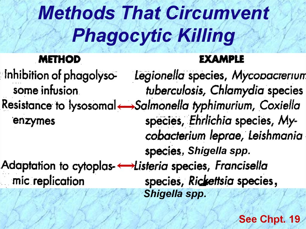
Methods That CircumventPhagocytic Killing
, Shigella spp.
,
Shigella spp.
See Chpt. 19
33.
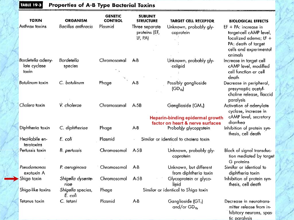
Pathogenesis and Virulence Factors (cont.)Characteristics of Shiga Toxin
Enterotoxic, neurotoxic and cytotoxic
Encoded by chromosomal genes
Two domain (A-5B) structure
Similar to the Shiga-like toxin of
enterohemorrhagic E. coli (EHEC)
NOTE: except that Shiga-like toxin is encoded by
lysogenic bacteriophage
34.
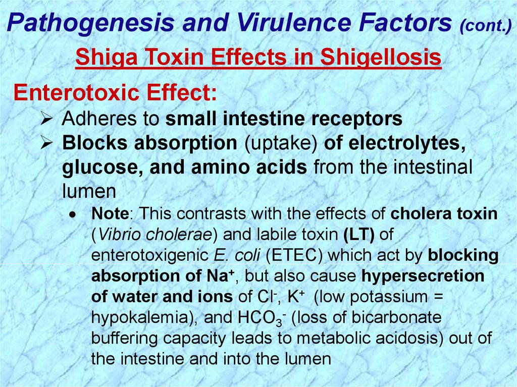
Pathogenesis and Virulence Factors (cont.)Shiga Toxin Effects in Shigellosis
Enterotoxic Effect:
Adheres to small intestine receptors
Blocks absorption (uptake) of electrolytes,
glucose, and amino acids from the intestinal
lumen
Note: This contrasts with the effects of cholera toxin
(Vibrio cholerae) and labile toxin (LT) of
enterotoxigenic E. coli (ETEC) which act by blocking
absorption of Na+, but also cause hypersecretion
of water and ions of Cl-, K+ (low potassium =
hypokalemia), and HCO3- (loss of bicarbonate
buffering capacity leads to metabolic acidosis) out of
the intestine and into the lumen
35.
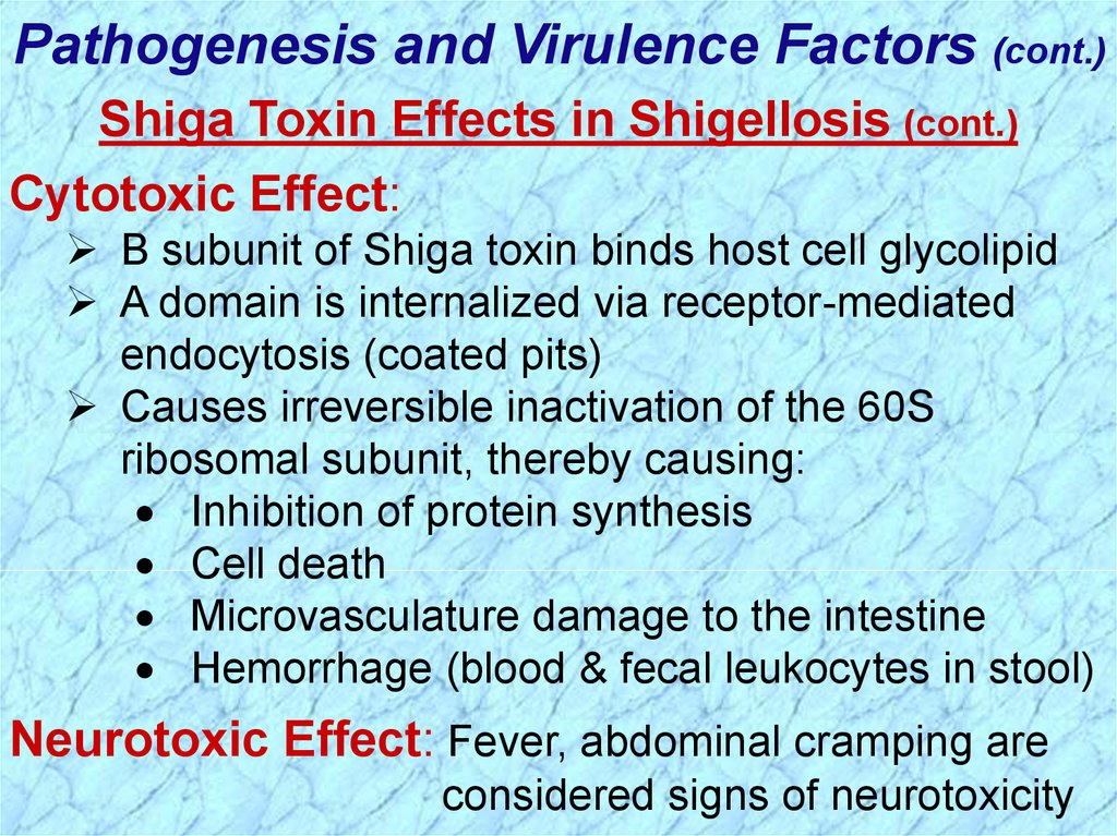
Pathogenesis and Virulence Factors (cont.)Shiga Toxin Effects in Shigellosis (cont.)
Cytotoxic Effect:
B subunit of Shiga toxin binds host cell glycolipid
A domain is internalized via receptor-mediated
endocytosis (coated pits)
Causes irreversible inactivation of the 60S
ribosomal subunit, thereby causing:
Inhibition of protein synthesis
Cell death
Microvasculature damage to the intestine
Hemorrhage (blood & fecal leukocytes in stool)
Neurotoxic Effect: Fever, abdominal cramping are
considered signs of neurotoxicity
36.
Heparin-binding epidermal growthfactor on heart & nerve surfaces
37.
38.
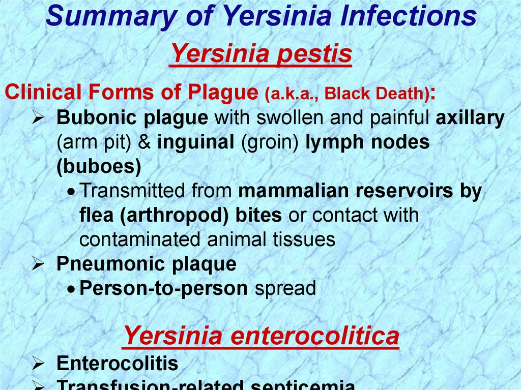
Summary of Yersinia InfectionsYersinia pestis
Clinical Forms of Plague (a.k.a., Black Death):
Bubonic plague with swollen and painful axillary
(arm pit) & inguinal (groin) lymph nodes
(buboes)
Transmitted from mammalian reservoirs by
flea (arthropod) bites or contact with
contaminated animal tissues
Pneumonic plaque
Person-to-person spread
Yersinia enterocolitica
Enterocolitis
39.
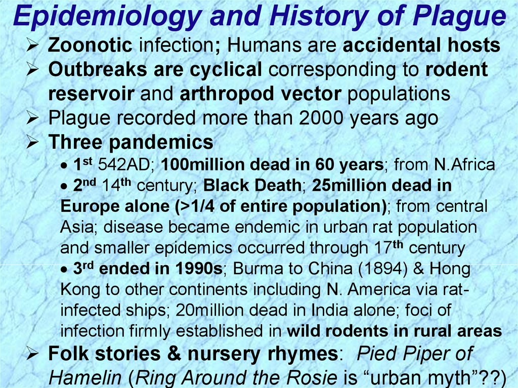
Epidemiology and History of PlagueZoonotic infection; Humans are accidental hosts
Outbreaks are cyclical corresponding to rodent
reservoir and arthropod vector populations
Plague recorded more than 2000 years ago
Three pandemics
1st 542AD; 100million dead in 60 years; from N.Africa
2nd 14th century; Black Death; 25million dead in
Europe alone (>1/4 of entire population); from central
Asia; disease became endemic in urban rat population
and smaller epidemics occurred through 17th century
3rd ended in 1990s; Burma to China (1894) & Hong
Kong to other continents including N. America via ratinfected ships; 20million dead in India alone; foci of
infection firmly established in wild rodents in rural areas
Folk stories & nursery rhymes: Pied Piper of
Hamelin (Ring Around the Rosie is “urban myth”??)
40.
Epidemiologyof Yersinia
Infection
41.
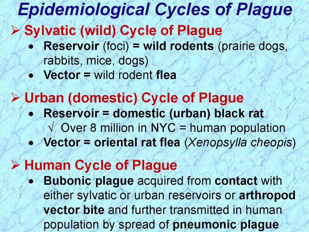
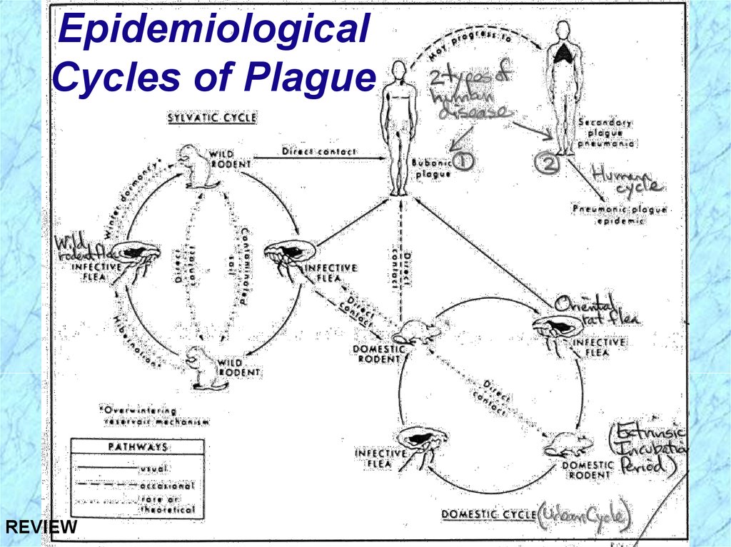
Epidemiological Cycles of PlagueSylvatic (wild) Cycle of Plague
Reservoir (foci) = wild rodents (prairie dogs,
rabbits, mice, dogs)
Vector = wild rodent flea
Urban (domestic) Cycle of Plague
Reservoir = domestic (urban) black rat
Over 8 million in NYC = human population
Vector = oriental rat flea (Xenopsylla cheopis)
Human Cycle of Plague
Bubonic plague acquired from contact with
either sylvatic or urban reservoirs or arthropod
vector bite and further transmitted in human
population by spread of pneumonic plague
42.
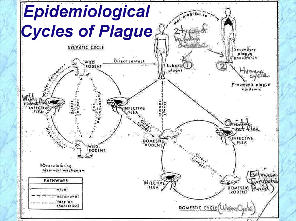
EpidemiologicalCycles of Plague
43.
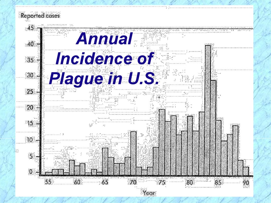
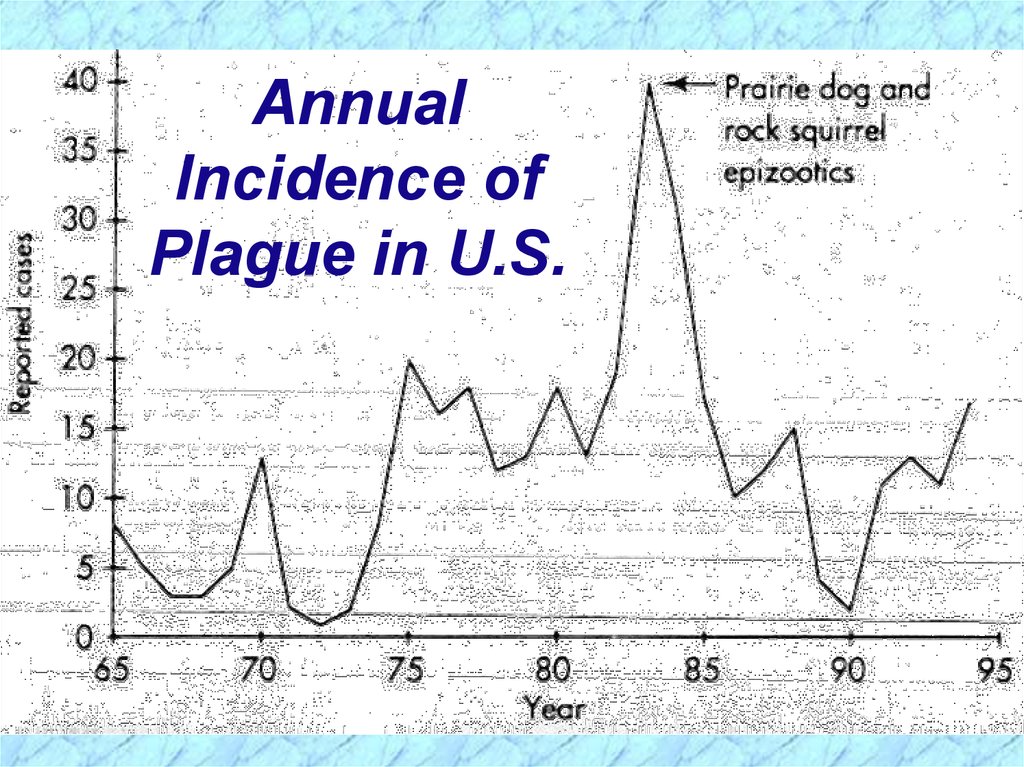
AnnualIncidence of
Plague in U.S.
44.
AnnualIncidence of
Plague in U.S.
45.
Arthropod-Borne Transmission of PlagueFleas required for perpetuation of plague vary
greatly in vector efficiency and host range
Organisms ingested during blood meal from
bacteremic host
Coagulase of flea may cause fibrin clot of
organism in stomach which fixes to spines of
proventriculus (throat parts of flea)
Organisms multiply causing blockage
Flea regurgitates infectious material into new
host during subsequent attempts at blood meal
Flea remains hungry & feeds more aggressively
Sudden eradication of rats could lead to outbreak
46.
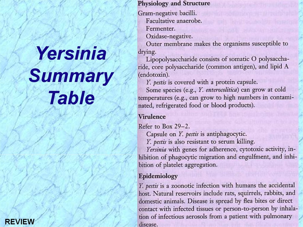
YersiniaSummary
Table
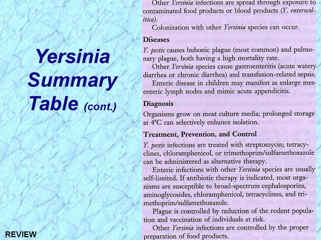
47.
YersiniaSummary
Table (cont.)
48.
49.
REVIEW50.
See HandoutsREVIEW
51.
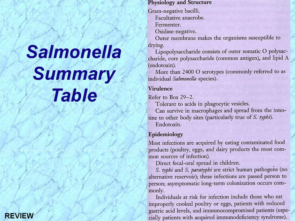
SalmonellaSummary
Table
REVIEW
52.
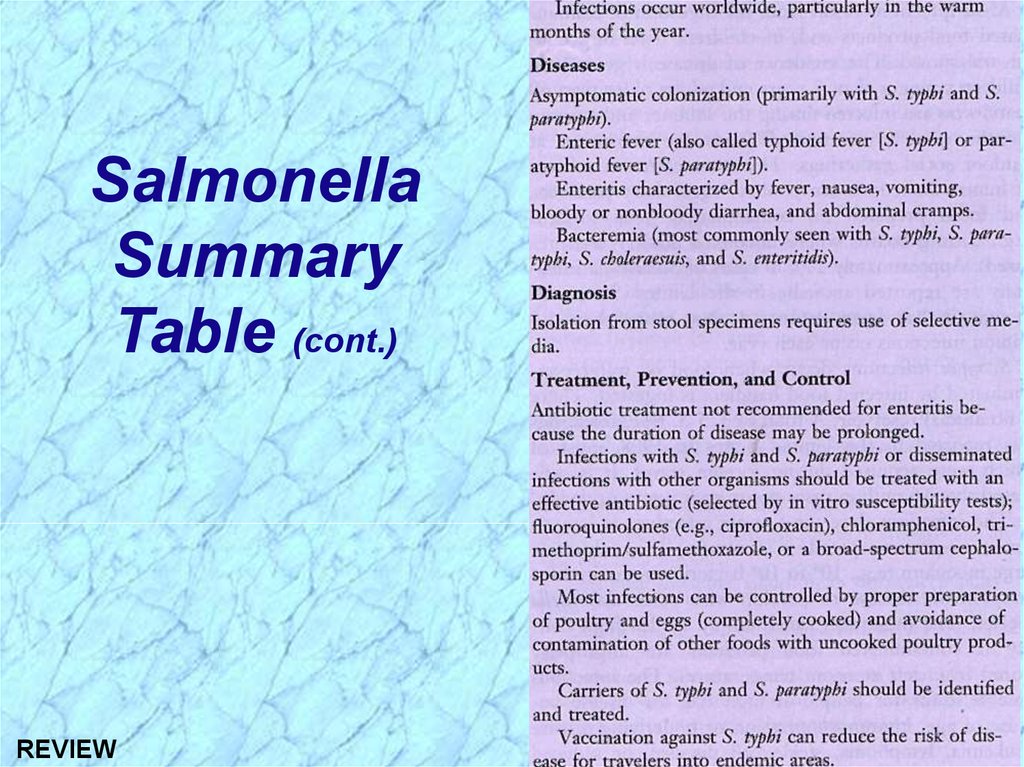
SalmonellaSummary
Table (cont.)
REVIEW
53.
Clinical Syndromes of SalmonellaSalmonellosis = Generic term for disease
Clinical Syndromes
Enteritis (acute gastroenteritis)
Enteric fever (prototype is typhoid fever and
less severe paratyphoid fever)
Septicemia (particularly S. choleraesuis, S. typhi,
and S. paratyphi)
Asymptomatic carriage (gall bladder is the
reservoir for Salmonella typhi)
REVIEW
54.
Epidemiology and Clinical Syndromesof Salmonella (cont.)
Enteritis
Most common form of salmonellosis with major
foodborne outbreaks and sporadic disease
High infectious dose (108 CFU)
Poultry, eggs, etc. are sources of infection
6-48h incubation period
Nausea, vomiting, nonbloody diarrhea, fever,
cramps, myalgia and headache common
S. enteritidis bioserotypes (e.g., S. typhimurium)
REVIEW
55.
Pathogenesis of SalmonellaEnteritis (cont.)
Virulence attributable to:
Invasiveness
Intracellular survival & multiplication
Endotoxin
Exotoxins: Effects in host have not been identified
Several Salmonella serotypes produce enterotoxins
similar to both the heat-labile (LT) and heat-stable
enterotoxins (ST), but their effect has not been identified
A distinct cytotoxin is also produced and may be involved
in invasion and cell destruction
REVIEW
56.
ClinicalProgression
of Salmonella
Enteritis
Lamina propria = thin
membrane between
epithelium & basement layer
Hyperplasia = abnormal
increase in # of normal cells
Hypertrophy = abnormal
increase in normal
tissue/organ size
Prostaglandins = potent
mediators of diverse set of
physiologic processes
REVIEW
57.
ClinicalProgression
of Enteric
Fever
(Typhoid fever)
Lumen (intraluminal);
ileocecal area = see
above - Anatomy of
Digestive Tract
(RES)
Liver, spleen, bone marrow
(10-14 days)
REVIEW
RES = sum total of
strongly phagocytic
Gastrointestinal cells; primarily found in
lymph nodes, blood,
Symptoms
liver, spleen and bone
marrow
Hyperplastic changes
= see hyperplasia
above - Clinical
Progression of Enteritis
58.
59.
ShigellaSummary
Table
REVIEW
60.
ShigellaSummary
Table (cont.)
REVIEW
61.
Epidemiology and Clinical Syndromesof Shigella
Shigellosis = Generic term for disease
Low infectious dose (102-104 CFU)
Humans are only reservoir
Transmission by fecal-oral route
Incubation period = 1-3 days
Watery diarrhea with fever; changing to dysentery
Major cause of bacillary dysentery (severe 2nd stage)
in pediatric age group (1-10 yrs) via fecal-oral route
Outbreaks in daycare centers, nurseries, institutions
Estimated 15% of pediatric diarrhea in U.S.
Leading cause of infant diarrhea and mortality
(death) in developing countries
REVIEW
62.
DEFINITIONSEnterotoxin = an exotoxin with enteric activity, i.e.,
affects the intestinal tract
Dysentery = inflammation of intestines (especially
the colon (colitis) of the large intestine) with
accompanying severe abdominal cramps,
tenesmus (straining to defecate), and frequent, lowvolume stools containing blood, mucus, and
fecal leukocytes (PMN’s)
Bacillary dysentery = dysentery caused by
bacterial infection with invasion of host cells/tissues
and/or production of exotoxins
REVIEW
63.
Pathogenesis of ShigellaShigellosis
Two-stage disease:
Early stage:
Watery diarrhea attributed to the enterotoxic
activity of Shiga toxin following ingestion and
noninvasive colonization, multiplication, and
production of enterotoxin in the small intestine
Fever attributed to neurotoxic activity of toxin
Second stage:
Adherence to and tissue invasion of large
intestine with typical symptoms of dysentery
Cytotoxic activity of Shiga toxin increases
severity
REVIEW
64.
Pathogenesis and Virulence Factors (cont.)Virulence attributable to:
Invasiveness
Attachment (adherence) and internalization
with complex genetic control
Large multi-gene virulence plasmid regulated by
multiple chromosomal genes
Exotoxin (Shiga toxin)
Intracellular survival & multiplication
REVIEW
65.
Pathogenesis and Virulence Factors (cont.)Characteristics of Shiga Toxin
Enterotoxic, neurotoxic and cytotoxic
Encoded by chromosomal genes
Two domain (A-5B) structure
Similar to the Shiga-like toxin of
enterohemorrhagic E. coli (EHEC)
NOTE: except that Shiga-like toxin is encoded by
lysogenic bacteriophage
REVIEW
66.
67.
YersiniaSummary
Table
REVIEW
68.
YersiniaSummary
Table (cont.)
REVIEW
69.
Summary of Yersinia InfectionsYersinia pestis
Clinical Forms of Plague (a.k.a., Black Death):
Bubonic plague with swollen and painful axillary
(arm pit) & inguinal (groin) lymph nodes
(buboes)
Transmitted from mammalian reservoirs by
flea (arthropod) bites or contact with
contaminated animal tissues
Pneumonic plaque
Person-to-person spread
Yersinia enterocolitica
Enterocolitis
REVIEW
70.
Epidemiology and History of PlagueREVIEW
Zoonotic infection; Humans are accidental hosts
Outbreaks are cyclical corresponding to rodent
reservoir and arthropod vector populations
Plague recorded more than 2000 years ago
Three pandemics
1st 542AD; 100million dead in 60 years; from N.Africa
2nd 14th century; Black Death; 25million dead in
Europe alone (>1/4 of entire population); from central
Asia; disease became endemic in urban rat population
and smaller epidemics occurred through 17th century
3rd ended in 1990s; Burma to China (1894) & Hong
Kong to other continents including N. America via ratinfected ships; 20million dead in India alone; foci of
infection firmly established in wild rodents in rural areas
Folk stories & nursery rhymes: Pied Piper of
Hamelin (Ring Around the Rosie is “urban myth”??)
71.
Epidemiological Cycles of PlagueSylvatic (wild) Cycle of Plague
Reservoir (foci) = wild rodents (prairie dogs,
rabbits, mice, dogs)
Vector = wild rodent flea
Urban (domestic) Cycle of Plague
Reservoir = domestic (urban) black rat
Over 8 million in NYC = human population
Vector = oriental rat flea (Xenopsylla cheopis)
Human Cycle of Plague
Bubonic plague acquired from contact with
either sylvatic or urban reservoirs or arthropod
vector bite and further transmitted in human
REVIEW population by spread of pneumonic plague
72.
EpidemiologicalCycles of Plague
REVIEW