


 chemistry
Similar presentations:


")


Lesson 2 and 3. The chemistry of life
1.
The chemistry of life:carbohydrates and lipids
2.
Carbohydrates are large biological molecules, or macromolecules, consisting of carbon (C),hydrogen (H), and oxygen (O) atoms, usually with a hydrogen:oxygen atom ratio of 2:1 (as in water).
The empirical formula Cm(H2O)n (where m could be different from n, m normally > than 3).
Carbohydrates include both sugars and polymers of sugars.
Monosaccharides are the most basic units of carbohydrates. Depending on the number of carbon atoms, several types
of monosaccharides can be distinguished.
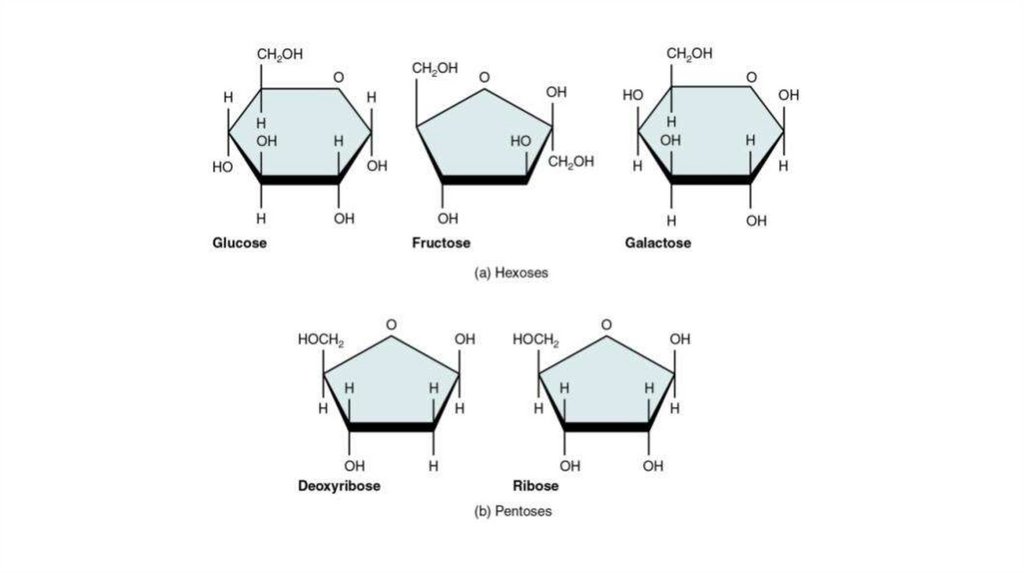
Glucose, fructose, galactose and other sugars that have six carbons are called hexoses.
Trioses (three-carbon sugars) and pentoses (five-carbon sugars) are also common. The most important pentoses are
ribose and deoxyribose.
Monosaccharides are major nutrients for cells.
In the process known as cellular respiration, cells extract energy in a series of reactions starting with glucose
molecules.
Simple-sugar molecules are not only a major fuel for cellular work, but their carbon skeletons also serve as raw
material for the synthesis of other types of small organic molecules, such as amino acids (mostly ribose and
deoxyribose) and fatty acids.
Sugar molecules that are not immediately used in these ways are generally incorporated as monomers into
disaccharides or polysaccharides.
3.
4.
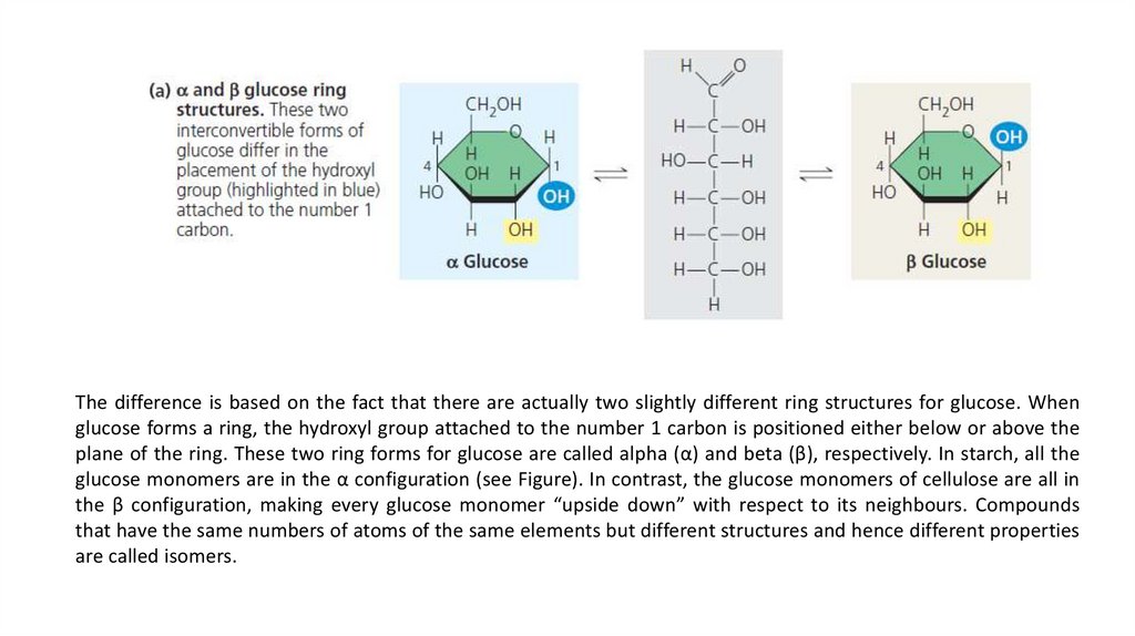
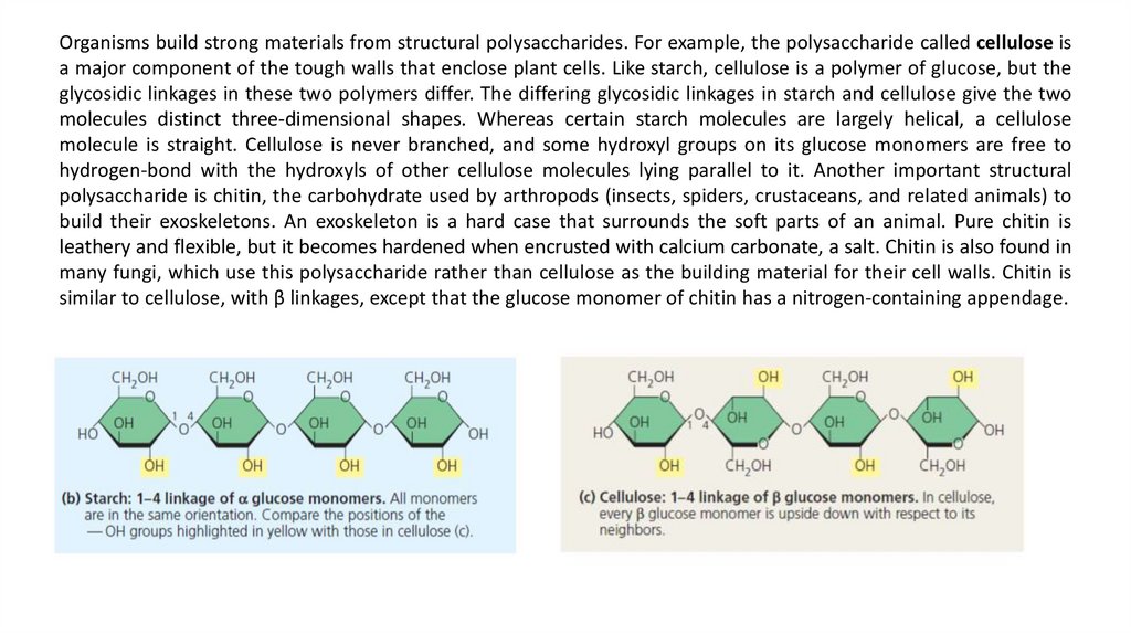
The difference is based on the fact that there are actually two slightly different ring structures for glucose. Whenglucose forms a ring, the hydroxyl group attached to the number 1 carbon is positioned either below or above the
plane of the ring. These two ring forms for glucose are called alpha (α) and beta (β), respectively. In starch, all the
glucose monomers are in the α configuration (see Figure). In contrast, the glucose monomers of cellulose are all in
the β configuration, making every glucose monomer “upside down” with respect to its neighbours. Compounds
that have the same numbers of atoms of the same elements but different structures and hence different properties
are called isomers.
5.
Disaccharides consist of two monosaccharides joined by a glycosidic linkage, a covalent bond formed between twomonosaccharides by a dehydration reaction. For example, maltose is a disaccharide formed by the linking of two
molecules of glucose. The most prevalent disaccharide is sucrose, which is table sugar. Its two monomers are glucose
and fructose. Lactose, the sugar present in milk, is another disaccharide, in this case a glucose molecule joined to a
galactose molecule. The functions of disaccharides are similar to those of monosaccharides.
6.
Polysaccharides are macromolecules, polymers with a few hundred to a few thousand monosaccharides joined byglycosidic linkages. Some polysaccharides serve as storage material, hydrolyzed as needed to provide sugar for cells.
Other polysaccharides serve as building material for structures that protect the cell or the whole organism. The
architecture and function of a polysaccharide are determined by its sugar monomers and by the positions of its
glycosidic linkages.
Both plants and animals store sugars for later use in the form of storage polysaccharides. Plants store starch, a
polymer of glucose monomers, as granules within cellular structures known as plastids, which include chloroplasts.
Synthesizing starch enables the plant to stockpile surplus glucose. Because glucose is a major cellular fuel, starch
represents stored energy. The sugar can later be withdrawn from this carbohydrate “bank” by hydrolysis, which
breaks the bonds between the glucose monomers. Animals store a polysaccharide called glycogen, a polymer of
glucose which is extensively branched. Humans and other vertebrates store glycogen mainly in liver and muscle
cells. Hydrolysis of glycogen in these cells releases glucose when the demand for sugar increases. This stored fuel
cannot sustain an animal for long, however. In humans, for example, glycogen stores are depleted in about a day
unless they are replenished by consumption of food. This is an issue of concern in low-carbohydrate diets.
7.
Organisms build strong materials from structural polysaccharides. For example, the polysaccharide called cellulose isa major component of the tough walls that enclose plant cells. Like starch, cellulose is a polymer of glucose, but the
glycosidic linkages in these two polymers differ. The differing glycosidic linkages in starch and cellulose give the two
molecules distinct three-dimensional shapes. Whereas certain starch molecules are largely helical, a cellulose
molecule is straight. Cellulose is never branched, and some hydroxyl groups on its glucose monomers are free to
hydrogen-bond with the hydroxyls of other cellulose molecules lying parallel to it. Another important structural
polysaccharide is chitin, the carbohydrate used by arthropods (insects, spiders, crustaceans, and related animals) to
build their exoskeletons. An exoskeleton is a hard case that surrounds the soft parts of an animal. Pure chitin is
leathery and flexible, but it becomes hardened when encrusted with calcium carbonate, a salt. Chitin is also found in
many fungi, which use this polysaccharide rather than cellulose as the building material for their cell walls. Chitin is
similar to cellulose, with β linkages, except that the glucose monomer of chitin has a nitrogen-containing appendage.
8.
Lipids are the one class of large biological molecules that does not include true polymers, and they are generally not bigenough to be considered macromolecules. The compounds called lipids are grouped together because they share one
important trait: They mix poorly, if at all, with water. The hydrophobic behavior of lipids is based on their molecular
structure. Although they may have some polar bonds associated with oxygen, lipids consist mostly of hydrocarbon
regions. Lipids are varied in form and function. The most biologically important types of lipids: fats, phospholipids, and
steroids.
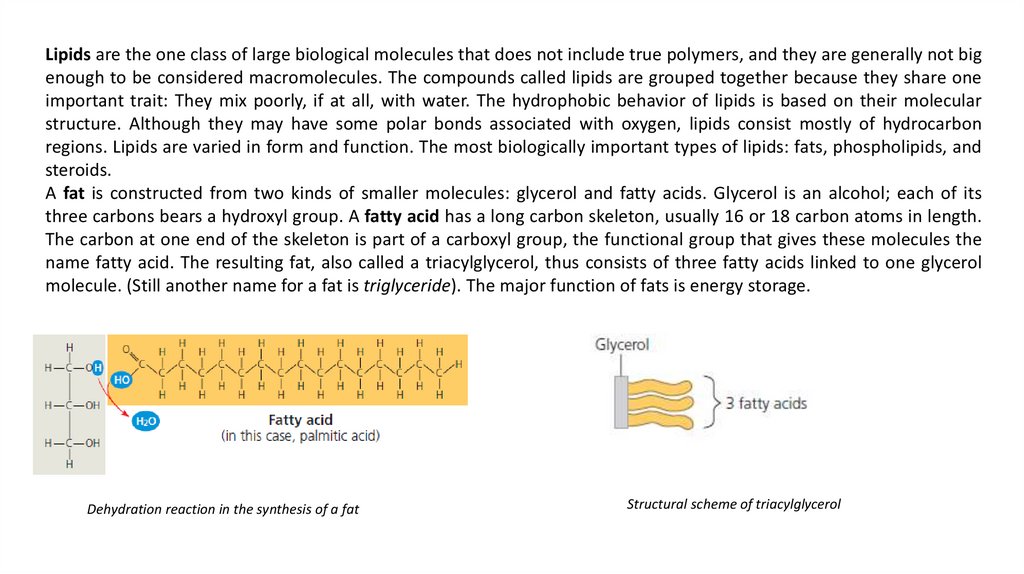
A fat is constructed from two kinds of smaller molecules: glycerol and fatty acids. Glycerol is an alcohol; each of its
three carbons bears a hydroxyl group. A fatty acid has a long carbon skeleton, usually 16 or 18 carbon atoms in length.
The carbon at one end of the skeleton is part of a carboxyl group, the functional group that gives these molecules the
name fatty acid. The resulting fat, also called a triacylglycerol, thus consists of three fatty acids linked to one glycerol
molecule. (Still another name for a fat is triglyceride). The major function of fats is energy storage.
Dehydration reaction in the synthesis of a fat
Structural scheme of triacylglycerol
9.
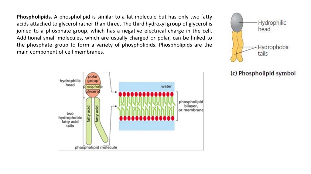
Phospholipids. A phospholipid is similar to a fat molecule but has only two fattyacids attached to glycerol rather than three. The third hydroxyl group of glycerol is
joined to a phosphate group, which has a negative electrical charge in the cell.
Additional small molecules, which are usually charged or polar, can be linked to
the phosphate group to form a variety of phospholipids. Phospholipids are the
main component of cell membranes.
10.
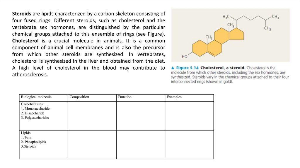
Steroids are lipids characterized by a carbon skeleton consisting offour fused rings. Different steroids, such as cholesterol and the
vertebrate sex hormones, are distinguished by the particular
chemical groups attached to this ensemble of rings (see Figure).
Cholesterol is a crucial molecule in animals. It is a common
component of animal cell membranes and is also the precursor
from which other steroids are synthesized. In vertebrates,
cholesterol is synthesized in the liver and obtained from the diet.
A high level of cholesterol in the blood may contribute to
atherosclerosis.
Biological molecule
Carbohydrates
1. Monosaccharide
2. Disaccharide
3. Polysaccharides
Lipids
1. Fats
2. Phospholipids
3.Steroids
Composition
Function
Examples
11.
The chemistry of life:proteins and nucleic acids
12.
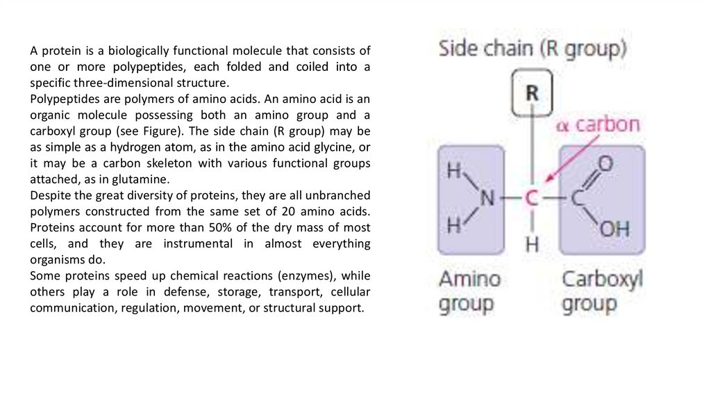
A protein is a biologically functional molecule that consists ofone or more polypeptides, each folded and coiled into a
specific three-dimensional structure.
Polypeptides are polymers of amino acids. An amino acid is an
organic molecule possessing both an amino group and a
carboxyl group (see Figure). The side chain (R group) may be
as simple as a hydrogen atom, as in the amino acid glycine, or
it may be a carbon skeleton with various functional groups
attached, as in glutamine.
Despite the great diversity of proteins, they are all unbranched
polymers constructed from the same set of 20 amino acids.
Proteins account for more than 50% of the dry mass of most
cells, and they are instrumental in almost everything
organisms do.
Some proteins speed up chemical reactions (enzymes), while
others play a role in defense, storage, transport, cellular
communication, regulation, movement, or structural support.
13.
Рeptide bond. Read the text below, draw the scheme of apolypeptide chain and mark the peptide bond (for more read
Campbell et al. 2009:80-81).
When two amino acids are positioned so that the carboxyl group of
one is adjacent to the amino group of the other, they can become
joined by a dehydration reaction, with the removal of a water
molecule. The resulting covalent bond is called a peptide bond.
Repeated over and over, this process yields a polypeptide, a polymer
of many amino acids linked by peptide bonds. Polypeptides range in
length from a few amino acids to a thousand or more. Each specific
polypeptide has a unique linear sequence of amino acids.
14.
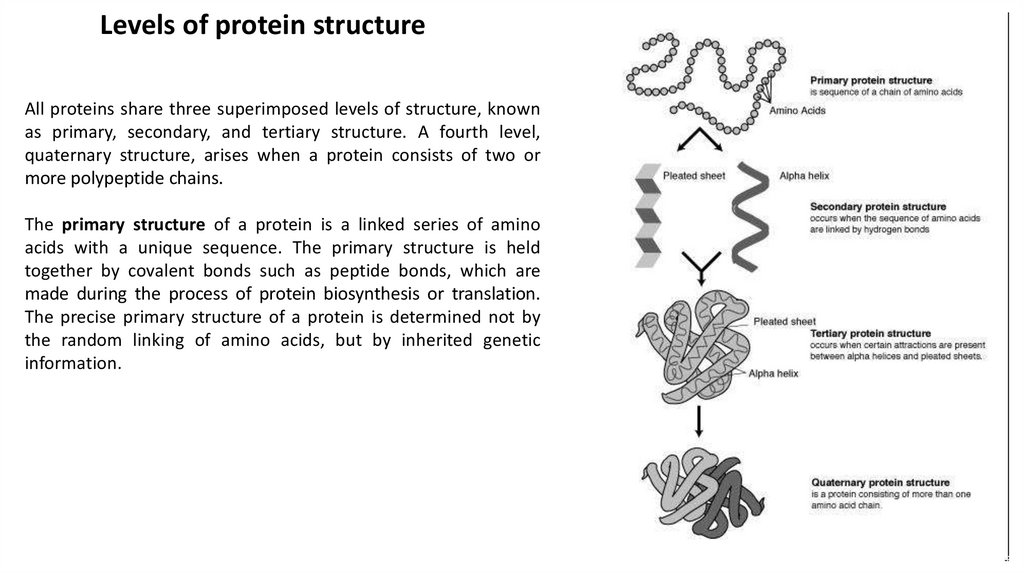
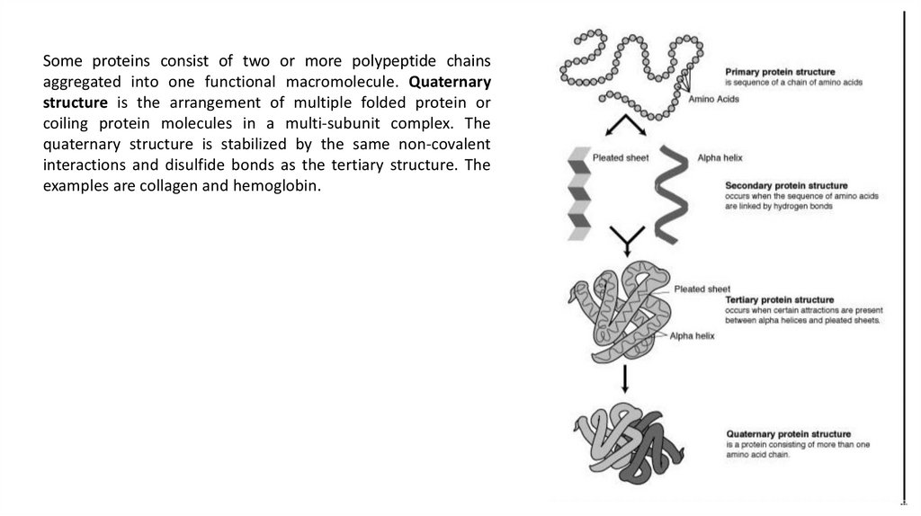
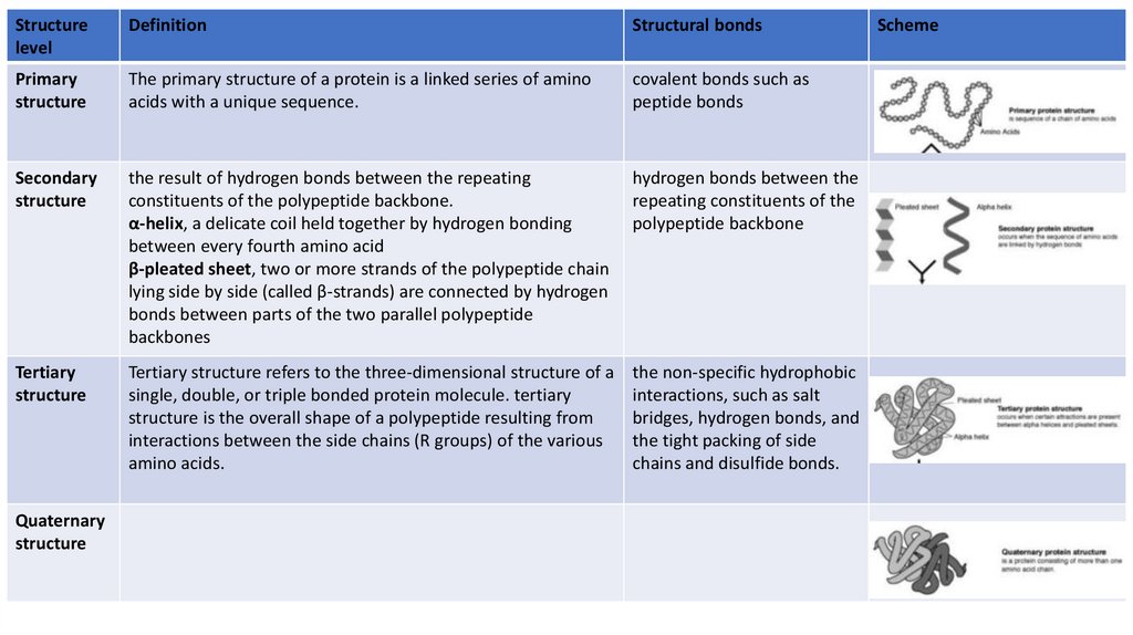
Levels of protein structureAll proteins share three superimposed levels of structure, known
as primary, secondary, and tertiary structure. A fourth level,
quaternary structure, arises when a protein consists of two or
more polypeptide chains.
The primary structure of a protein is a linked series of amino
acids with a unique sequence. The primary structure is held
together by covalent bonds such as peptide bonds, which are
made during the process of protein biosynthesis or translation.
The precise primary structure of a protein is determined not by
the random linking of amino acids, but by inherited genetic
information.
15.
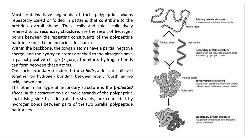
Most proteins have segments of their polypeptide chainsrepeatedly coiled or folded in patterns that contribute to the
protein’s overall shape. These coils and folds, collectively
referred to as secondary structure, are the result of hydrogen
bonds between the repeating constituents of the polypeptide
backbone (not the amino acid side chains).
Within the backbone, the oxygen atoms have a partial negative
charge, and the hydrogen atoms attached to the nitrogens have
a partial positive charge (Figure); therefore, hydrogen bonds
can form between these atoms.
One such secondary structure is the α-helix, a delicate coil held
together by hydrogen bonding between every fourth amino
acid, shown above.
The other main type of secondary structure is the β-pleated
sheet. In this structure two or more strands of the polypeptide
chain lying side by side (called β-strands) are connected by
hydrogen bonds between parts of the two parallel polypeptide
backbones.
16.
Tertiary structure refers to the three-dimensional structure of asingle, double, or triple bonded protein molecule. The alphahelixes and beta pleated-sheets are folded into a compact
globular structure. While secondary structure involves
interactions between backbone constituents, tertiary structure
is the overall shape of a polypeptide resulting from interactions
between the side chains (R groups) of the various amino acids.
The folding is driven by the non-specific hydrophobic
interactions, the burial of hydrophobic residues from water, but
the structure is stable only when the parts of a protein domain
are locked into place by specific tertiary interactions, such as
salt bridges, hydrogen bonds, and the tight packing of side
chains and disulfide bonds.
17.
Some proteins consist of two or more polypeptide chainsaggregated into one functional macromolecule. Quaternary
structure is the arrangement of multiple folded protein or
coiling protein molecules in a multi-subunit complex. The
quaternary structure is stabilized by the same non-covalent
interactions and disulfide bonds as the tertiary structure. The
examples are collagen and hemoglobin.
18.
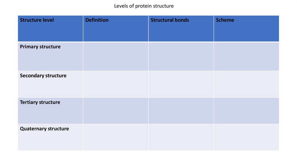
Levels of protein structureStructure level
Primary structure
Secondary structure
Tertiary structure
Quaternary structure
Definition
Structural bonds
Scheme
19.
Structurelevel
Definition
Structural bonds
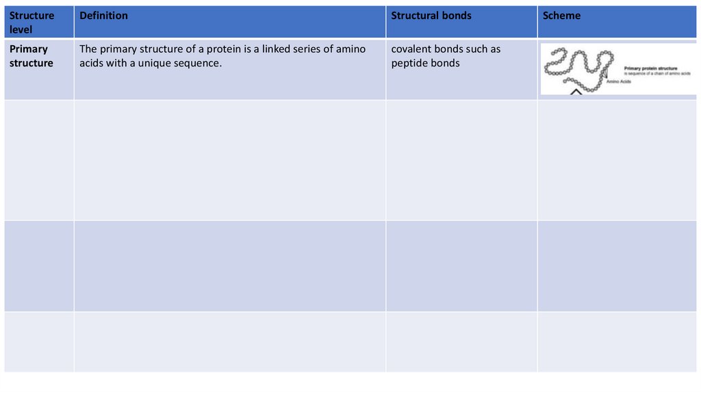
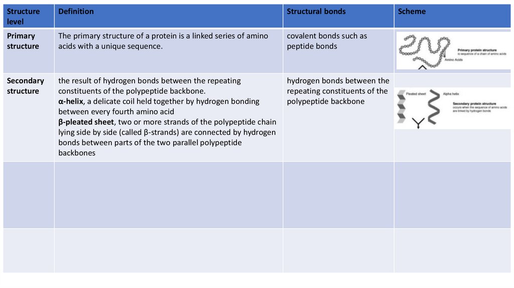
Primary
structure
The primary structure of a protein is a linked series of amino
acids with a unique sequence.
covalent bonds such as
peptide bonds
Scheme
20.
Structurelevel
Definition
Structural bonds
Primary
structure
The primary structure of a protein is a linked series of amino
acids with a unique sequence.
covalent bonds such as
peptide bonds
Secondary
structure
the result of hydrogen bonds between the repeating
constituents of the polypeptide backbone.
α-helix, a delicate coil held together by hydrogen bonding
between every fourth amino acid
β-pleated sheet, two or more strands of the polypeptide chain
lying side by side (called β-strands) are connected by hydrogen
bonds between parts of the two parallel polypeptide
backbones
hydrogen bonds between the
repeating constituents of the
polypeptide backbone
Scheme
21.
Structurelevel
Definition
Structural bonds
Primary
structure
The primary structure of a protein is a linked series of amino
acids with a unique sequence.
covalent bonds such as
peptide bonds
Secondary
structure
the result of hydrogen bonds between the repeating
constituents of the polypeptide backbone.
α-helix, a delicate coil held together by hydrogen bonding
between every fourth amino acid
β-pleated sheet, two or more strands of the polypeptide chain
lying side by side (called β-strands) are connected by hydrogen
bonds between parts of the two parallel polypeptide
backbones
hydrogen bonds between the
repeating constituents of the
polypeptide backbone
Tertiary
structure
Tertiary structure refers to the three-dimensional structure of a
single, double, or triple bonded protein molecule. tertiary
structure is the overall shape of a polypeptide resulting from
interactions between the side chains (R groups) of the various
amino acids.
the non-specific hydrophobic
interactions, such as salt
bridges, hydrogen bonds, and
the tight packing of side
chains and disulfide bonds.
Quaternary
structure
Scheme
22.
Structurelevel
Definition
Structural bonds
Primary
structure
The primary structure of a protein is a linked series of amino
acids with a unique sequence.
covalent bonds such as
peptide bonds
Secondary
structure
the result of hydrogen bonds between the repeating
constituents of the polypeptide backbone.
α-helix, a delicate coil held together by hydrogen bonding
between every fourth amino acid
β-pleated sheet, two or more strands of the polypeptide chain
lying side by side (called β-strands) are connected by hydrogen
bonds between parts of the two parallel polypeptide
backbones
hydrogen bonds between the
repeating constituents of the
polypeptide backbone
Tertiary
structure
Tertiary structure refers to the three-dimensional structure of a
single, double, or triple bonded protein molecule. tertiary
structure is the overall shape of a polypeptide resulting from
interactions between the side chains (R groups) of the various
amino acids.
the non-specific hydrophobic
interactions, such as salt
bridges, hydrogen bonds, and
the tight packing of side
chains and disulfide bonds.
Quaternary
structure
Quaternary structure is the arrangement of multiple folded
protein or coiling protein molecules in a multi-subunit
complex. The examples are collagen and hemoglobin.
The quaternary structure is
stabilized by the same noncovalent interactions and
disulfide bonds as the
tertiary structure.
Scheme
23.
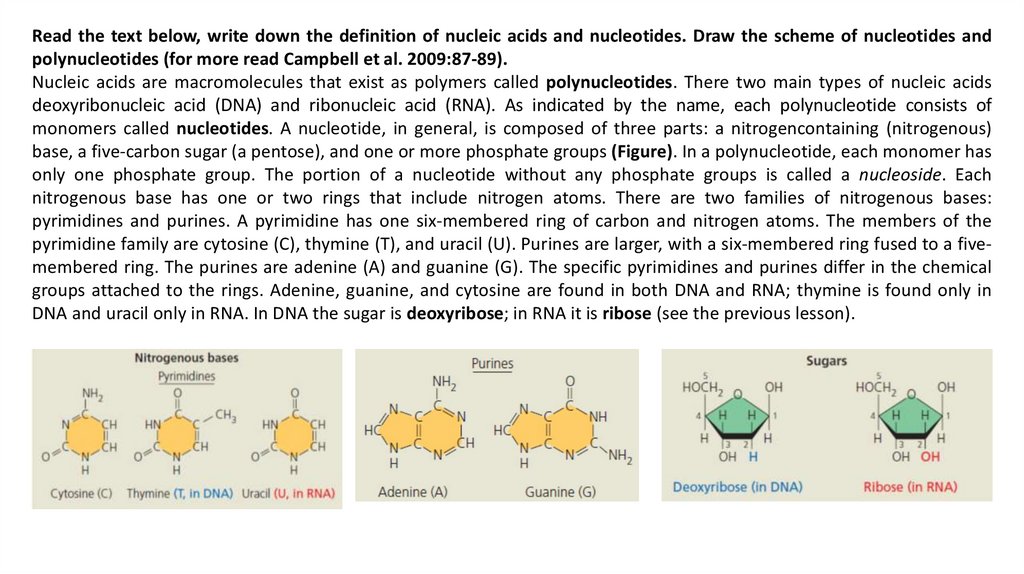
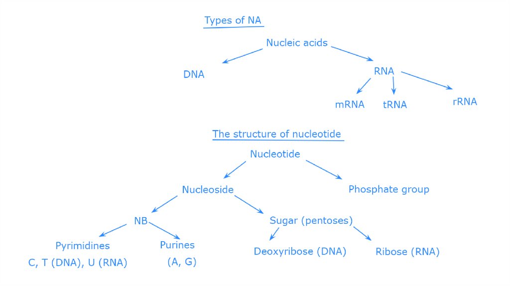
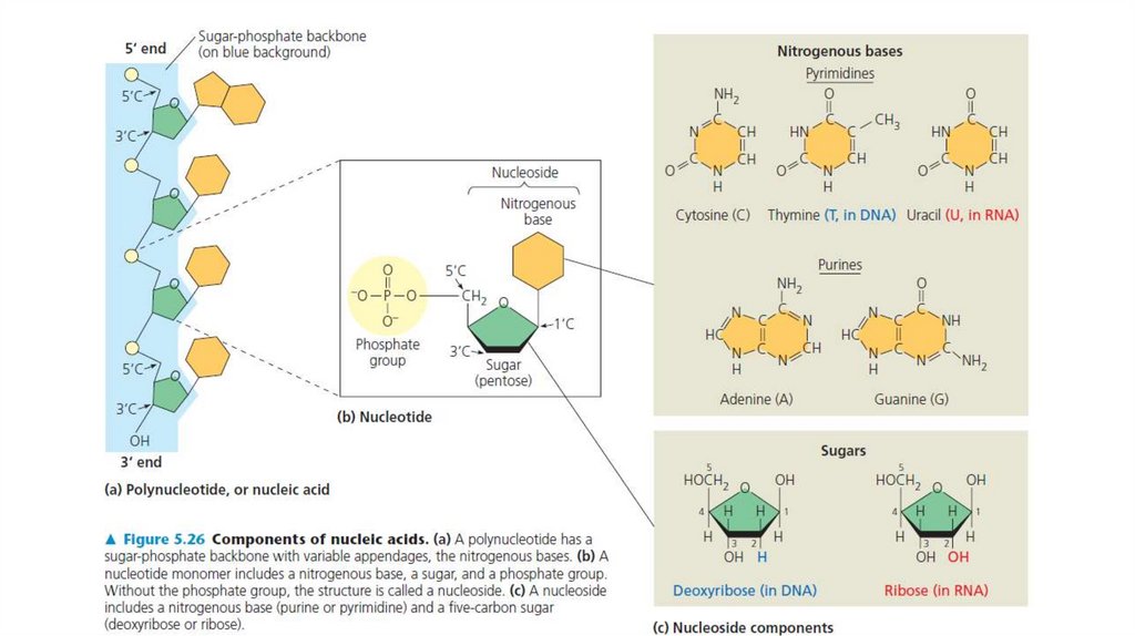
Read the text below, write down the definition of nucleic acids and nucleotides. Draw the scheme of nucleotides andpolynucleotides (for more read Campbell et al. 2009:87-89).
Nucleic acids are macromolecules that exist as polymers called polynucleotides. There two main types of nucleic acids
deoxyribonucleic acid (DNA) and ribonucleic acid (RNA). As indicated by the name, each polynucleotide consists of
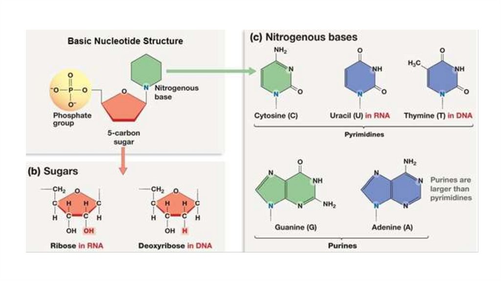
monomers called nucleotides. A nucleotide, in general, is composed of three parts: a nitrogencontaining (nitrogenous)
base, a five-carbon sugar (a pentose), and one or more phosphate groups (Figure). In a polynucleotide, each monomer has
only one phosphate group. The portion of a nucleotide without any phosphate groups is called a nucleoside. Each
nitrogenous base has one or two rings that include nitrogen atoms. There are two families of nitrogenous bases:
pyrimidines and purines. A pyrimidine has one six-membered ring of carbon and nitrogen atoms. The members of the
pyrimidine family are cytosine (C), thymine (T), and uracil (U). Purines are larger, with a six-membered ring fused to a fivemembered ring. The purines are adenine (A) and guanine (G). The specific pyrimidines and purines differ in the chemical
groups attached to the rings. Adenine, guanine, and cytosine are found in both DNA and RNA; thymine is found only in
DNA and uracil only in RNA. In DNA the sugar is deoxyribose; in RNA it is ribose (see the previous lesson).
24.
25.
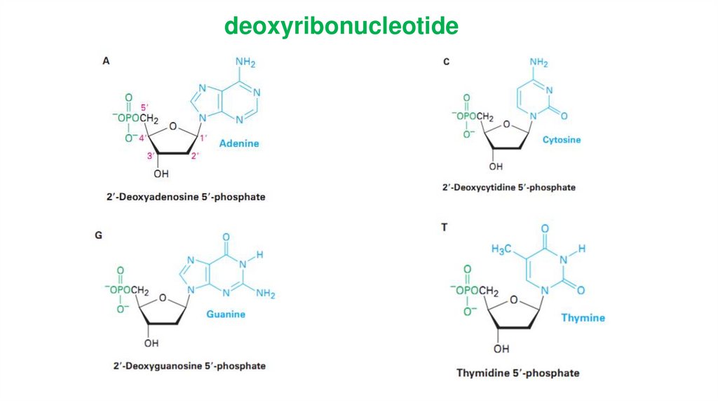
deoxyribonucleotide25
26.
Bases attached to a sugar is callednucleoside.
Sugar + phosphate + base =
nucleotide.
DNA only : Tymine, 2-deoxyribose
RNA only : Uracil, ribose
DNA and RNA : adenine, guanine,
cytosine
26
27.
28.
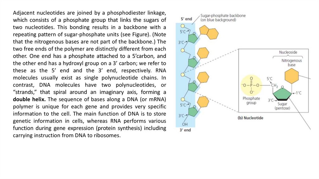
Adjacent nucleotides are joined by a phosphodiester linkage,which consists of a phosphate group that links the sugars of
two nucleotides. This bonding results in a backbone with a
repeating pattern of sugar-phosphate units (see Figure). (Note
that the nitrogenous bases are not part of the backbone.) The
two free ends of the polymer are distinctly different from each
other. One end has a phosphate attached to a 5’carbon, and
the other end has a hydroxyl group on a 3’ carbon; we refer to
these as the 5’ end and the 3’ end, respectively. RNA
molecules usually exist as single polynucleotide chains. In
contrast, DNA molecules have two polynucleotides, or
“strands,” that spiral around an imaginary axis, forming a
double helix. The sequence of bases along a DNA (or mRNA)
polymer is unique for each gene and provides very specific
information to the cell. The main function of DNA is to store
genetic information in cells, whereas RNA performs various
function during gene expression (protein synthesis) including
carrying instruction from DNA to ribosomes.
29.
According to the Watson-Crick model of aDNA molecule consists of two polynucleotide
chains forming a double helix with diameter of
1.8 - 2.0 nm. At each turn of the helix are ten
base pairs.
The sugar– phosphate backbone runs along
the outside of the helix, and the amine bases
hydrogen bond to one another on the inside. Both
major and minor grooves are visible.
Two polynucleotide strands are antiparallel to
each other, so direction of phosphodiester
formation is opposite: one chain is 5' - 3' end and
the other of 3' – 5' end.
.
DNA double helix fragment in space-filling
29
30.
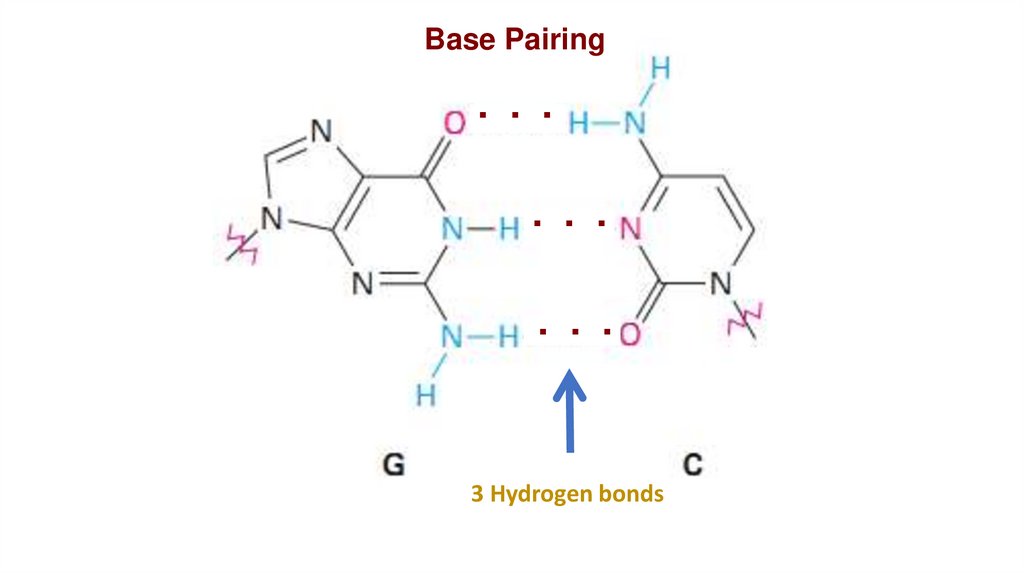
Base Pairing. . .
. . .
. . .
3 Hydrogen bonds
30
31.
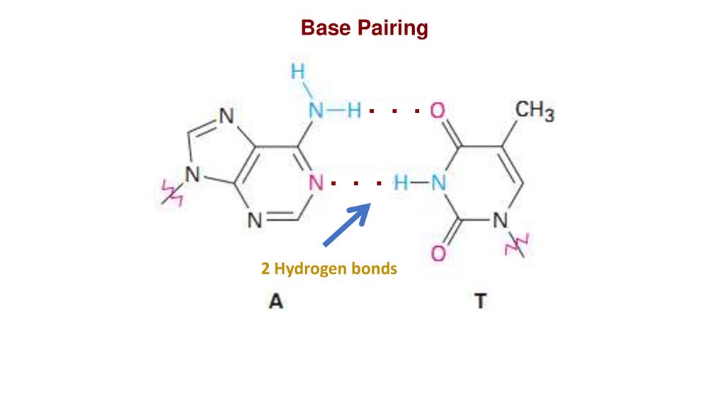
Base Pairing. . .
. . .
2 Hydrogen bonds
31
32.
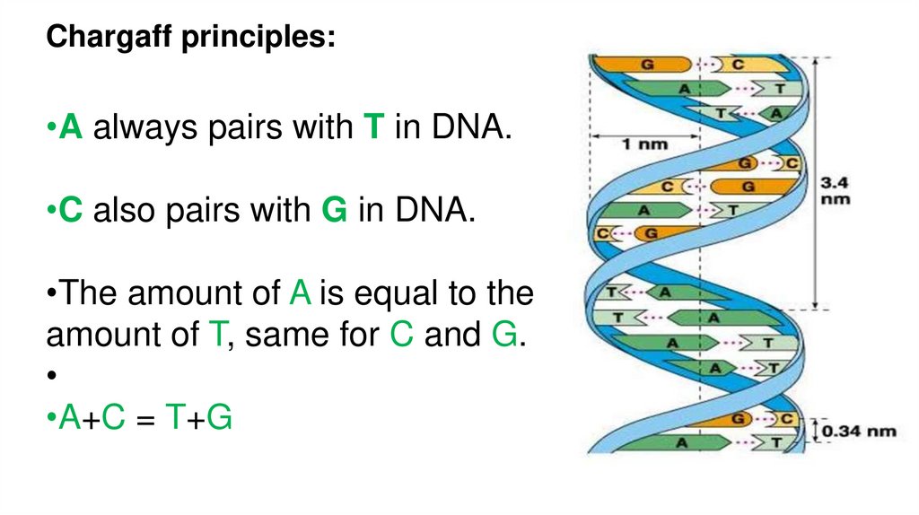
Chargaff principles:•A always pairs with T in DNA.
•C also pairs with G in DNA.
•The amount of A is equal to the
amount of T, same for C and G.
•A+C = T+G
32
33.
34.
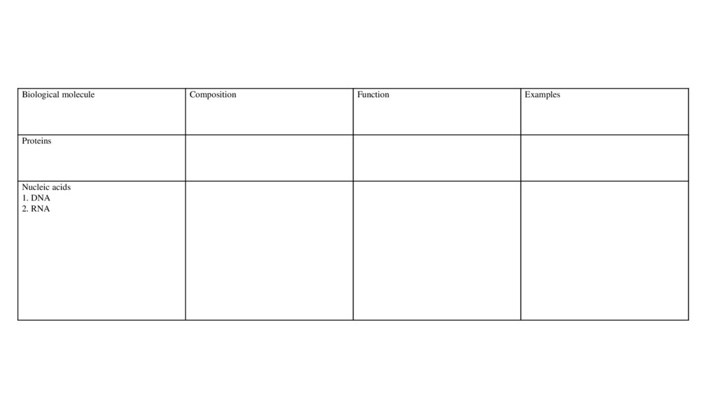
Biological moleculeProteins
Nucleic acids
1. DNA
2. RNA
Composition
Function
Examples