


 medicine
Similar presentations:


Syndrome of acute inflammation of mucous membranes of respiratory tracts. Tonsillitises
1.
SYNDROME OF ACUTEINFLAMMATION OF MUCOUS
MEMBRANES of
RESPIRATORY TRACTS
TONSILLITISES
2.
SYNDROME OF ACUTE INFLAMMATION OFMUCOUS MEMBRANES of RESPIRATORY
TRACTS
Among diseases developing with inflammation of upper
respiratory tracts (URT), acute respiratory diseases
(ARD) that is characterized by inflammation of mucous
membranes of respiratory tracts are most widespread.
On occasion term of ARD is concrete disease, but this
nosology form does not exist although. Undifferentiated
ARD is syndromal diagnosis and widespread in pediatric
practice.
In every case we must set etiology if it is possible, that it
is very important for adequate therapy.
Laboratory serologic researches allow to put a correct
diagnosis retrospectively.
3.
Term «acute respiratory viral infections» (ARVI) isa group of viral diseases without concrete
nosology.
ARD include not only viral but also mycoplasm
and bacterial diseases.
Consequently, the term «ARD the
undifferentiated» includes diagnosis
undeciphered etiology during special researches
or without it.
ARD are united by:
common way of transmission (respiratory),
primary damage of respiratory tracts.
4.
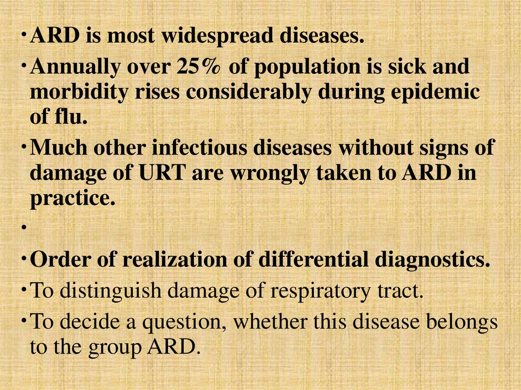
ARD is most widespread diseases.Annually over 25% of population is sick and
morbidity rises considerably during epidemic
of flu.
Much other infectious diseases without signs of
damage of URT are wrongly taken to ARD in
practice.
Order of realization of differential diagnostics.
To distinguish damage of respiratory tract.
To decide a question, whether this disease belongs
to the group ARD.
5.
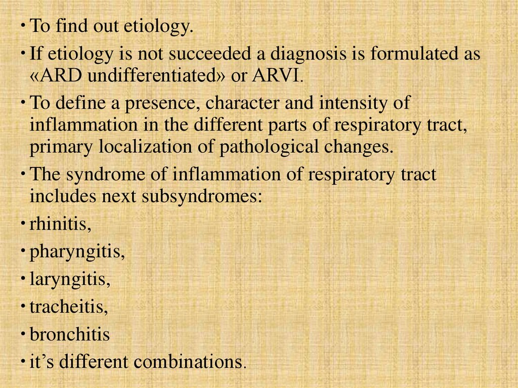
To find out etiology.If etiology is not succeeded a diagnosis is formulated as
«ARD undifferentiated» or ARVI.
To define a presence, character and intensity of
inflammation in the different parts of respiratory tract,
primary localization of pathological changes.
The syndrome of inflammation of respiratory tract
includes next subsyndromes:
rhinitis,
pharyngitis,
laryngitis,
tracheitis,
bronchitis
it’s different combinations.
6.
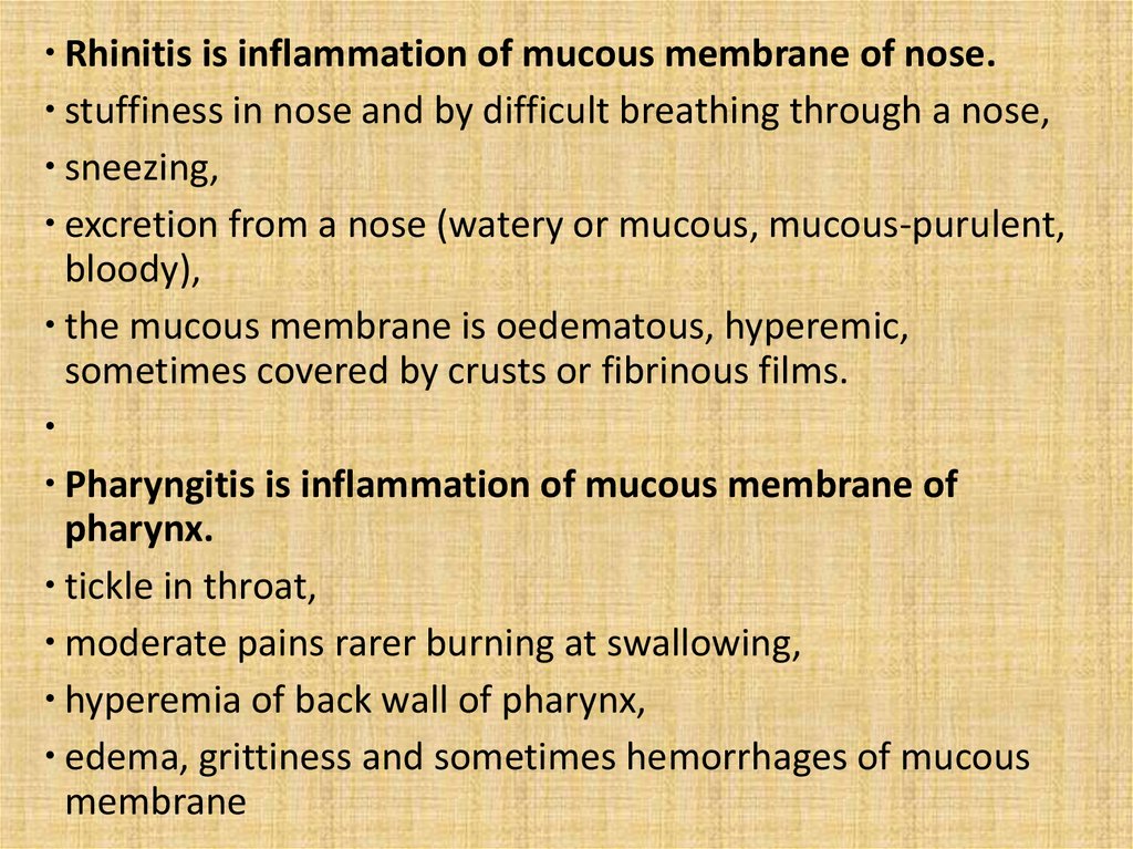
Rhinitis is inflammation of mucous membrane of nose.stuffiness in nose and by difficult breathing through a nose,
sneezing,
excretion from a nose (watery or mucous, mucous-purulent,
bloody),
the mucous membrane is oedematous, hyperemic,
sometimes covered by crusts or fibrinous films.
Pharyngitis is inflammation of mucous membrane of
pharynx.
tickle in throat,
moderate pains rarer burning at swallowing,
hyperemia of back wall of pharynx,
edema, grittiness and sometimes hemorrhages of mucous
membrane
7.
Character of exudate is mucous, mucouspurulent, purulent.good light is important.
White films are possible (diphtheria, burning of
mucous membrane, candidiasis).
Laryngitis is inflammation of mucous membrane
of larynx.
It is frequent component of ARD in combination
with inflammation of other parts of URT, rarely −
isolated.
It pesents at infectious diseases (flu,
parainfluenza, adenoviral diseases, measles,
whooping-cough and other)
8.
It appears under influence of somenoninfectious factors − overcooling,
chemical irritation and other
tickle in throat, cough.
Change of voice (hoarse or aphonia).
Sometimes − small pain at swallowing.
Special «barking» cough.
Laryngoscopy shows hyperemia and edema
of mucous membrane of larynx and vocal
cords.
Stenosing laryngitis can be false croup or
true croup (diphtheria of larynx).
9.
4 degrees of stenosis :I − short breathing difficulty, no respiratory
insufficiency;
II − attacks of breathing difficulty are frequent,
indrawing of areas of thorax (jugular fossa, supraand subclavicular space, epigastric area), noisy
breathing, moderate respiratory insufficiency.
III − II + cyanosys of lips, extremities, uneasiness
of patient, acutely expressed respiratory
insufficiency.
IV − asphyxia.
Stenosing laryngitis is observed mainly at
children.
10.
Chronic laryngitis at noninfectiouspathology − hoarseness and rapid
fatigueability of voice, tickle in throat,
dryness.
Tracheitis.
dyscomfort behind breastbone,
objectively − at fibrobronchoscopia.
Bronchitis and bronchiolitis.
11.
As a component of ARVI if it combines with the damageof URT.
For ARD an acute bronchitis is characteristic only.
Exacerbation of chronic bronchitis is not ARD.
At ARD a bronchitis combines with the manifestations of
inflammation of URT.
At some ARD (RS-virus) symptoms of bronchitis are on
the first plan.
Clinical manifestations of acute bronchitis are cough in
the beginning usually dry, mucous or mucopurulent
sputum, general intoxication; objectively: dry or moist
rales.
Disorders of bronchial passableness manifest by
lengthening of exhalation.
12.
A bronchiolitis is more severe form of acutebronchitis.
Bronchioles are involved in a process.
The signs of general intoxication and disorder of
bronchial passableness are more expressed:
breathlessness, development of obstructive
emphysema, respiratory insufficiency, painful
cough with a scanty sputum.
For differential diagnostics of syndrome of acute
inflammation of respiratory tracts it is very
important to educe carefully a prevalence of
inflammatory changes in one or another part.
13.
Next groups of diseases with inflammatorychanges of upper respiratory tracts are possible:
ARD;
inflammation of respiratory tracts as sign of other
infectious disease;
inflammation of respiratory tracts as a result of chemical
influence;
exacerbation of chronic inflammatory diseases of
respiratory tracts (chronic rhinitis, nasal asthma, chronic
laryngitis, chronic bronchitis of and other).
For an infectiologist first two groups of diseases have
main practical value.
14.
Acute respiratory diseases:adenoviral diseasees;
bacterial nasopharyngitis
(streptococcus, staphylococcus and
other);
herpetic respiratory diseasees;
flu;
coronaviral respiratory diseasees;
meningococcal nasopharyngitis;
15.
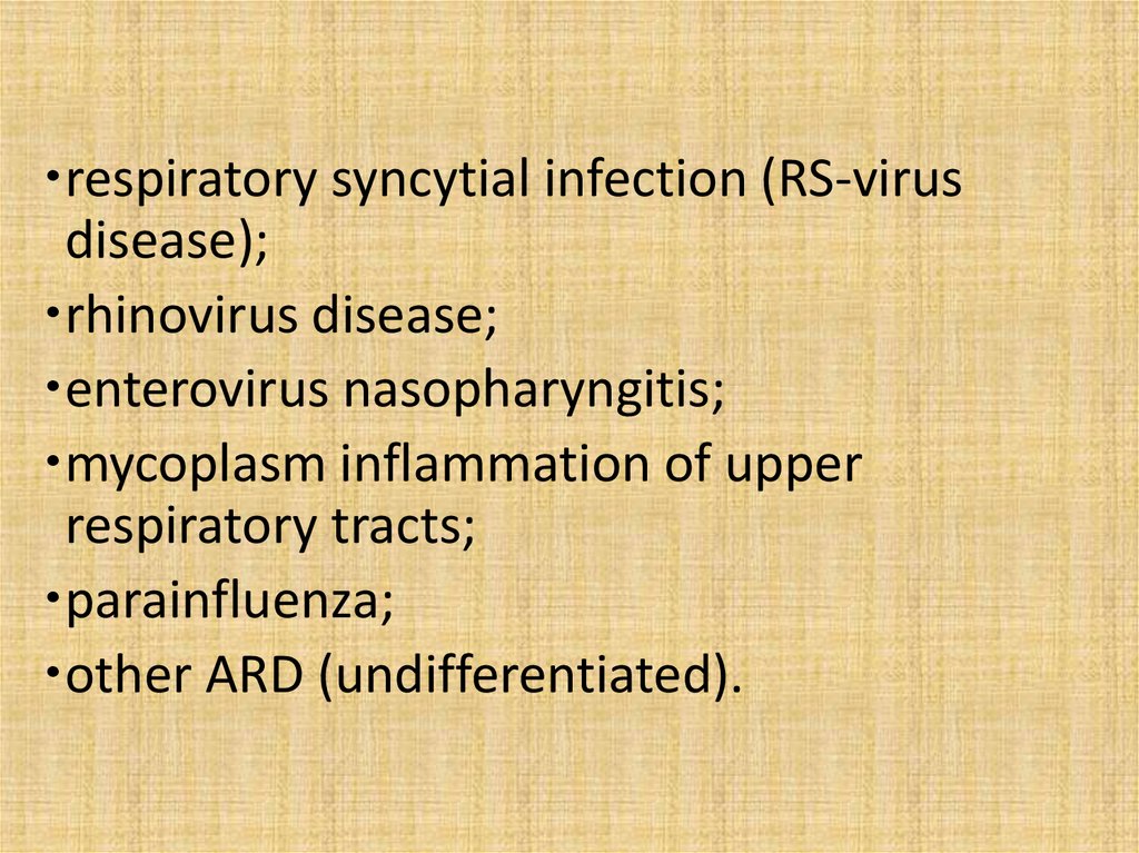
respiratory syncytial infection (RS-virusdisease);
rhinovirus disease;
enterovirus nasopharyngitis;
mycoplasm inflammation of upper
respiratory tracts;
parainfluenza;
other ARD (undifferentiated).
16.
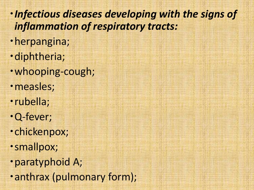
Infectious diseases developing with the signs ofinflammation of respiratory tracts:
herpangina;
diphtheria;
whooping-cough;
measles;
rubella;
Q-fever;
chickenpox;
smallpox;
paratyphoid A;
anthrax (pulmonary form);
17.
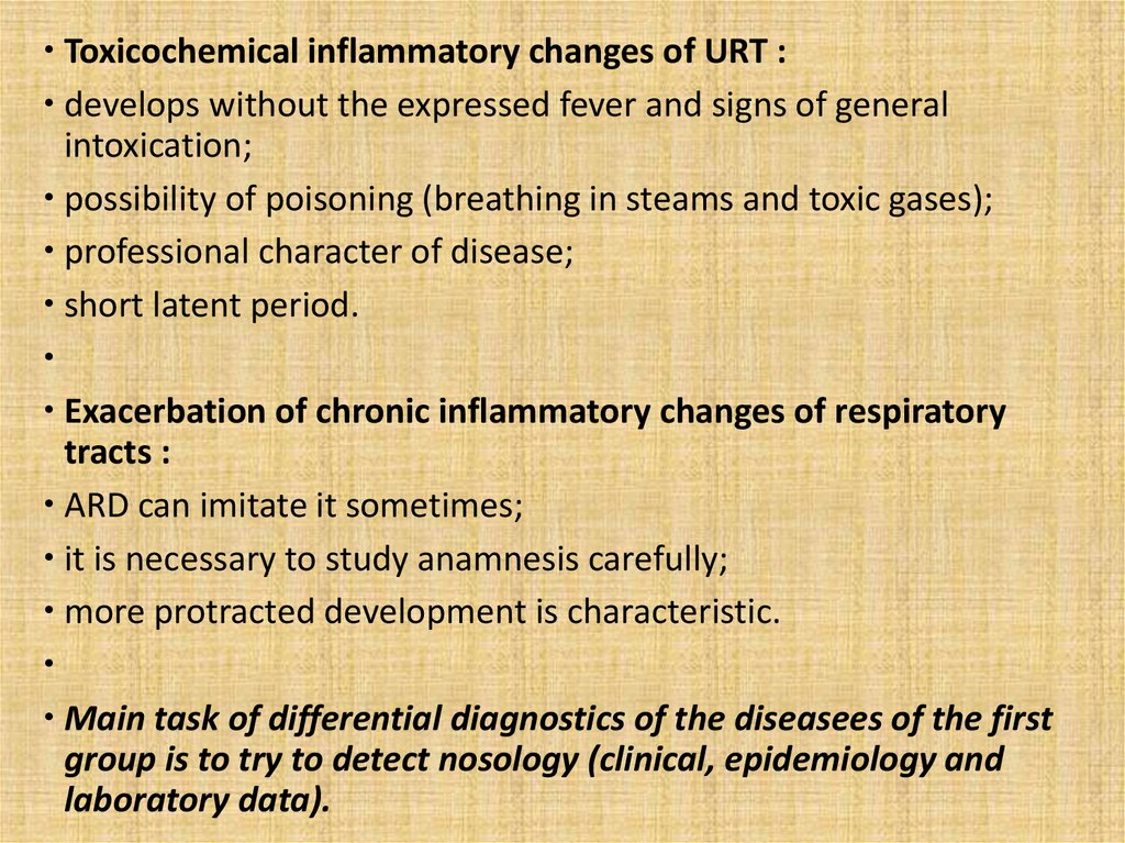
Toxicochemical inflammatory changes of URT :develops without the expressed fever and signs of general
intoxication;
possibility of poisoning (breathing in steams and toxic gases);
professional character of disease;
short latent period.
Exacerbation of chronic inflammatory changes of respiratory
tracts :
ARD can imitate it sometimes;
it is necessary to study anamnesis carefully;
more protracted development is characteristic.
Main task of differential diagnostics of the diseasees of the first
group is to try to detect nosology (clinical, epidemiology and
laboratory data).
18.
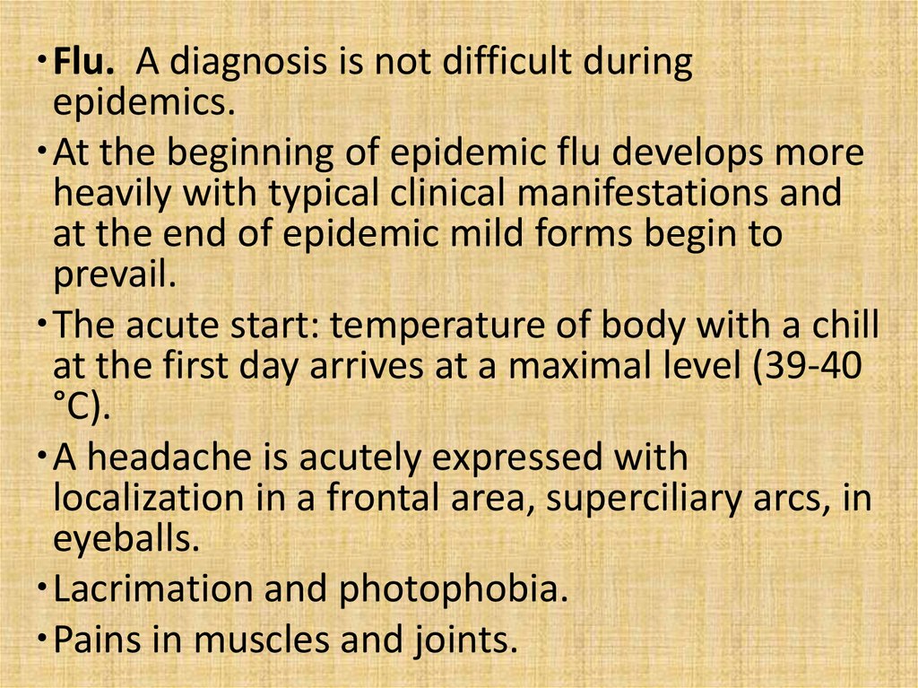
Flu. A diagnosis is not difficult duringepidemics.
At the beginning of epidemic flu develops more
heavily with typical clinical manifestations and
at the end of epidemic mild forms begin to
prevail.
The acute start: temperature of body with a chill
at the first day arrives at a maximal level (39-40
°С).
A headache is acutely expressed with
localization in a frontal area, superciliary arcs, in
eyeballs.
Lacrimation and photophobia.
Pains in muscles and joints.
19.
General intoxication is expressed(meningism, encephalopathy ).
Hypotension.
85% cases last 3-5 days.
The presence of the expressed tracheitis is
characteristic.
rhinitis, pharyngitis, laryngitis are possible.
Laboratory confirmation: PCR, IHT,
Immunofluorescence.
20.
ParainfluenzaUnlike a flu does not have such clear clinical
presentation.
There are not large epidemics.
Latent period 3-6 days.
20% of all ARD at adults and 30% at children
arecaused by viruses of parainfluenza.
Seasonality is cold period of year with an increase
at the end of winter and at the beginning of
spring.
It usually begins gradually, maximal expressed
clinical symptomatology arrives in 2-3 days.
21.
The temperature of body, as a rule, is subfebrile.The symptoms of general intoxication are
expressed poorly.
Rhinitis, pharyngitis and especially laryngitis are
typical. Inflammatory changes are most expressed
in a larynx at children (false croup).
A tracheobronchitis develops rarely.
Complication pneumonia arise up rarer than at
flu.
Conjunctivitis at 50% of patients.
Diagnostics: immunofluorescent method, PCR,
IHR.
22.
Adenoviral of diseaseFrequency is same as flu and parainfluenza.
Epidemic (up to 50% ARD) is possible.
Epidemic are marked during the first 3 months in
new formed collectives.
Latent period protracted (more frequently 5-7
days).
Seasonality is cold season.
Beginning of disease is usually acute, a
temperature rises to the feverish level (over
38°С).
23.
Duration of temperature is saved more protractedly (to10-20 days).
General condition is moderate, high fever, general
intoxication are expressed weaker than at flu.
Part of patients has a twowave temperature curve (even
in default of complications).
At adults disease usually develops in mild form.
Mainly rhinopharyngitis, sometimes is laryngitis (at
children) are typical.
The most frequent form is pharingoconjunctival.
Increase of peripheral lymphonoduses, conjunctivitis
(more frequent follicle).
Stomach-aches, diarrhea are possible.
24.
Pneumonias develop rarely.Diagnostics - immunofluorescent method, PCR, IHT.
Respiratory syncytial infection.
It meets mainly at children (30-70% bronchitis and
bronchiolitiss and 10-30% acute pneumonias).
At adults sporadic cases develop relatively easily but
are frequently (about 25%) complicated by
pneumonias at elderly.
A characteristic feature is a rapid damage of
bronchial tubes and bronchioles, other parts of URT
are damaged mildly.
25.
Development is more protracted than at otherARD.
Diagnostics - immunofluorescent method, PCR,
IHT.
Rhinovirus disease.
Absence or poorly expressed general intoxication
and expressed rhinitis with rhinorrhea are typical.
Complications from the side of ENT-organs only.
Diagnostics - immunofluorescent method, PCR,
IHR.
26.
Enterovirus (EVI) nasopharyngitis.Among the different manifestations of EVI it can develop
as rhinopharyngitis.
General intoxication and signs of inflammation of URT.
The inflammatory changes of mucous membranes of
respiratory tracts show up at most patients as rhinitis or
rhinopharyngitis.
20% − hyperemia of conjunctiva.
Rarely − laryngitis.
A tracheitis and bronchitis do not almost develop.
A peak of morbidity is on the end of summer and
beginning of autumn.
Along with the typical manifestations of ARD other
clinical forms are marked: epidemic myalgia, enterovirus
exanthema, herpangina and other
27.
These manifestations are original indicators of EVI. Sporadic casesis difficult to recognize clinically.
Diagnostics - immunofluorescent method, PCR, IHT, ELISA.
Herpetic respiratory diseases.
HSV-1 causes from 5 till 7% all ARD.
Children are ill mainly.
Signs of inflammation of URT are present at a primary infection; in
future virus is saved as a latent infection and at relapse skin
damages appear.
It develops as usual pharyngitis.
Intoxication is expressed poorly.
Diagnostics - immunofluorescent method, PCR, IHT, ELISA.
A herpetic rash does not have a substantial differential diagnostic
value.
28.
Mycoplasm inflammation of upper respiratorytracts.
М. рпеитоniе, experiments on volunteers show
possibility of exsudative pharyngitis
About 5% all ARD.
Epidemic are marked during the first 3 months in
new formed collectives (educational centers, recruits
of and other).
Clinically it have a lot in common with ARD of other
etiology, therefore differential diagnostics is difficult.
Some features – start is subacute or gradual, more
long development and most expressed toxicosis on
the 4-11th day of disease are typical.
29.
Bacterial ARD.Streptococci, staphylococci and other
cause isolated damages of some part of
URT.
Antibiotic therapy is very effective.
Severe generalised forms can develop
after meningococcal nasopharyngitis
and rhinitisatism, myocarditis, nephritis
and other - after streptococcal
pharyngitis.
30.
Streptococcal pharyngitis.Вeta-haemolytic streptococcustococcus group А is
characteristic for pharyngitis and other
streptococcus diseases (scarlatina, quinsies,
erysipelas).
In a acute period bright hyperemia of pharynx,
soft palate, amygdales presents. Mildly expressed
rhinitis, laryngitis, hemorrhages and oedema can
appear.
Antibiotic therapy already results by
normalization of temperature and reduction of
general intoxication during 24 hours and during
72 hours- by changes in pharynx. CBC − moderate
leucocytosis
31.
Meningococcal nasopharyngitis (rhinopharyngitis).Inflammation of mucous membrane of nose and
pharynx.
At some patients the signs of meningitis or
meningococcemia can appear in a few days.
It is very important to recognize disease and begin
therapy to avoid development of generalised forms in
good time (expresstests and microscopy).
It has a lot of common with viral nasopharyngitis.
General weakness, a moderate headache, stuffiness in a
nose, excretions from a nose (from the beginning
mucous-purulent, quickly become purulent, sometimes
with the admixture of blood) are typical.
Cough and moderate pains at swallowing can appear.
32.
The temperature is normal (40%) or subfebrile.Edema and hyperemia of mucous membrane of
epipharynx, mucous-purulent exudation.
CBC − small neutrophilic leucocytosis.
Penicillins quickly result in the improvement of
the general state.
Etiologic diagnosis is bacteriologicexamination of
mucus from epipharynx before use of antibiotics.
It is necessary to take into account epidemiology
data (cases in a collective).
There are no laryngitises, tracheitiss, bronchitis,
but specific meningococcal pneumonias are
possible.
33.
INFLAMMATION of MUCOUS MEMBRANES ofRESPIRATORY TRACTS AT the DISEASEES not INCLUDED
In GROUP of ACUTE RESPIRATORY DISEASES – one of
other manifestations of disease.
Diphtheria of nose.
It is manifested by ichor excretions from nose with
maceration of skin around and upper lip.
Hyperemia, bulge of mucous membrane, fibrinous tapes
with bleeding after removing of it.
Usually it combine with diphtheria of
pharynx(widespread diphtheria).
A diagnosis must be necessarily confirmed by
bacteriologicexamination.
34.
Whooping-cough and parapertussis.These diseases are differentiated only
bacteriologically.
Initial period – catarrhal (rhinitis and
laryngotracheitis).
Common state is satisfactory, temperature is usually
normal or subfebrile.
Rhinitis and cough in the beginning are small,
increase later, typical fits of the convulsive coughing
appear only at the end of 2th week.
Later − signs of bronchitis.
For diagnostics epidemiology data are important
(absence of vaccination, contact with a patient).
Specific diagnostics − bacteriologicexamination or
serologic.
35.
Measles.Before appearance of characteristic
exanthema it is frequently interpreted as
ARD.
Fever, pharyngitis.
Expressed conjunctivitis with bright
hyperemia.
Filatov-Koplik spots on the mucous
membrane of mouth.
Typical morbillous exanthema appears on
the 4th day.
36.
Rubella.Signs of acute rhinopharyngitis, small hyperemia
of conjunctiva, subfebrile temperature of body
are possible at atypical rubella (without an
exanthema).
Moderate increase of neck lymphatic nodes
(similar at a parainfluenza and adenoviral
disease).
Growth of titles of antibodies in 4 times and more
confirms rubella.
Atypical rubella takes 25-30% of all cases.
It is especially important at pregnancy.
Diagnostics - IHT, ELISA.
37.
Herpangina.Pharyngitis, intoxication.
Enanthema: hyperemia − small red
spots (2-3 mm) - vesiculas (4-5 mm) aphthae (to 5-7 mm).
Total about 20 elements of enanthema
develop one week and disappear
without trace.
Diagnostics bases on only clinical data.
38.
Q-Fever.Bronchial tubes are damaged mainly, but
moderate inflammation of upper respiratory
tracts as pharyngitis and laryngitis with the
expressed intoxication are marked.
Hyperemia of face, neck, injection of vessels of
sclera like at flu.
Seasonality is summer-autum while ARD meet
rarely.
Increase of liver and spleen appearss fom 3-4th
day (not characteristically for the flu and other
ARD).
Diagnostics − CFR with an antigen of Rickettsia
Burnetti.
39.
Chicken-pox.Pharyngitis, laryngitis, sometimes specific viral
pneumonia can develop.
A diagnosis is not difficult due to typical
polymorphic vesicular rash.
Paratyphoid А.
There are rhinopharyngitis, tracheobronchitis,
conjunctivitis at initial period.
It develops more gradually with growth of fever.
Intoxication fall short of to the inflammatory
changes of respiratory tracts.
40.
Diagnostics is bacteriological andserologic.
Anthrax (pulmonary form).
Acutely expressed rhinitis, pharyngitis,
laryngotracheitis, bronchitis and
conjunctivitis.
Symptoms of hemorragic pneumonia,
quickly developing shock.
Diagnostics is bacteriologic,
bacterioscopic and serologic.
41.
TONSILLITISES (inflammation of palatal tonsils).Tonsillitis can be manifestation of quinsy or one of signs
of other infectious disease.
Quinsy (acute bscterial tonsillitis) − inflammation of
palatal tonsils and regional lymphatic nodes is basic
manifestation of disease.
Tonsillitis is inflammation of tonsils at other infectious
and noninfectious diseases as one of signs of these
diseasees.
Chronic tonsillitis − is the chronic inflammation of
palatal tonsils and regional lymphatic nodes, quite
frequent with development of manifestations of chronic
general intoxication and specific complications.
42.
For diagnostic of tonsillitis it is necessary toobserve certain rules:
Good illumination at examination for correct
perception of color.
Medical careful examination of fauces (by
means of two spatulas).
Sizes, color, relief and state of palatal
handles, soft palate, tongue, back wall of
pharynx are estimated.
Presence of mucus, pus, fibrinous films is
marked
43.
Quinsy.It can be caused by haemolytic
streptococcus (85%), staphylococcus aureus
(10%), fusobacteria (necrotic Vincent's
quinsy).
Clinically the expressed intoxication
corresponding to the inflammatory changes
of palatal tonsils, pain at swallowing
present.
Diagnostics is bacteriological.
44.
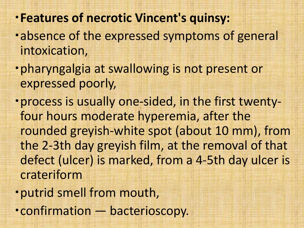
Features of necrotic Vincent's quinsy:absence of the expressed symptoms of general
intoxication,
pharyngalgia at swallowing is not present or
expressed poorly,
process is usually one-sided, in the first twentyfour hours moderate hyperemia, after the
rounded greyish-white spot (about 10 mm), from
the 2-3th day greyish film, at the removal of that
defect (ulcer) is marked, from a 4-5th day ulcer is
crateriform
putrid smell from mouth,
confirmation — bacterioscopy.
45.
The group of symptomatic tonsillitisesincludes a number of diseasees of
infectious and noninfectious nature :
Infectious:
infectious
adenoviral diseases; mononucleosis;
anginal-bubonic form candidiasis of fauces;
of rabbit-fever;
parainfluenza, flu and
anginal-septic form other ARD;
of listeriosis;
syphilis;
diphtheria of
scarlatina;
pharynx;
paratyphoid.
46.
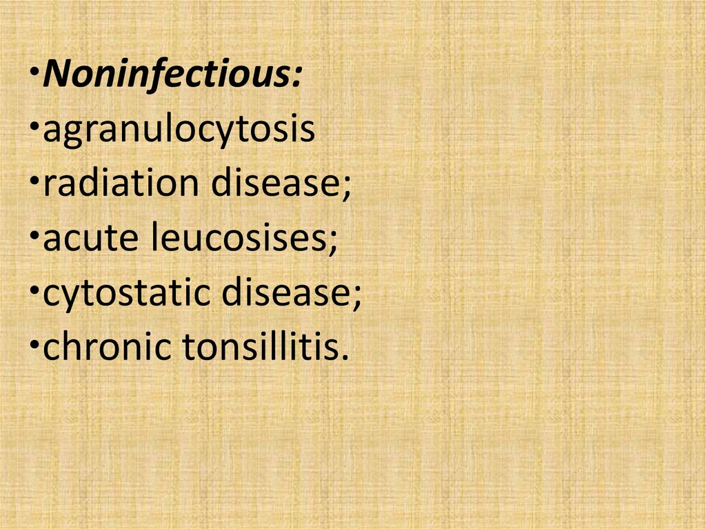
Noninfectious:agranulocytosis
radiation disease;
acute leucosises;
cytostatic disease;
chronic tonsillitis.
47.
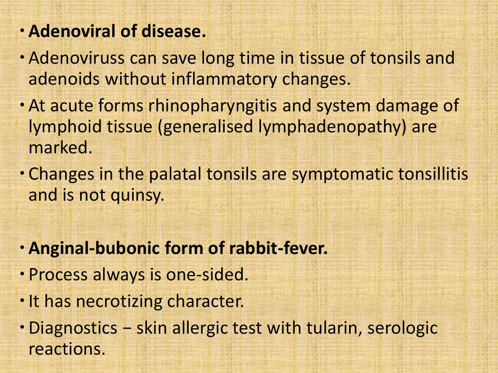
Adenoviral of disease.Adenoviruss can save long time in tissue of tonsils and
adenoids without inflammatory changes.
At acute forms rhinopharyngitis and system damage of
lymphoid tissue (generalised lymphadenopathy) are
marked.
Changes in the palatal tonsils are symptomatic tonsillitis
and is not quinsy.
Anginal-bubonic form of rabbit-fever.
Process always is one-sided.
It has necrotizing character.
Diagnostics − skin allergic test with tularin, serologic
reactions.
48.
Film on the tonsil has greyish color and deep ulcerappears after sloughing.
After repairing of ulcer scars on an tonsil appear.
Unlike necrotic Vincent's quinsy high fever and
intoxication are typical.
A characteristic feature is formation of bubo (upper and
anterior neck)
A bubo is not soldered with surrounding tissues,
movable, a skin above it is not changed, fluctuation and
fistula usually appear at the end of 3th week from the
beginning of illness.
A feverish period proceeds 10-15 days, but cicatrization
of ulcer on an tonsil and reverse development of bubo
take place considerably slower.
49.
Anginal-septic form of listeriosis.Tonsillitis develops on a background a
severe general (septic) disease with the
varied clinical manifestations.
Quite frequent rash (erythema with the
figure of «butterfly» on face).
Generalised lymphadenopathy, constantly −
hepatosplenomegaly, and at some cases
purulent meningitis.
Clinical diagnosis is difficult.
50.
Diagnostics − bacteriologic (from blood, neurolymph,pharynx), serologic (CFR and IHR).
A listerious allergen is produced for skin test − passing of
negative to positive.
Diphtheria of pharynx.
Damage of palatal tonsils is obligatory.
Differential diagnostics is especially laboured at
development of streptococcus quinsy at the bacillicarrier
of toxigenic C.diphtheriae.
The catarrhal form of diphtheria of pharynx is
differentiated with a catarrhal quinsy; island-like − with
lacunar, membranous − with a necrotizing quinsy, and
toxic – with quinsy complicated by abscess.
Diagnostics − bacteriologic examination (toxigenic
C.diphtheriae), serologic, bacterioscopy.
51.
Catarrhal form of diphtheria of pharynx.Atypical form, without fibrinous films on tonsils.
The temperature of body is normal or
subfebrile.
A pharyngalgia at swallowing is absent or
expressed poorly.
The mucous membrane of tonsils is mildly
hyperemic and on the 2-3th day small edema
and cyanochroic colouring is possible.
It can pass to membranous in future.
Specific characteristic complications are
possible.
52.
Island-like form of diphtheria of pharynx.The small areas of fibrinousого film (small
«islands») on tonsils are easily taken off by a
spatula, thin.
Lacunar quinsy develops with a high fever,
expressed symptoms of general intoxication,
great pains in a throat.
Island-like a form develops with subfebrileой
temperature of body, pains at swallowing are
absent or expressed poorly.
Without specific treatment illness passes to the
membranous form.
53.
Membranous diphtheria of pharynx.Expressed continuous fibrinous films keeping
indoors outside tonsils.
A film is taken off hardly, in place of it bleeding
surface appears. The taken off film sinks in water.
Intoxication is expressed and does not correlate
with the temperature of body.
High probability of specific complications
characteristic for diphtheria.
Widespread diphtheria of pharynx.
fibrinous film goes across and on the surrounding
areas of mucous membrane − back wall of
pharynx, soft palate, uvula.
54.
Toxic diphtheria is characterized byappearance of edema of neck cellular tissue.
At toxic diphtheria false diagnose of quinsy
complicated by abscess is possible.
The expressed pharyngalgia is not present
(diphtherin has anaesthetic action).
Grey-brown films can cover both palatal
tonsils and surrounding tissues.
Edema on a neck, high fever, expressed
symptoms of intoxication are typical.
55.
Appearance of paratonsillar abscess is characterizedby a rapid fervescence, chill expressed by a toxicosis.
Trismus of masticatory musculature limits opening of
mouth.
One-sided edema and infiltration are marked in
default of films on the mucous membrane of tonsils.
The diagnosis of diphtheria is confirmed by the
selection of causative agent.
Infectious mononucleosis.
Almost always develops with the damage of pharynx.
Acute beginning, high fever, intoxication.
56.
Appearance of tonsillitis is possible after ferver,general intoxication and other signs of
mononucleosis.
It can imitate lacunar quinsy, necrotizing changes
of tonsils and even fibrinous films.
A fever and toxicosis are saved protractedly, and
changes in a pharynx do not grow and keep
indoors outside tonsils.
Generalised lymphadenopathy appears early
(from the first days of illness).
Hepatosplenomegaly appears at the 3-5th day, a
spleen ise considerably increased and painful at
palpation.
57.
Acute hepatitis can develop.CBC − leucocytosis, neutropenia,
lymphomonocytosis, atypical mononuclears.
The changes of blood appear early and
saved long time (sometimes few months).
Diagnostics is an serologic, PCR.
58.
Candidiasis of fauces.Arises up as a result of dysbacteriosis
and immunodeficit.
The temperature of body remains
normal, rarer subfebrile.
Film related to the mycosis, greyishwhite color, usually by separate areas,
not only at tonsils but also mucous
membrane of cavity of mouth, back
walls of pharynx.
59.
Inflammation of tonsils is not actual it is nottonsillitis but widespread candida damage of
mucous membrane of fauces.
Diagnostics is mycologic research.
Flu, parainfluenza and other ARD – cataral
tonsillitis is included in clinic of inflammation of
respiratory tracts.
Syphilis.
At the primary syphilis original primary
syphiloma at gate of infection.
Increase of one tonsils, mucous membrane is
hyperemic.
60.
A regional lymphatic node is mildlyincreased, dense consistency, painless.
Pains at swallowing is not marked.
General condition is normal.
Small erosion on tonsils (5-10 mm is in a
diameter) with clear dense edges and clean
bottom.
Abundant exanthema and/or enanthema
present at second stage of disease.
A diagnosis is confirmed by specific
laboratory methods.
61.
ScarlatinaLike quinsy it is caused by haemolytic
streptococcustococcus group A.
Along with a clinic signs of quinsy, influence of
erythrogenic toxin manifests by skin hyperemia,
rash with, tachycardia.
Unusually bright hyperemia of mucous
membrane is a «blazing pharynx».
Clean hyperemic language with increased papillae
(«raspberry language»).
Laboratory confirmation of diagnosis is absent.
62.
Typhoid fever.Palatal tonsils and archs are edematous, small ulcers
covered by a greyish films are possible.
Other manifestations of main disease have
prevalence.
Diagnostics is bacteriological, serologic.
Noninfectious illnesses.
Tonsillitis presents at haematological diseases
(different forms of leucosises), neutropenia (acute
radiation illness, immune agranulocytosis and
other), changes of tonsils have necrotizing
character.
63.
Chronic tonsillitis.Character of development is decompensated and
subcompensated.
Periodic exacerbation and remissions are typical.
Exacerbation is provoked by supercooling or ARD.
In remission signs of chronic toxicosis (general weakness,
fatigueability, irritability, subfebrile fever), vasoneurosis
(lability of pulse, orthostatic hypotension, dyscomfort in
heart area) present.
Chemical burns can also cause the inflammatory
changes of mucous membrane of tonsils, sometimes
with formation of films and do not cause diagnostic
difficulties.