


 medicine
Similar presentations:


Lasik
1.
LASIK2.
IndicationsStable refraction
(no change over a period of 2 years)
Age ≥21 years
Adequate central corneal thickness
Myopia ≤–10.00D
Hyperopia ≤+4.00 to 5.00D
Astigmatism ≤6D
LASIK
Pallikaris in 1990
The excimer laser is used to ablate the corneal stromal
tissue to achieve the desired refractive change
Surgical procedure
The patient lies on a couch with the excimer laser
delivery system above the patient’s head.
The cornea of the eye to be operated is anaesthetized
with topical anaesthetic drops.
Absolute contraindications
A lid speculum is inserted after instilling topical
Keratoconus
anaesthesia to prevent blinking during the treatment.
Central corneal thickness <410μm
The patient is asked to fixate on the laser bream and
Unstable refraction
the cornea is marked with gentian violet to
Deep corneal dystrophy
help realign the flap.
Previous corneal melt (or systemic
A suction ring is applied to the limbus and the
conditions predisposing to corneal melt) pressure increased to more than 65mmHg to ensure a
History of herpetic keratitis
regular cut. transient loss of vision may occur
Amblyopia
An automated microkeratome is fitted on the track
and activated to pass across the cornea to create
Relative contraindications
stromal flap.
Cataract
The vacuum is released and the epithelial flap is
Selected occupations (e.g., commercial reflected back to expose the stromal bed.
pilots)
The hinge of the flap is made, either nasally or
Obsessive personality
at the 12 o’clock position.
3.
excimer laser ablation is carried out on the corneal stroma. usually takes less than 90 seconds.The patients are warned that they might experience a pungent smell during laser ablation.
The flap is washed with balanced salt solution and replaced.
Centration is checked and the edges are smoothed down. The endothelial pump mechanism
keeps it in place. After checking the adhesion, the speculum is removed.
Topical antibioticsand topical corticosteroid are prescribed for 1 week.
Post-operative care
The patient is directed to avoid swimming, dust or smoke and any contact sport for about 1
month after the surgery.
A clear eye shield is worn during sleep for 2 week to avoid trauma while sleeping. s
The patient is examined after 1 day, 1 week, 1 month, 3 months, 6 months and 1 year.
Complications
Non-refractive
Flap related complications
The intra-operative flap complications include incomplete or free (completely cut) flap, lost flap,
decentred flap, irregular flap and flap stria.
Late complications are epithelial ingrowth (epithelium within the stromal interface, one of the
most common cause of reduced visual acuity wrinkles or striae, interface infection and flap
dislocations. Central islands Interface debris
Infectious keratitis (rare) Diffuse interstitial keratitis (sands ofSahara) Endothelial cell loss
Haze Glare and haloes Night-vision problems Reduction in corneal sensitivity Dry eye
Posterior ectasia
Refractive complications
under- or overcorrection, regression, decentred ablation and induced irregular astigmatism
caused by folds or microstriae of flaps
4.
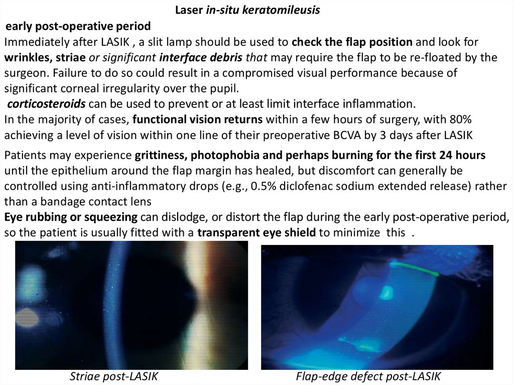
Laser in-situ keratomileusisearly post-operative period
Immediately after LASIK , a slit lamp should be used to check the flap position and look for
wrinkles, striae or significant interface debris that may require the flap to be re-floated by the
surgeon. Failure to do so could result in a compromised visual performance because of
significant corneal irregularity over the pupil.
corticosteroids can be used to prevent or at least limit interface inflammation.
In the majority of cases, functional vision returns within a few hours of surgery, with 80%
achieving a level of vision within one line of their preoperative BCVA by 3 days after LASIK
Patients may experience grittiness, photophobia and perhaps burning for the first 24 hours
until the epithelium around the flap margin has healed, but discomfort can generally be
controlled using anti-inflammatory drops (e.g., 0.5% diclofenac sodium extended release) rather
than a bandage contact lens
Eye rubbing or squeezing can dislodge, or distort the flap during the early post-operative period,
so the patient is usually fitted with a transparent eye shield to minimize this .
Striae post-LASIK
Flap-edge defect post-LASIK
5.
At 1 weekThe epithelium should fully cover the flap margin by 1 week post-LASIK.
Epithelial defects should be monitored carefully, since they increase the risk of epithelial
ingrowth and diffuse lamellar keratitis (DLK).
Interface debris should also be watched as it may lead to focal infiltrates that require flap refloatation.
At 1 month
By this stage, the vision tends to be very good.
some patients start to notice visual problems such as reduced-quality night vision and haloes
around lights.
The refractive error may have stabilized in those treated for lower degrees of myopia (<6.00DS),
although regression of approximately 15% of the pre-operative error is not uncommon20 (e.g., –
0.25D after LASIK for –1.50D, and –1.50D after LASIK for –10.00D), and is associated with an
increase in corneal thickness and central corneal steepening.
If epithelial ingrowth is going to develop it tends to do so within the first month. The clinician
must then decide whether the location and extent of the ingrowth warrant intervention.
At 3 months
After LASIK, healing is limited to the region around the lamellar interface and haze occurs
around the flap margin only.
Most LASIK patients demonstrate a stable refractive error by 3 months, with the exception of
those treated for very high myopia. The possibility of an enhancement procedure can be
discussed if the refractive outcome is poor,( error is greater than 1.00DS.) usually performed
between 3 and 6 months after the first procedure , both the refractionand corneal topography
must be stableand there must be sufficient residual corneal thickness. cycloplegic refraction
is essential to minimize the risk of overcorrection.
6.
At 6 months and beyond90–100% of eyes achieve±1.00D of emmetropia at 6 months post-LASIK for corrections of
–8.00D or less. Hyperopic LASIK has proved slightly more successful than hyperopic PRK, but the
stabilization rate is approximately four times longer than for myopic treatments.
Complications specific to LASIK
Non-refractive
Flap related complications
The intra-operative flap complications include incomplete or free (completely cut) flap, lost flap,
decentred flap, irregular flap and flap stria.
Late complications are epithelial ingrowth (epithelium within the stromal interface, one of the
most common cause of reduced visual acuity wrinkles or striae, interface infection and flap
dislocations. Central islands Interface debris
Infectious keratitis (rare) Diffuse interstitial keratitis (sands ofSahara) Endothelial cell loss
Haze Glare and haloes Night-vision problems Reduction in corneal sensitivity Dry eye
Posterior ectasia
Refractive complications
under- or overcorrection, regression, decentred ablation and induced irregular astigmatism
caused by folds or microstriae of flaps
The vast majority of complications manifest themselves within 6–8 weeks of LASIK surgery.
Most can be treated and have a minimal effect on the final outcome after surgery, if managed
properly. Serious adverse complications that lead to a significant permanent visual loss, such as
infections and corneal ectasia, are very rare, but side effects such as dry eyes, night-time
starbursts and reduced contrast sensitivity are relatively common for the first few months.
7.
Epithelial ingrowthEpithelial ingrowth occurs when nests of epithelial cells trapped beneath the flap begin to
proliferate Ingrowth presents as a milky deposit in the interface and is more common after
enhancement than after the initial procedure.
The extent should be measured since growth less than 1.0mm from the flap margin is
acceptable, as it is usually self-limiting.
Ingrowth greater than 1.0mm, invading the visual axis or progressing rapidly requires surgical
management, particularly if the flap margin is rolled or eroded, as it can lead to significant
irregularity and flap melt. Although a small degree of ingrowth is common (approximately 15%
of eyes), few cases require management. Untreated ingrowth can lead to corneal irregularity
and glare, and very occasionally to corneal melt.
Nests of proliferating epithelial cells
trapped beneath the flap can result
in epithelial ingrowth.
Ingrowth often presents as a milky
deposit in the interface.
8.
MicrostriaeFine grey lines that are related to crinkles in Bowman’s membrane are not uncommon in those
treated for moderate or high myopia, as the flap does not fit the remodelled stromal bed. Such
cases are difficult to manage and are usually left alone unless vision is compromised
Interface debris
Some debris is seen in virtually all eyes post-LASIK. Sources include dust from the atmosphere,
meibomian secretions(Figure5.5), metallic deposits and oils from the microkeratome blade and
fibres (Figure 5.6).
Debris is usually inert and causes no problems, but it can be associated with stromal infiltrates
or DLK, in which case it requires treatment with topical corticosteroids.
.
Post-LASIK interference debris.
Fibres trapped in the interface post-LASIK.
9.
Diffuse lamellar keratitis (Sands of the Sahara)DLK is a sterile, diffuse inflammation at the level of the interface that may be accompanied
by anterior chamber activity
It looks a little like post-PRK haze, but is very obviously confined to the interface.
It is thought to be an immune response to interface debris or perhaps bacterial toxins.
The onset tends to occur within a day or two of the LASIK procedure, with symptoms such as
pain and photophobia, and additional signs of ciliary hyperaemia and lacrimation.
Visual quality may be reduced because of the increase in forward light scatter, although Snellen
acuity is unaffected generally.
Referral back to the operating surgeon is required for treatment with topical corticosteroids
such as fluorometholone, antibiotics and cycloplegics. The flap may be lifted and irrigated in
some cases.
10.
A number of systems are used to grade DLK, including one that divides cases into one of fourcategories
Cases with central involvement (type II) took significantly longer (12.1 days) to resolve than
cases with central sparing(type I – 3.5 days). Not surprisingly, central involvement carries a much
higher risk of a reduction in BCVA. The majority of cases were sporadic rather than part of
a cluster.
DLK can also present many months after LASIK in association with an epithelial defect.
White blood cells migrate from the limbal blood vessels into the interface, since it is the easiest
path for them to take.
Central corneal sparing is much more likely if the DLK is related to an epithelial defect.
There is also a reported case of DLK that occurred 10 months post-LASIK in association with
acute iritis, 40 which suggests that DLK is a nonspecific corneal inflammatory response rather
than a condition caused by a particular agent. Appropriate management of patients with DLK
generally results in complete resolution of the condition.
11.
Corneal integrityhealing does not appear to lead to the growth of collagen fibres between the corneal flap
and the ablated stromal bed. The flap is attached to the underlying cornea only at its margins,
by the corneal epithelium, and therefore does not contribute significantly to the strength of the
cornea. This can be attributed to the endothelial pump and the multiple layers of corneal
epithelium that cover the flap margin.
flap dislocation can occur at any time if the trauma is discrete and from such an angle that it
catches the edge of the flap. Patient’s who report with flap dislocation should be referred
urgently to the operating surgeon for irrigation and refloating of the flap, followed by a course
of topical antibiotics and corticosteroids, since DLK and epithelial ingrowth are common after
such an occurrence.
Keratectasia
Keratectasia is a rare condition in which surgically induced corneal thinning leads to protrusion
of the corneal tissue, an increase in myopia and irregular astigmatism, and consequently to a
reduction in visual performance. Some cases require a corneal graft to achieve functional
vision.
This is a severe complication that may not present for a year or more post-surgery
Most cases of keratectasia can be attributed to miscalculation of the remaining corneal
thickness. The general consensus is that keratectasia can be avoided by ensuring that the
residual stromal bed after creation of the flap is at least 250μm in thickness.
Unless the thickness of the stromal bed is measured intra-operatively, it is not always possible to
ensure that adequate thickness remains because of the limited accuracy of microkeratomes,
Iatrogenic
ectasia is of
most
commonly associated with the treatment of high myopia (>–15.00DS),
(standard deviation
±30μm).
since a deeper ablation is required and residualcorneal thickness calculations become much
more critical. The risk of inducing a corneal ectasia is small if a minimum of 30% of the corneal
12.
A recent study of 2873 eyes reported ectasia in 0.66%.The authors noted that ectasia did not occur in those treated for myope less than –8.00DS
or those with a residual corneal bed thickness of 325μm or more.
The anterior 100–120μm of the corneal stroma is known to have a more tightly interwoven
anterior lamellae than the underlying stroma, which makes this part of the stroma stronger and
more resistant to swelling than the deeper layers.
Examination of the biomechanics of the cornea after severance of anterior lamellae during the
creation of the flap and the reshaping of the underlying stroma suggests that the whole cornea,
including the posterio rsurface, bows forwards as a result of surgery.
This movement, which has also been implicated in the refractive regression seen post-LASIK,
suggests that the anterior lamellae play an important structural role.
Retinal complications
The risk of retinal detachment increases with increasing myopia above –3.00D, and highly
myopic eyes (greater than –10D) also have an increased risk of primary open angle glaucoma,
pigment dispersion syndrome, cataracts and myopic maculopathy.
In theory, creation of the corneal flap could lead to retinal complications, such as retinal tears
or rhegmatogenous retinal detachment, particularly in susceptible individuals.
This highlights the importance both of a thorough retinal examination with scleral indentation
(to allow the identification and treatment of retinal lesions prior to surgery) and of the
education of all patients in the importance of regular eye examinations post-surgery.
13.
Complications common to all forms of excimer laser surgeryUndercorrection
Residual myopia is usually the result of an inaccurate pre-operative refraction or an insufficient
period free of contact lenses prior to surgery. Enhancement can be considered once the
refraction has stabilized.
Overcorrection
An initial hyperopic result is to be expected after PRK, but if hyperopia greater than1.00D with
minimal haze formation is still present 6 weeks post-surgery, the patient may be an ‘underhealer’ and require a hyperopic enhancement. Hyperopic treatments are not as successful as
myopic procedures, with a relatively high risk of regression, irregularity and a long stabilization
period.
Regression
Regression is the loss of refractive effect over time and is more common following larger
refractive corrections, particularly after PRK. A degree of regression is expected during the first 6
weeks post-PRK and the first 3 weeks post-LASIK, and is associated with stromal remodelling,
thickening of the epithelium and corneal biomechanics.
Severe regression associated with intense haze is very rare now that PRK is limited to the
treatment of low myopia. The risk of regression is much higher in all people exposed to high
levels of ultraviolet radiation (natural sunlight ), and in females who take oral contraceptives.
Dry eye
Grittiness and asthenopia associated with dry eye are relatively common during the first 6
months post-excimer laser surgery. A number of possible causes include damage to the
conjunctival goblet cells by the lid speculum and impaired corneal sensitivity.
Preservative-free ocular lubricants throughout the day and an ointment at night suffice.
Punctal plugs can be useful in more severe cases .
14.
Intraocular pressure elevationIf corticosteroids are used to treat the intense haze of DLK, for example, a small proportion of
patients will demonstrate a significant rise in intraocular pressure (IOP).
Steroid responders require immediate referral for cessation of topical corticosteroids and
possible beta-blocker treatment. When assessing IOP post-surgery, clinicians should note that all
excimer laser techniques lead to an artificiallylow IOP reading by about 2mmHg, which is related
to the reduced thickness of the central cornea
Stromal infiltrates
Infiltrates, both sterile and infectious, can occur in the presence of a bandage contact lens (postPRK or -LASEK) or interface debris (post-LASIK). Sterile infiltrates are also associated with the use
of nonsteroidal anti-inflammatory eye drops.
These must be assumed to be infectious until proved otherwise and the patient referred back to
the surgeon for topical antibiotics (infectious) or topical corticosteroids (sterile).
Corneal infections
Cases of infectious keratitis are rare, but both fungal and bacterial infections have been reported
in the early post-operative period.
These can take the form of a corneal ulcer with epithelial staining, infiltrates and stromal
oedema, or be confined to the interface (LASIK). Rapid referral is necessary to identify the
cultures and for intensive treatment, but a penetrating keratoplasty may be the only solution.
Excimer laser procedures have also been known to reactivate the herpes simplex virus, of which
the classic dendritic pattern should be a warning. Those at risk should have been screened out
prior to surgery.
15.
Visual outcomeUnaided vision
PRK
For low and medium degrees of myopia (<–6.00D), 88–99% achieve 6/12 or better (uncorrected
vision), and 58–78% achieve 6/6 or better by 12 months post- PRK.
LASIK
For eyes treated for –9.50D or less, the percentage of eyes that achieve 6/6 or better has been
quoted as 83%, with 6/12 vision or better achieved by 86–100% at 6 months post-LASIK
LASEK
For a range of myopia up to –11.25D, an unaided vision of 6/4.5 was achieved by 19% of eyes,
6/6 by 63% of eyes and 6/7.5 by 18% of eyes
Visual complications
high percentage of patients (7.3%) treated for hypermetropia >+4.00D were found to lose two
or more lines of best-corrected acuity,
spherical aberration and coma are highly dependent on pupil size and, on average, there is
between a five- and seven-fold increase in total aberrations as the pupil dilates from 3 to 7mm.
Coma is associated with decentration of the ablation zone in relation to the pupil centre and
increases with increasing preoperative refractive error
night-vision problems were associated with high levels of stromal haze, which caused stray
light, and with treatment zones significantly smaller than the average pupil, which led to
extreme aberrations. Nowadays, haze is less common because high myopes are no longer
treated with PRK. LASIK and LASEK cause little or no haze in the majority of cases and ablation
zone diameters for all techniques have increased from around 4 or 5mm upto 6 or 6.5mm,
which makes them larger than or the same size as the average pupil under low illumination.
16.
Assessment of corneal profileParticular topographical features that may signal problems include central islands, decentred
ablations and irregularities.
A central island is defined as a 2–4mm area with 1.5–3.5D of corneal steepening associated
with undercorrection, more common after treatment with a broad-beam laser.
Decentrations of the ablation in the region of 0.5mm are very common and tend to cause a
slight reduction in visual quality, related to an increase in higher order aberrations (coma), but
rarely a reduction in high-contrast acuity.
Larger decentration scan cause monocular diplopia, irregular astigmatism and a loss of BCVA.
Such cases require retreatment, ideally using a laser with a topographic or wavefront link.
Decentrations may be symptom free if the pupil itself is slightly decentred.
Slit-lamp examination
Retroillumination is particularly useful in revealing complications such as flap microstriae,
interface debris and ectasia.
Haloes
Haloes are more common in eyes that have undergone small-diameter ablations, and in patients
with naturally large pupils (6.5–7.0mm in diameter).
Ablation diameters of 6.0mm or more have significantly reduced halo problems in the majority
of patients