


 medicine
 biology
Similar presentations:


")


RBC’s count by haemocytometer
1.
Pre-pharmacy2.
1- RBC’s count by haemocytometer⦿ Aim:
● The number of RBC’s is counted by
haemocytometer in a given volume.
3.
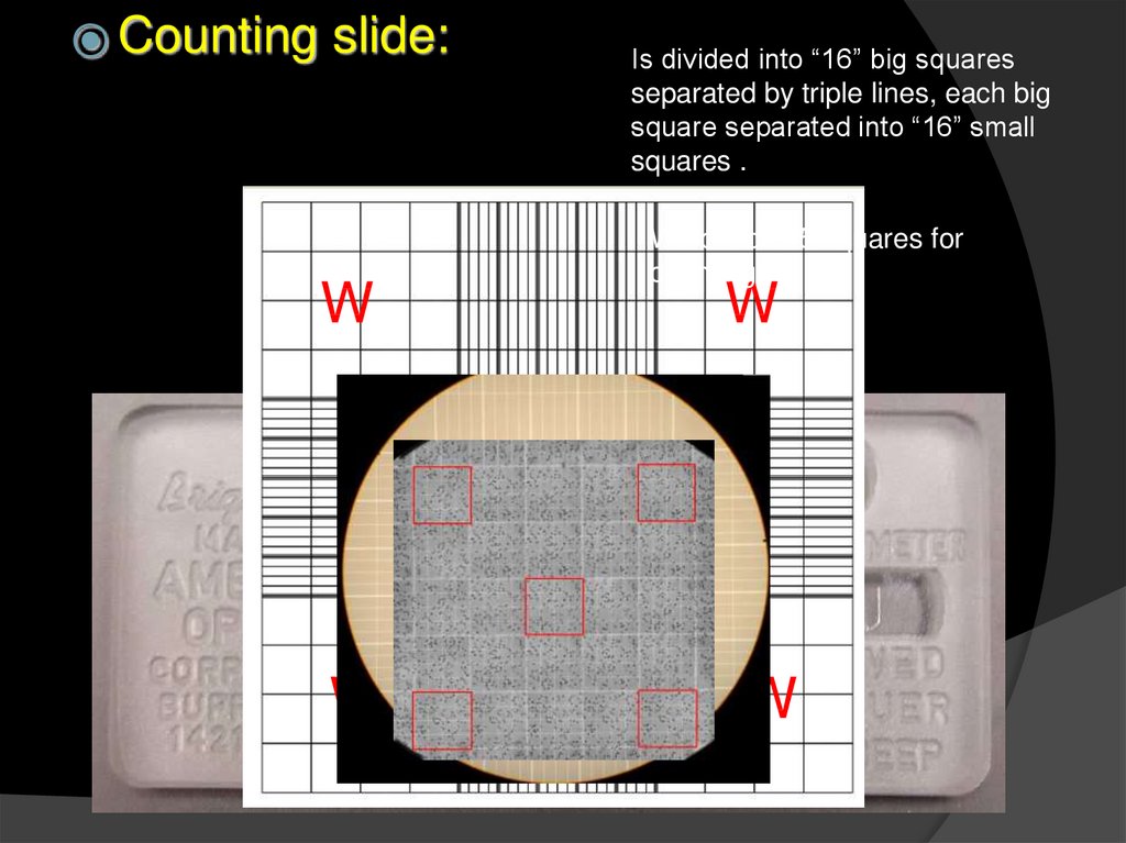
⦿ Countingslide:
W
W
Is divided into “16” big squares
separated by triple lines, each big
square separated into “16” small
squares .
We choose 5 squares for
counting!
W
W
4.
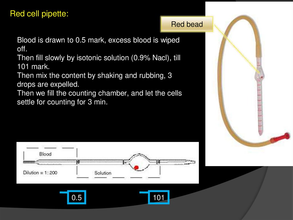
Red cell pipette:Red bead
Blood is drawn to 0.5 mark, excess blood is wiped
off.
Then fill slowly by isotonic solution (0.9% Nacl), till
101 mark.
Then mix the content by shaking and rubbing, 3
drops are expelled.
Then we fill the counting chamber, and let the cells
settle for counting for 3 min.
0.5
101
5.
Shake well tomix with the
hose end
sealed with
your finger.
6.
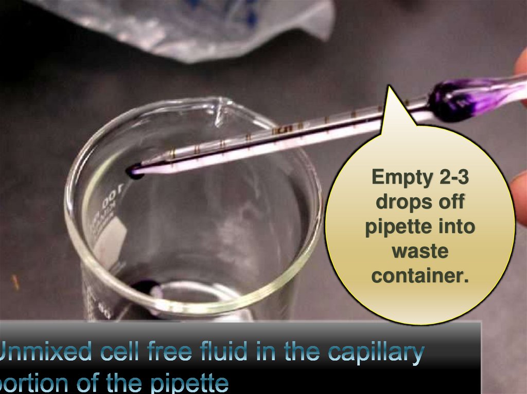
Empty 2-3drops off
pipette into
waste
container.
7.
A special device will be used for countingprocedures called haemocytometer
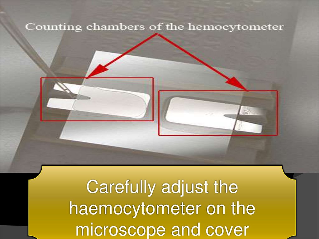
8.
Carefully adjust thehaemocytometer on the
microscope and cover
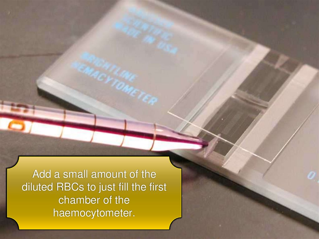
9.
Add a small amount of thediluted RBCs to just fill the first
chamber of the
haemocytometer.
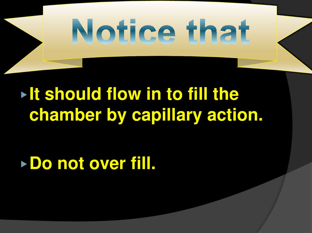
10.
▶ Itshould flow in to fill the
chamber by capillary action.
▶ Do
not over fill.
11.
To improve yourskill, repeat the
dilution a
second time and
fill the second
chamber.
12.
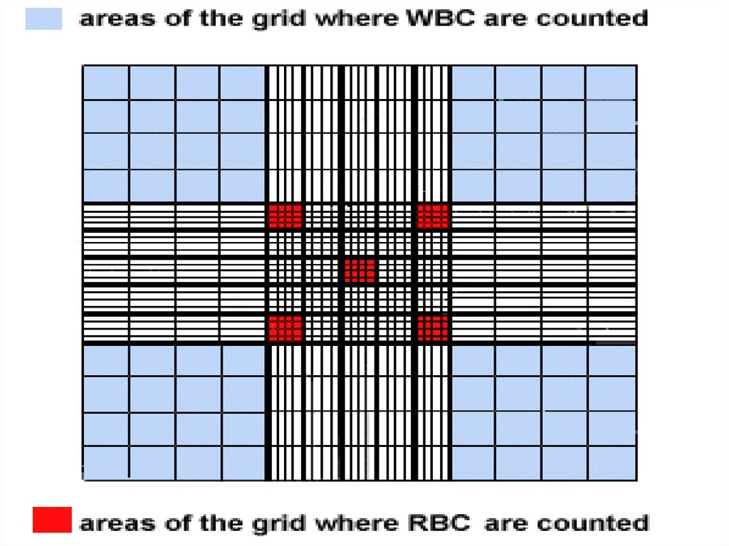
13.
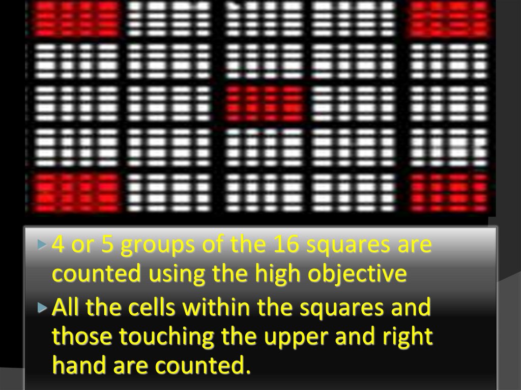
▶ 4 or 5 groups of the 16 squares arecounted using the high objective
▶ All the cells within the squares and
those touching the upper and right
hand are counted.
14.
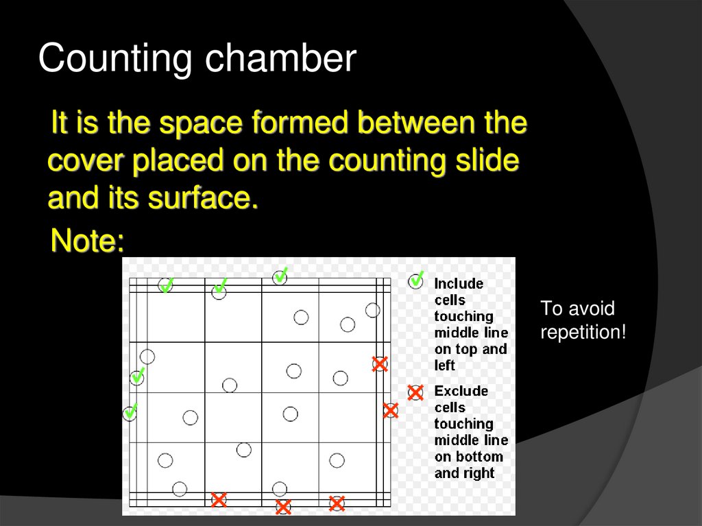
Counting chamberIt is the space formed between the
cover placed on the counting slide
and its surface.
Note:
To avoid
repetition!
15.
16.
⦿ Calculations:● No, of RBC’s
/ small square =
● Volume of small square =
No, of RBC’s
=
Diluting factor
17.
⦿ Normal●♂
value:
= 4.8 -5.6 million cell/
mm3
●♀ = 4.6 – 5.2 million cell
/ mm3
18.
2- colorimetric determination of “Hb”by haemometer
⦿ Aim:
● Determination of amount of “Hb” by
change in color using haemometer.
● Hb → hemoglobin : it’s formed in the
bone marrow, and consists of:
→ “haem + globin”
○ Haem : Iron + protoporphrin
○ Globin : Amino acid + ribonucleic acid
19.
⦿ Principle:● The haemolysis of RBC’s by using
the acid “HCl” to get a free “Hb” in
the medium .
● During this the color changes form
red
brown
20.
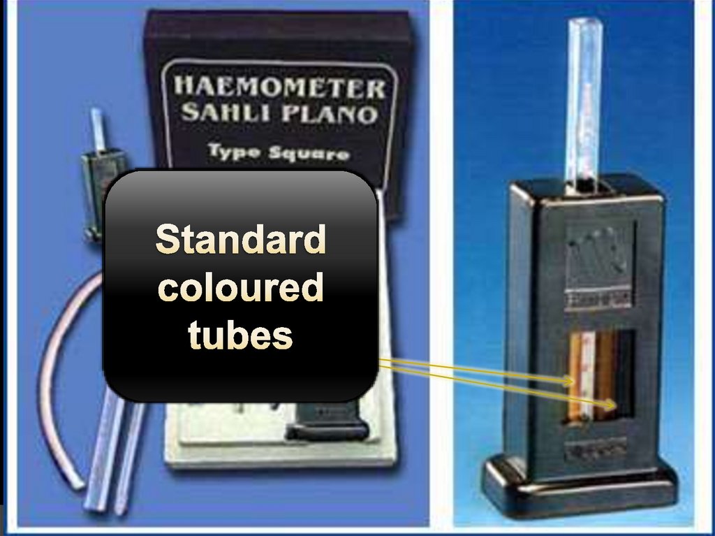
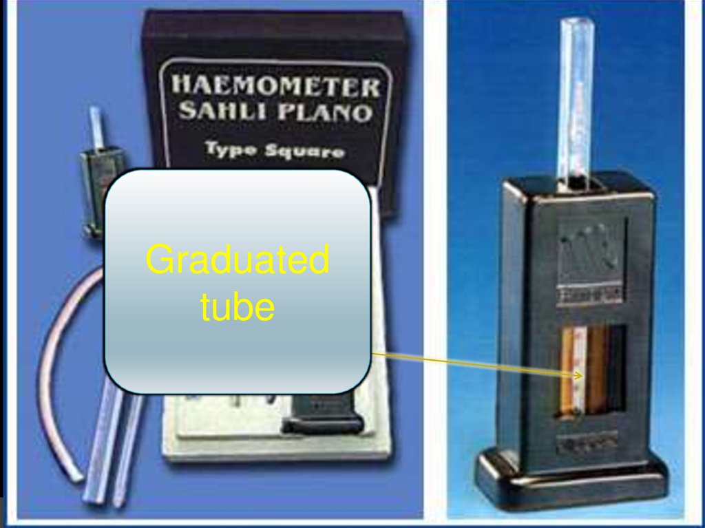
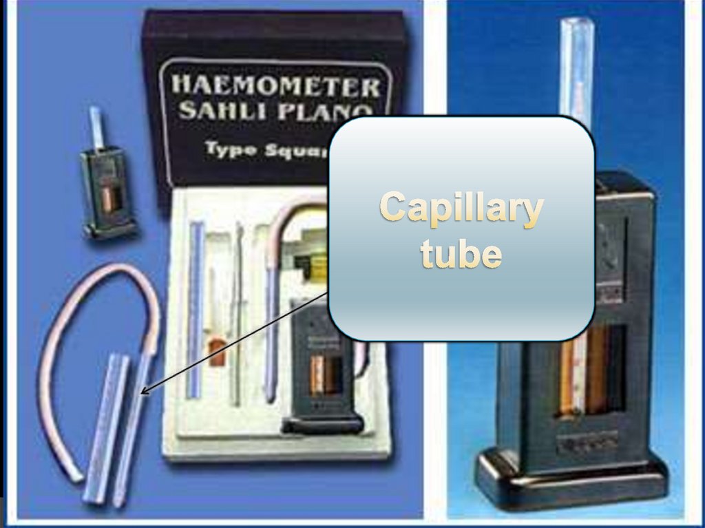
⦿ Haemometer:● Consists of:
○ “2” standard colored tubes
○ Graduated tube
○ Capillary tube
21.
22.
Graduatedtube
23.
24.
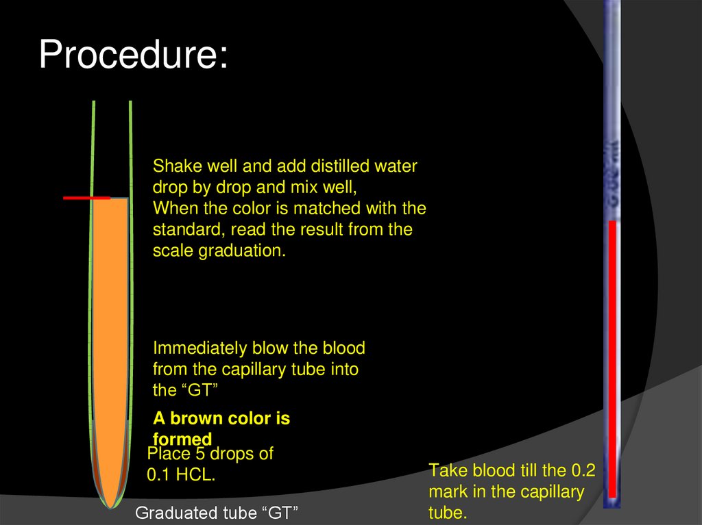
Procedure:Shake well and add distilled water
drop by drop and mix well,
When the color is matched with the
standard, read the result from the
scale graduation.
Immediately blow the blood
from the capillary tube into
the “GT”
A brown color is
formed
Place 5 drops of
0.1 HCL.
Graduated tube “GT”
Take blood till the 0.2
mark in the capillary
tube.
25.
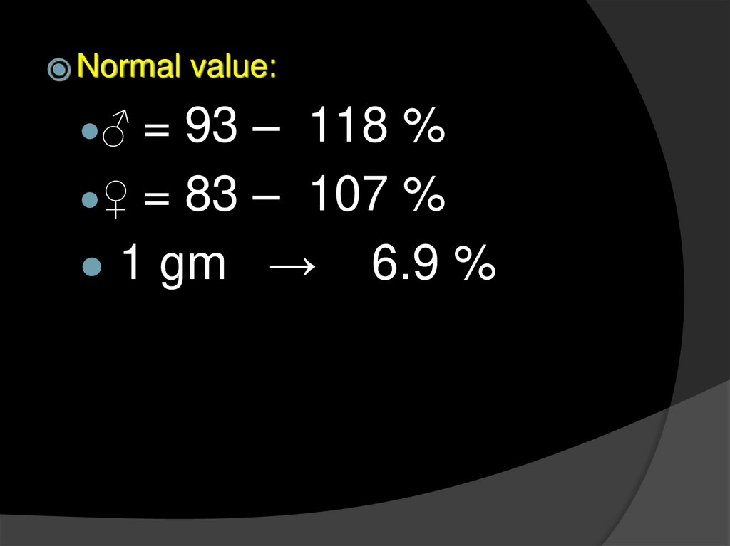
⦿ Normalvalue:
= 93 – 118 %
●♀ = 83 – 107 %
● 1 gm → 6.9 %
●♂
26.
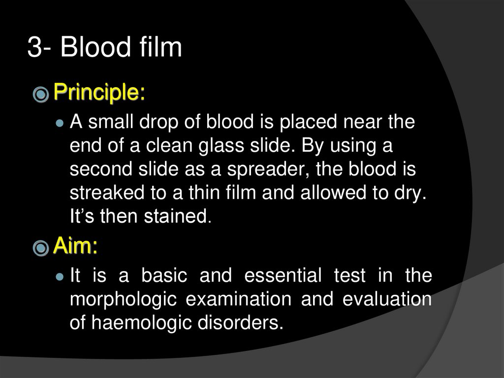
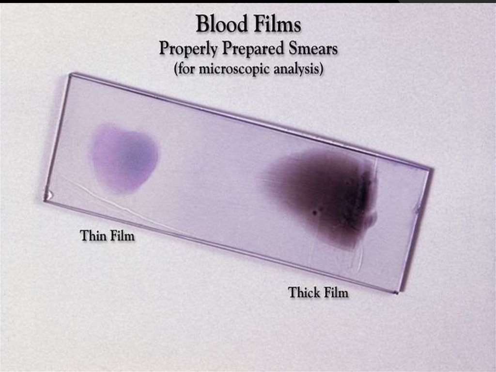
3- Blood film⦿ Principle:
● A small drop of blood is placed near the
end of a clean glass slide. By using a
second slide as a spreader, the blood is
streaked to a thin film and allowed to dry.
It’s then stained.
⦿ Aim:
● It is a basic and essential test in the
morphologic examination and evaluation
of haemologic disorders.
27.
Method:A finger puncture in
made, and a small
drop of blood is placed
on the end of a slide.
Spreader slide is held 30-40 .. We
approach the drop of blood , then we
push smooth and tight towards the
opposite side.
Let the blood to air dry , then
stain.
28.
▶1.
2.
29.
SpeedAngle
30.
31.
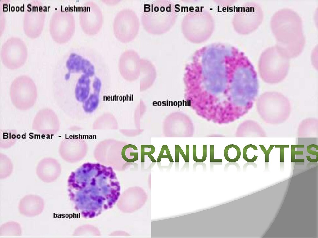
⦿ Stainsused:
Leishman Stain : and it consists of
○ Methylene blue :
● It stains nuclear DNA
○ Eosin in methyl alcohol :
● Eosin stains the more basic
compounds as “Hb” with “pinkish”
color
● Methyl alcohol acts as a “Fixative”
32.
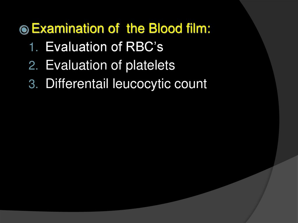
⦿ Examinationof the Blood film:
1. Evaluation of RBC’s
2. Evaluation of platelets
3. Differentail leucocytic count
33.
34.
35.
4- Determination of bloodgroups
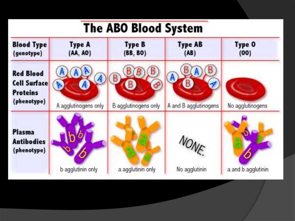
⦿ Principle
:
● The blood consists of plasma and cells
(RBC’s- WBC’s- Platelets), the RBC’s
express specific Antigens on their
membrane, “Agglutinogens” and the
plasma contain Antibodies “Agglutinins)
● Agglutination: it’s a process in which the
antigens on the RBC’s are clumped by
the their antibodies in the plasma.
36.
37.
⦿ Thisdiagram shows the possible
ways of blood transfusion without
causing agglutination to the blood:
O: is a universal “Donor”
AB: is a universal “Acceptor”
38.
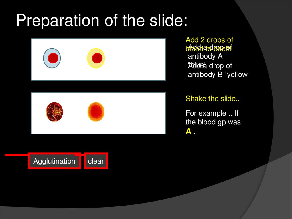
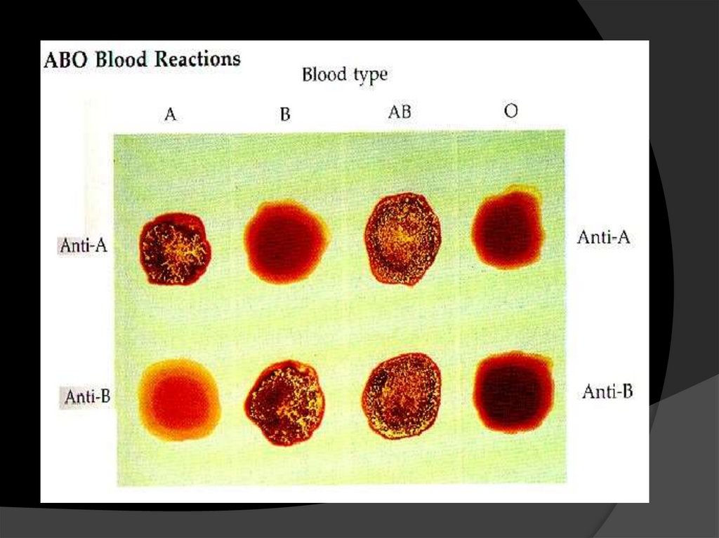
Preparation of the slide:Add 2 drops of
Add atodrop
of
blood
each!
antibody A
“blue”
Add a drop of
antibody B “yellow”
Shake the slide..
For example .. If
the blood gp was
A.
Agglutination
clear
39.
40.
41.
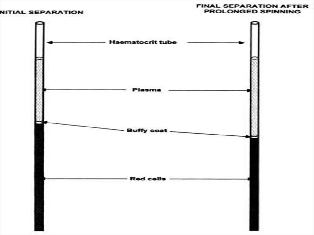
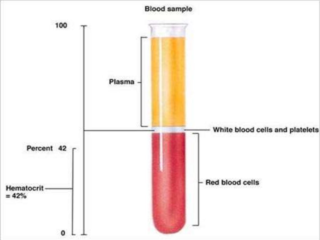
Define:⦿hematocrit (Ht ), also known
as packed cell volume (PCV)
or erythrocyte volume
fraction (EVF), is the volume
percentage (%) of red blood cells
in blood.
⦿ It
is normally about 45% for men and
40% for women
42.
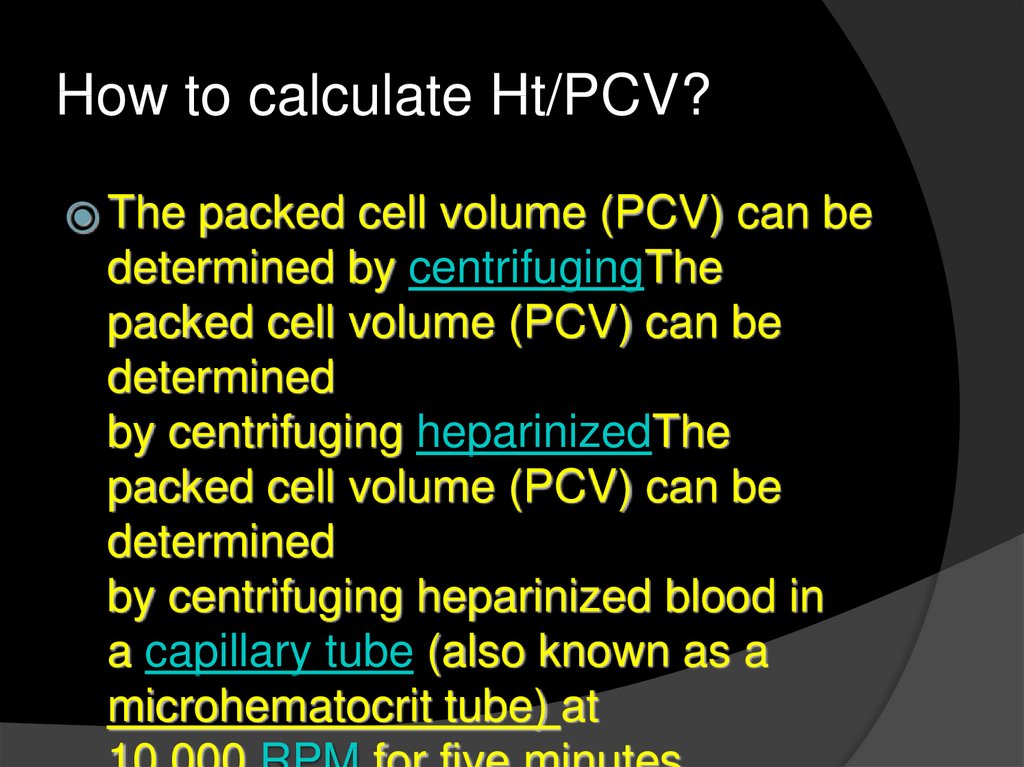
How to calculate Ht/PCV?⦿ The
packed cell volume (PCV) can be
determined by centrifugingThe
packed cell volume (PCV) can be
determined
by centrifuging heparinizedThe
packed cell volume (PCV) can be
determined
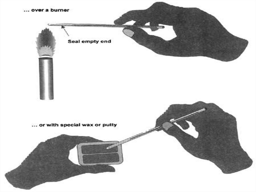
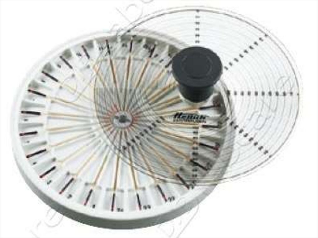
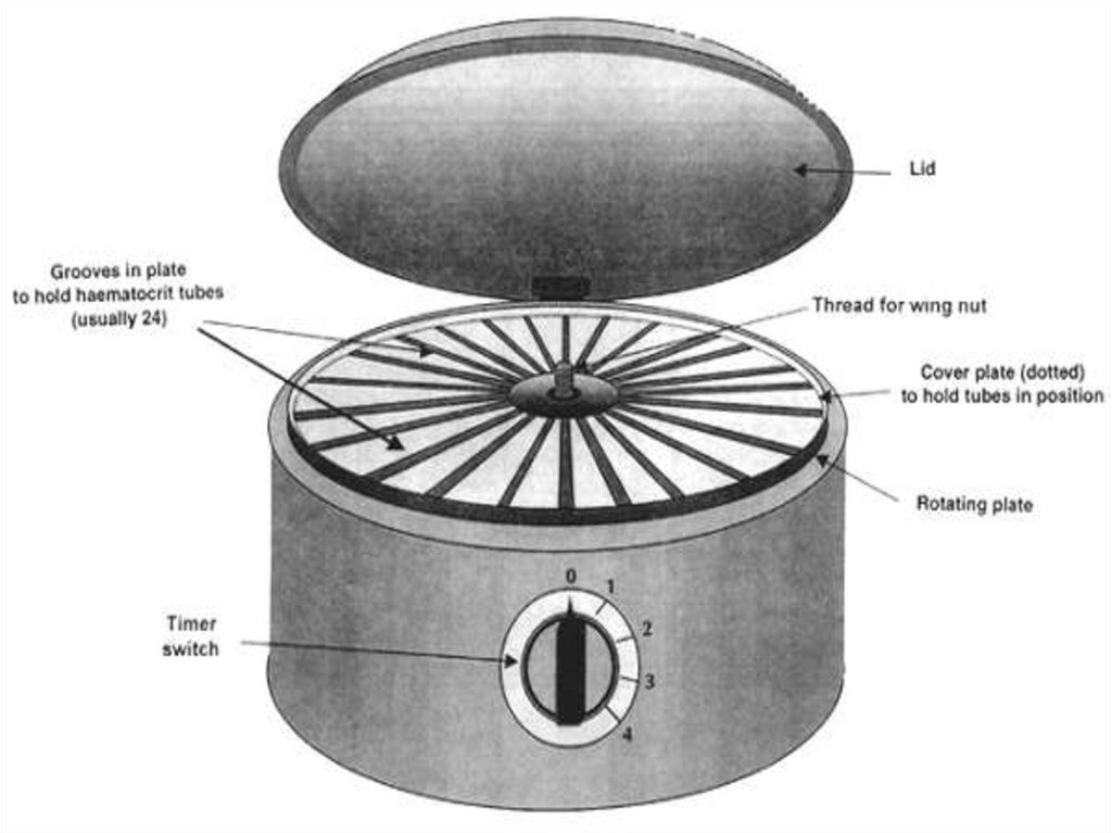
by centrifuging heparinized blood in
a capillary tube (also known as a
microhematocrit tube) at
43.
44.
45.
46.
47.
48.
49.
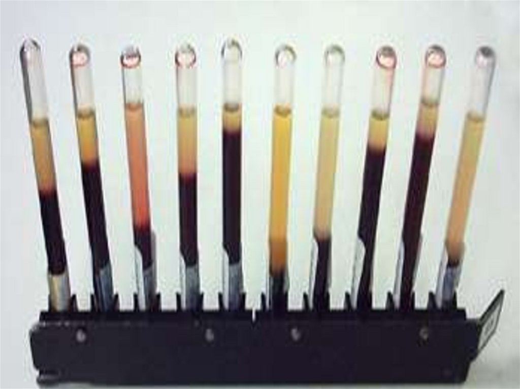
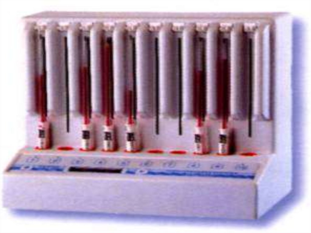
(ESR=Erythrocytesedimentation rate)
50.
Define:⦿ Rateof sedimentation is the rate at
which red blood cells sediment in a
period of one hour.
51.
How To perform the test?⦿ Anticoagulated
blood is placed in an
upright tube, known as a Westergren
tube, and the rate at which the red
blood cells fall is measured and
reported in mm/h
52.
53.
54.
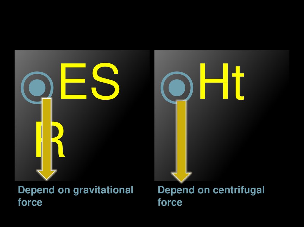
⦿ES ⦿HtR
Depend on gravitational
force
Depend on centrifugal
force