


")


")


")

 biology
Similar presentations:

")
. Recent developments of biotechnology in veterinary medicine and animal husbandry")


Antigen-antibody reactions and selected tests
1. ANTIGEN-ANTIBODY REACTIONS AND SELECTED TESTS
ANTIGEN-ANTIBODY REACTIONSAND SELECTED TESTS
• TEACHING OBJECTIVES
1.To describe the nature of Ag-Ab reactions
2.To compare and contrast antibody affinity
and avidity
3.To delineate the basis for antibody
specificity and cross reactivity
4. To discuss the principles of commonly used
tests for antigen/antibody reactions
2.
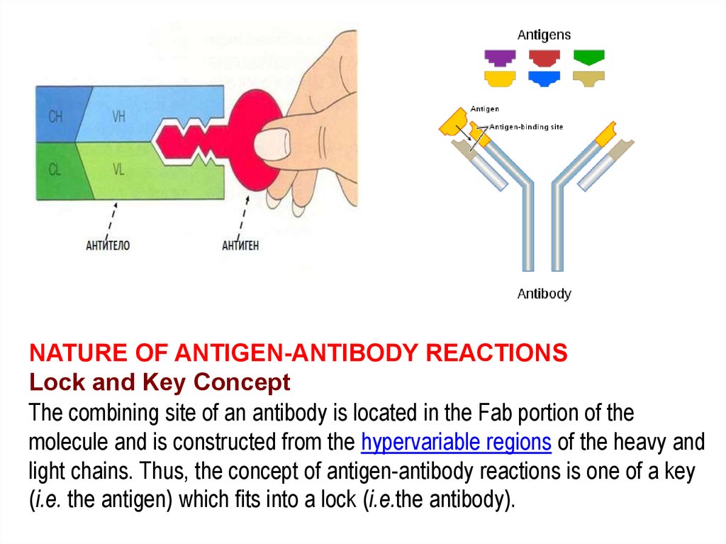
NATURE OF ANTIGEN-ANTIBODY REACTIONSLock and Key Concept
The combining site of an antibody is located in the Fab portion of the
molecule and is constructed from the hypervariable regions of the heavy and
light chains. Thus, the concept of antigen-antibody reactions is one of a key
(i.e. the antigen) which fits into a lock (i.e.the antibody).
3.
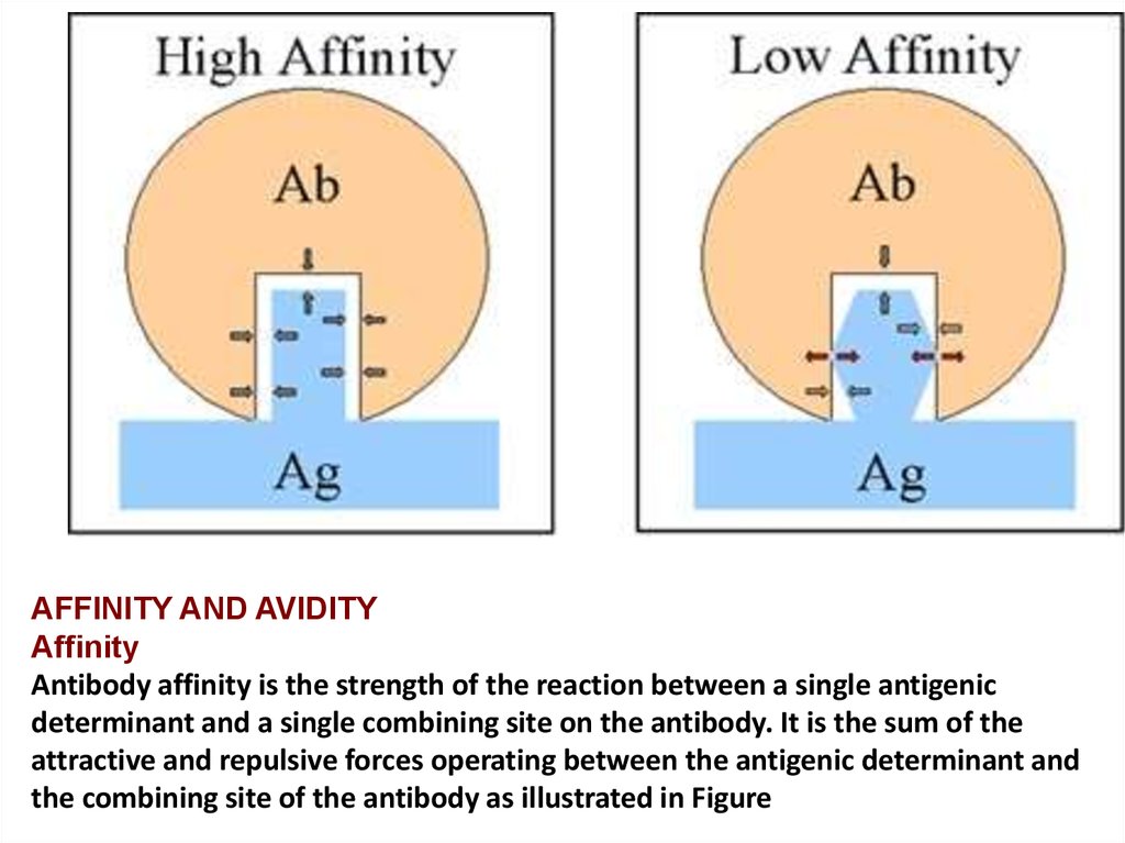
AFFINITY AND AVIDITYAffinity
Antibody affinity is the strength of the reaction between a single antigenic
determinant and a single combining site on the antibody. It is the sum of the
attractive and repulsive forces operating between the antigenic determinant and
the combining site of the antibody as illustrated in Figure
4.
5.
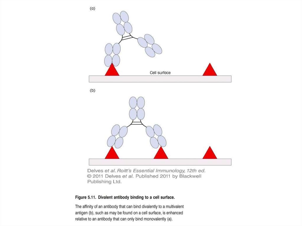
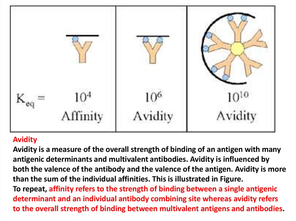
AvidityAvidity is a measure of the overall strength of binding of an antigen with many
antigenic determinants and multivalent antibodies. Avidity is influenced by
both the valence of the antibody and the valence of the antigen. Avidity is more
than the sum of the individual affinities. This is illustrated in Figure.
To repeat, affinity refers to the strength of binding between a single antigenic
determinant and an individual antibody combining site whereas avidity refers
to the overall strength of binding between multivalent antigens and antibodies.
6.
7.
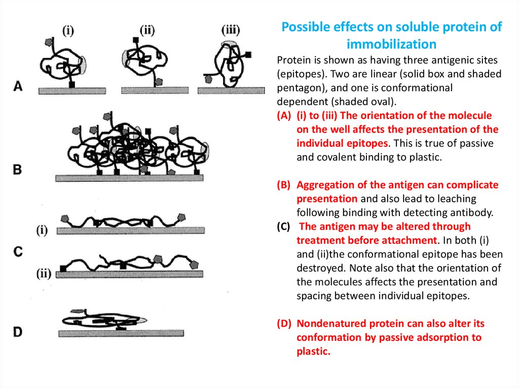
Possible effects on soluble protein ofimmobilization
Protein is shown as having three antigenic sites
(epitopes). Two are linear (solid box and shaded
pentagon), and one is conformational
dependent (shaded oval).
(A) (i) to (iii) The orientation of the molecule
on the well affects the presentation of the
individual epitopes. This is true of passive
and covalent binding to plastic.
(B) Aggregation of the antigen can complicate
presentation and also lead to leaching
following binding with detecting antibody.
(C) The antigen may be altered through
treatment before attachment. In both (i)
and (ii)the conformational epitope has been
destroyed. Note also that the orientation of
the molecules affects the presentation and
spacing between individual epitopes.
(D) Nondenatured protein can also alter its
conformation by passive adsorption to
plastic.
8.
SPECIFICITY AND CROSS REACTIVITYSpecificity
Specificity refers to the ability of an individual antibody
combining site to react with only
one antigenic determinant or the ability of a population
of antibody molecules to react with only one antigen.
In general, there is a high degree of specificity in
antigen-antibody reactions.
Antibodies can distinguish differences in:
•The primary structure of an antigen
•Isomeric forms of an antigen
•Secondary and tertiary structure of an antigen
9.
APLICATION OF ANTIGEN-ANTIBODY REACTIONS1. Diagnosis of infectious and parasitic diseases and the
establishment of detection antibody titers (serodiagnosis);
2. Diagnosis of diseases to identify antigens of pathogens in
the body;
3. Identification of cultures of bacteria and viruses isolated
from humans and animals;
4. Determination of the composition and characteristics of
human tissue: blood group, Rh factor, transplantation
antigens;
5. Identification of the human body and in the environment
of any substances having antigenicity (hormones, enzymes,
toxins, drugs, drugs, etc.).
6. Assessment of immune status to determine the
quantitative and functional characteristics of immune
system cells and their products.
7. Identification of immunopathological conditions, allergies,
transplant and anti-tumor responses.
10.
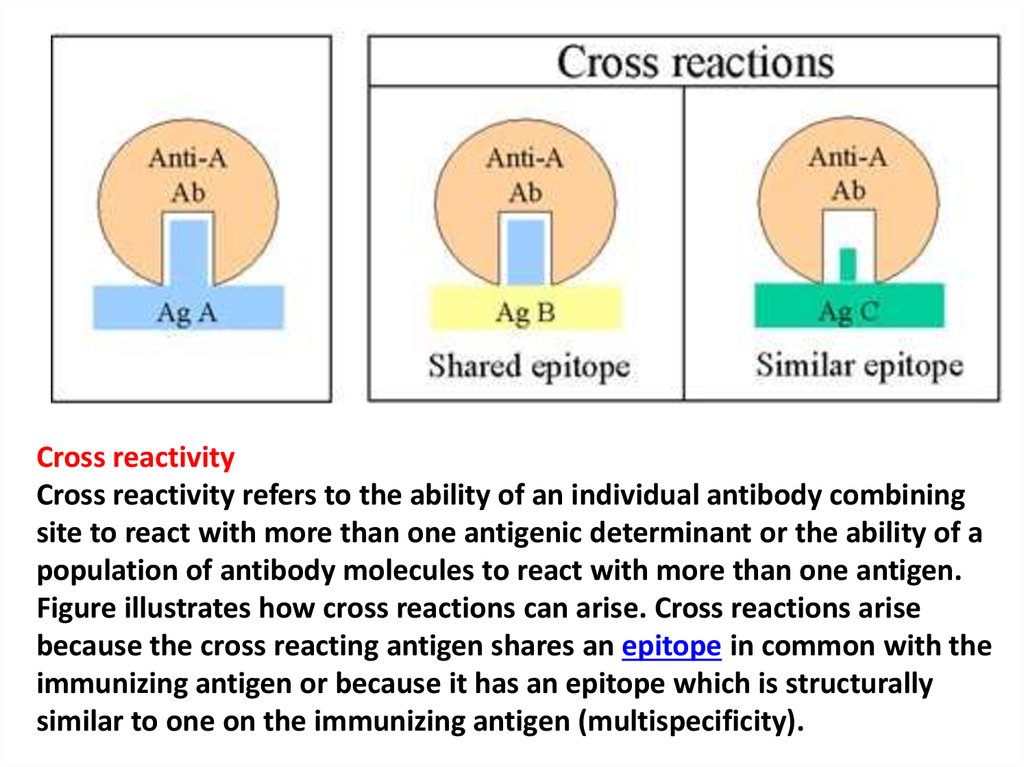
Cross reactivityCross reactivity refers to the ability of an individual antibody combining
site to react with more than one antigenic determinant or the ability of a
population of antibody molecules to react with more than one antigen.
Figure illustrates how cross reactions can arise. Cross reactions arise
because the cross reacting antigen shares an epitope in common with the
immunizing antigen or because it has an epitope which is structurally
similar to one on the immunizing antigen (multispecificity).
11. Agglutination test
Agglutination test(agglutinacio - склеивание)
- gluing and precipitation of
the bacteria under the
influence of antibodies in
an environment with the
electrolyte.
12. STATEMENT OF MICROAGGLUTINATION TEST
13. THE RESULTS OF MICROAGGLUTINATION TEST
14.
15.
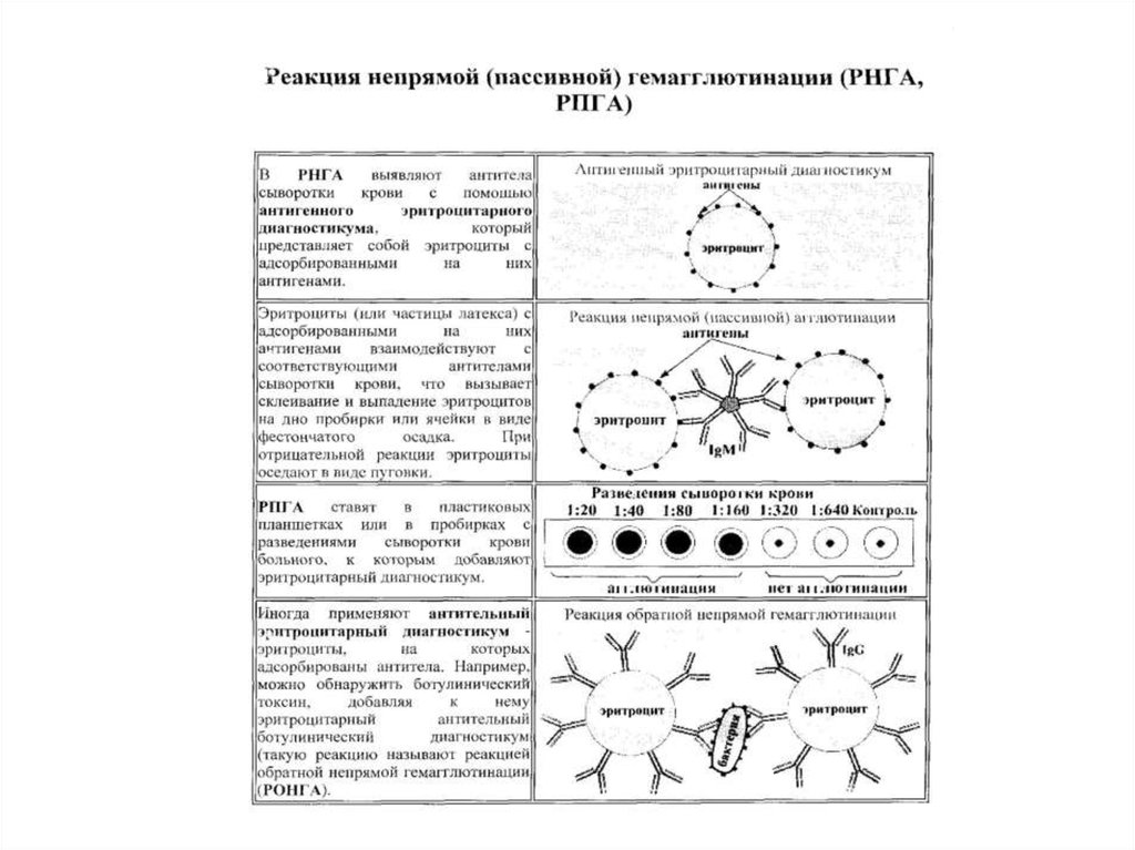
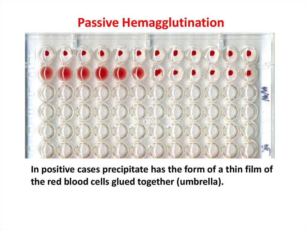
Passive HemagglutinationIn positive cases precipitate has the form of a thin film of
the red blood cells glued together (umbrella).
16.
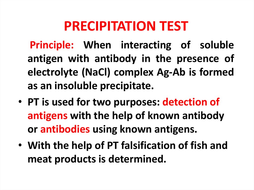
PRECIPITATION TESTPrinciple: When interacting of soluble
antigen with antibody in the presence of
electrolyte (NaCl) complex Ag-Ab is formed
as an insoluble precipitate.
• PT is used for two purposes: detection of
antigens with the help of known antibody
or antibodies using known antigens.
• With the help of PT falsification of fish and
meat products is determined.
17. THE PRINCIPLE OF PRECIPITATION TEST
18. Ring-precipitation test
The test is carriedout by layering the
antigen on the
immune serum
Formation of the Ag-Ab
complex
-
+
19. THE PRINCIPLE OF RADIAL IMMUNODIFFUSION TEST
The test is carried out in agar gel plates or in Petri dishes. Holes are cut out In the frozengel at some distance from each other, and filled with the solutions of antigen and antisera.
In the case of optimally selected ratio of antigens to antibodies, precipitation bands are
formed in the gel between the wells. Since the reagents diffuse from the wells
concentrically, the method allows several simultaneous reactions to be carried out by
placing several wells with antigens around the antiserum well.
20. Radial Immunodiffusion (RID)
21.
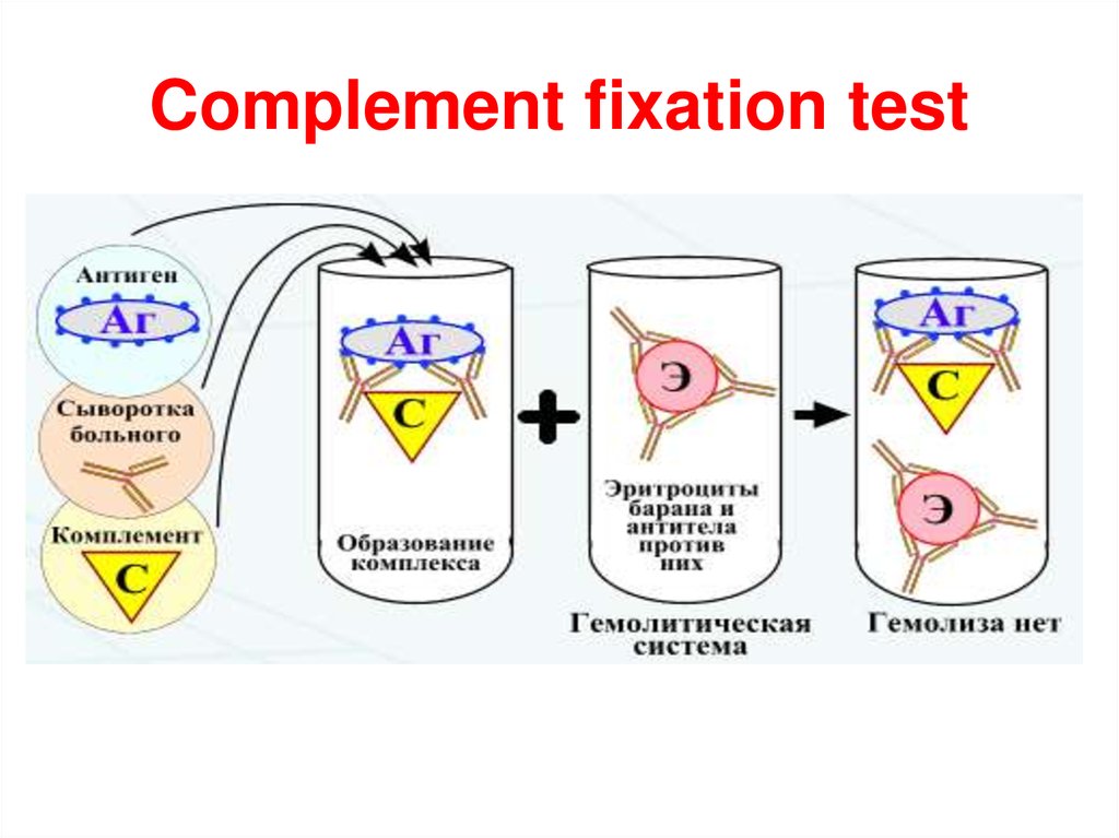
Complement fixation test22.
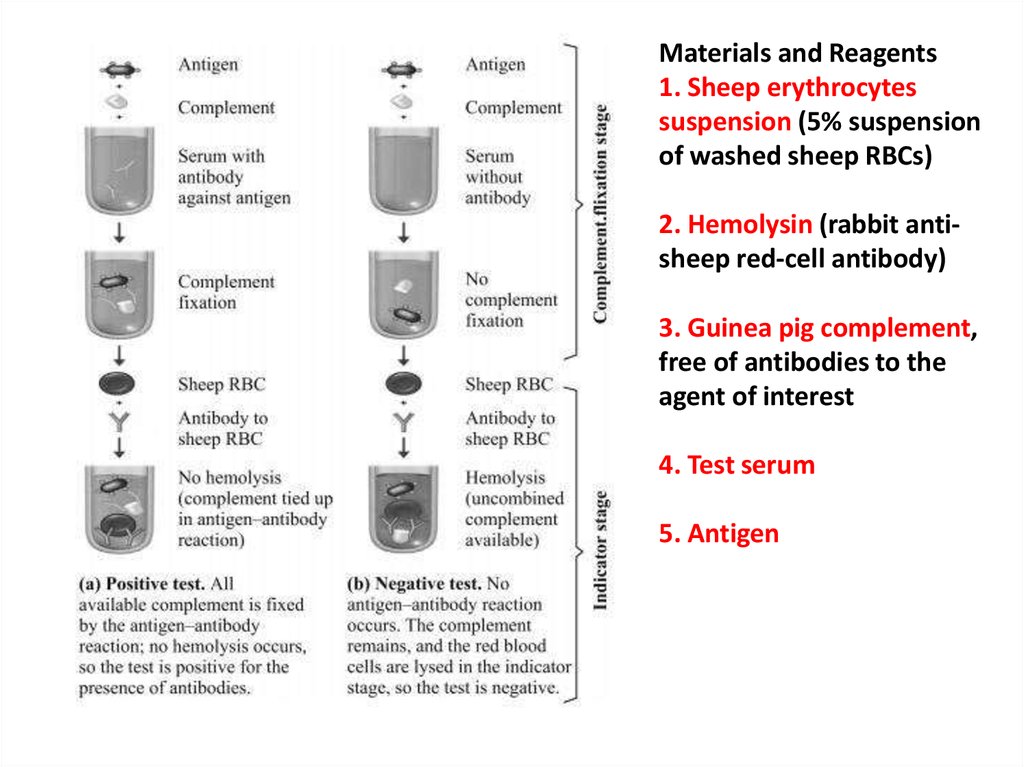
Materials and Reagents1. Sheep erythrocytes
suspension (5% suspension
of washed sheep RBCs)
2. Hemolysin (rabbit antisheep red-cell antibody)
3. Guinea pig complement,
free of antibodies to the
agent of interest
4. Test serum
5. Antigen
23.
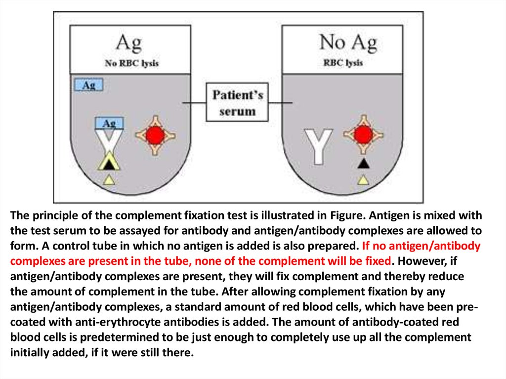
The principle of the complement fixation test is illustrated in Figure. Antigen is mixed withthe test serum to be assayed for antibody and antigen/antibody complexes are allowed to
form. A control tube in which no antigen is added is also prepared. If no antigen/antibody
complexes are present in the tube, none of the complement will be fixed. However, if
antigen/antibody complexes are present, they will fix complement and thereby reduce
the amount of complement in the tube. After allowing complement fixation by any
antigen/antibody complexes, a standard amount of red blood cells, which have been precoated with anti-erythrocyte antibodies is added. The amount of antibody-coated red
blood cells is predetermined to be just enough to completely use up all the complement
initially added, if it were still there.
24.
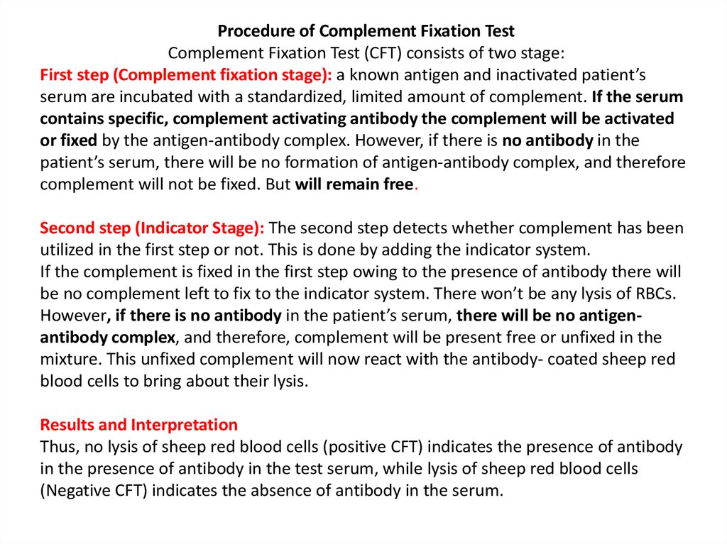
Procedure of Complement Fixation TestComplement Fixation Test (CFT) consists of two stage:
First step (Complement fixation stage): a known antigen and inactivated patient’s
serum are incubated with a standardized, limited amount of complement. If the serum
contains specific, complement activating antibody the complement will be activated
or fixed by the antigen-antibody complex. However, if there is no antibody in the
patient’s serum, there will be no formation of antigen-antibody complex, and therefore
complement will not be fixed. But will remain free.
Second step (Indicator Stage): The second step detects whether complement has been
utilized in the first step or not. This is done by adding the indicator system.
If the complement is fixed in the first step owing to the presence of antibody there will
be no complement left to fix to the indicator system. There won’t be any lysis of RBCs.
However, if there is no antibody in the patient’s serum, there will be no antigenantibody complex, and therefore, complement will be present free or unfixed in the
mixture. This unfixed complement will now react with the antibody- coated sheep red
blood cells to bring about their lysis.
Results and Interpretation
Thus, no lysis of sheep red blood cells (positive CFT) indicates the presence of antibody
in the presence of antibody in the test serum, while lysis of sheep red blood cells
(Negative CFT) indicates the absence of antibody in the serum.
25.
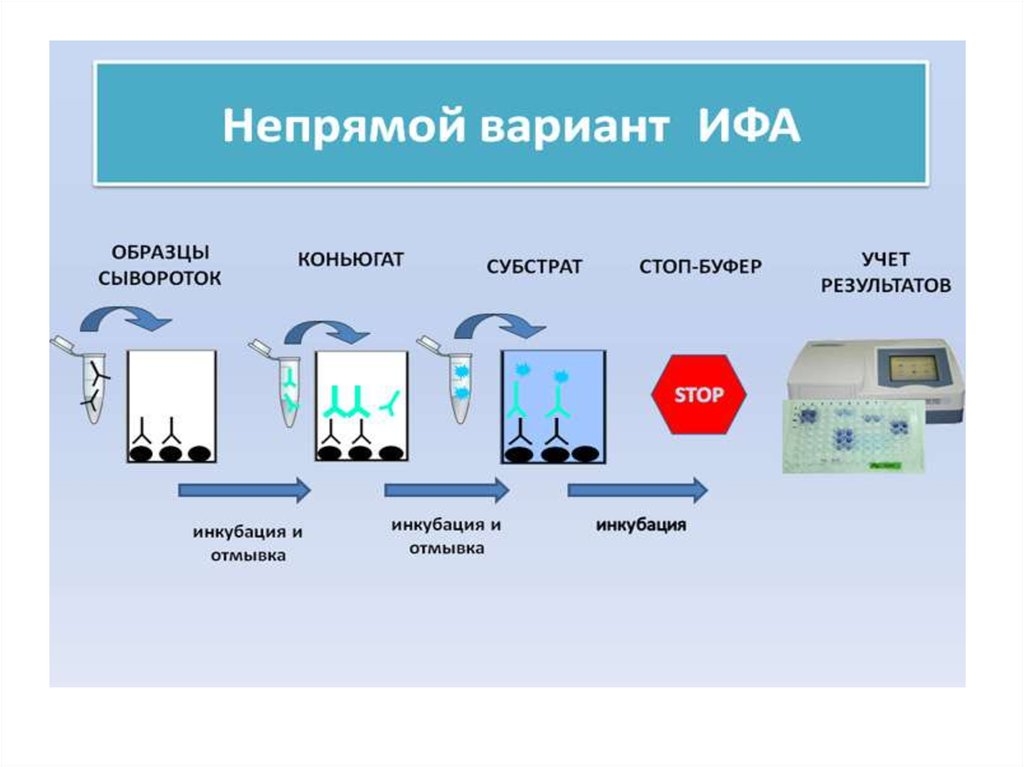
Enzyme linkedimmunosorbent assay
ELISA - it is serological test in which for
the visualization of the formed antigenantibody complex enzyme labels
(horseradish peroxidase or alkaline
phosphatase) are used . These marker
(indicator) enzymes are able to cleave
the substrate and cause a color change.
26.
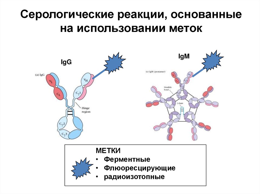
Серологические реакции, основанныена использовании меток
IgM
IgG
МЕТКИ
• Ферментные
• Флюоресцирующие
• радиоизотопные
27.
28.
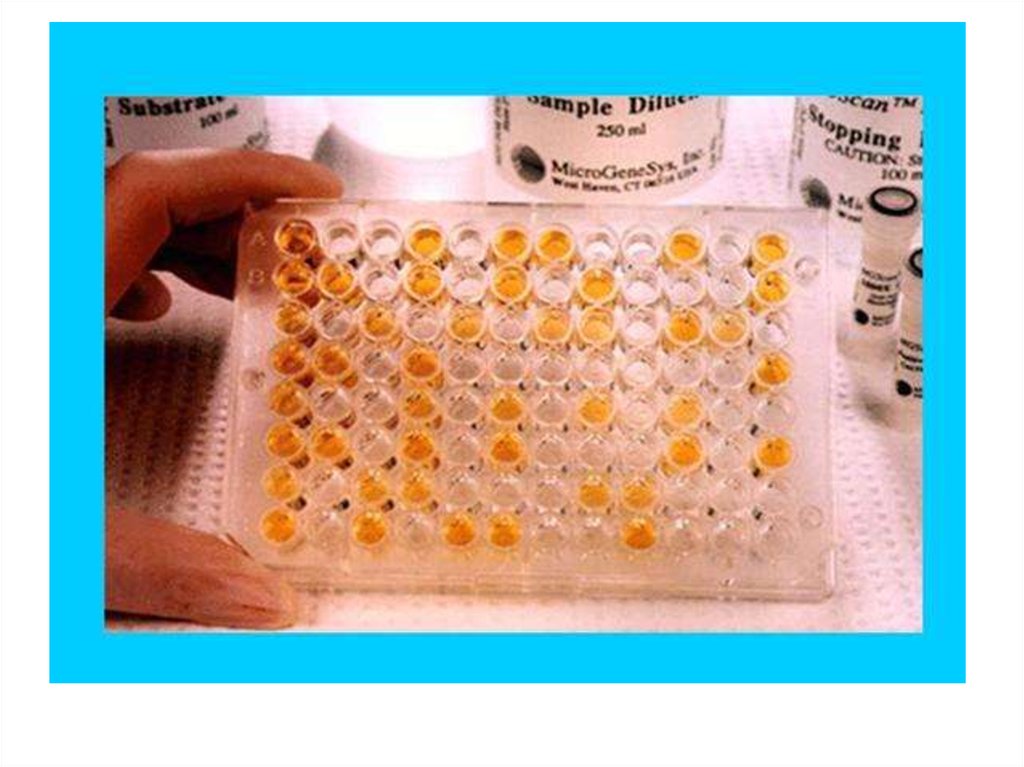
29. 96-луночный планшет для ИФА
30.
31. Спектрофотометр для ИФА
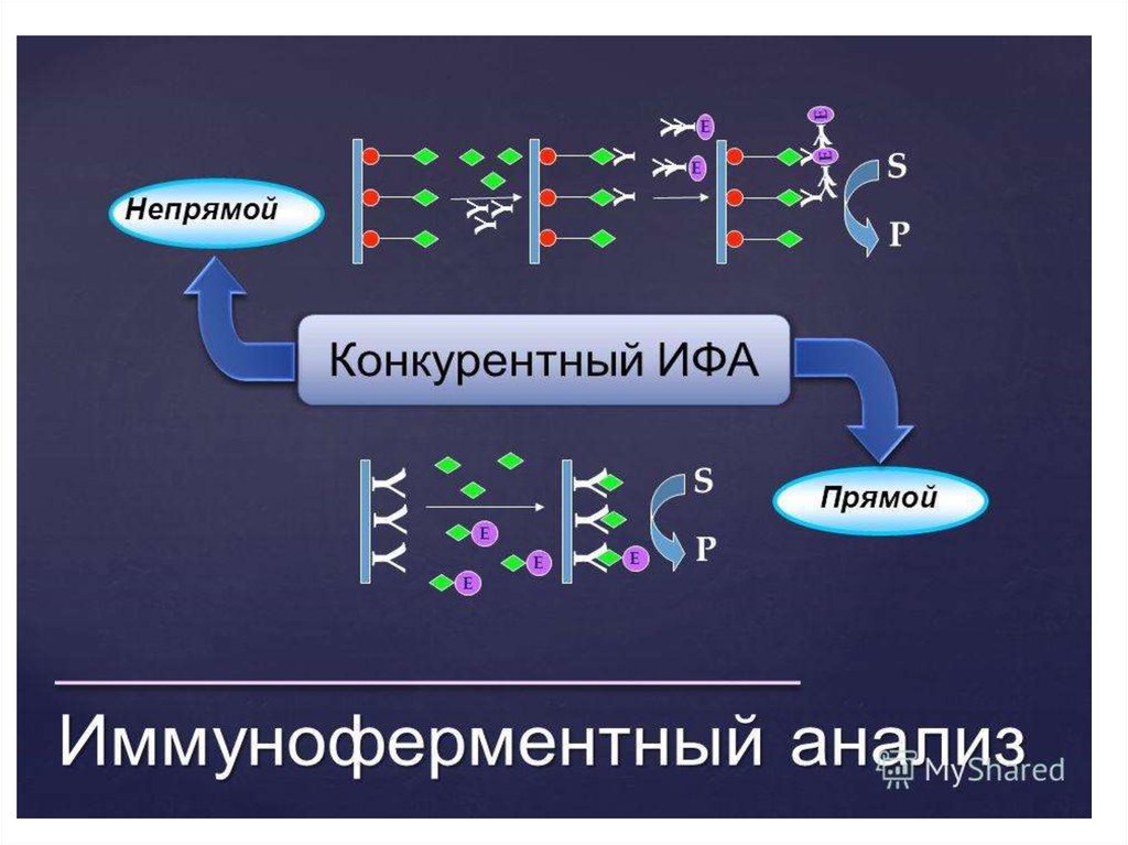
32. ПРИНЦИП НЕПРЯМОГО ИФА
33.
34. Sandwich ELISA
This variant of ELISA is extremely common for the
determination of antigens possessing more than
one determinant.
In the process of analysis, the antigen is
"squeezed" between antibody molecules, which
led to the name of the "sandwich" method. This
name is now used practically in all literature as an
official term.
35. Принцип сэндвич-ИФА
36.
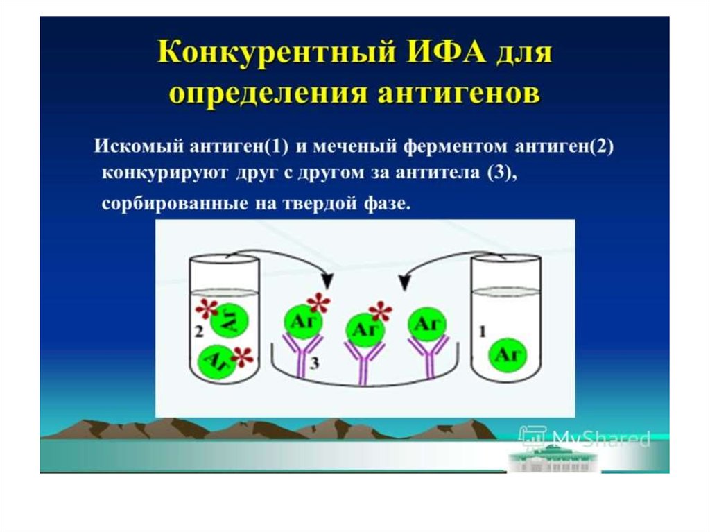
37. Конкурентный ИФА для определения антигена (антибиотиков)
СубстратАнтибиотик
Специфические АТ
Антивидовой
конъюгат
Конъюгат
АБ-носитель
38.
39.
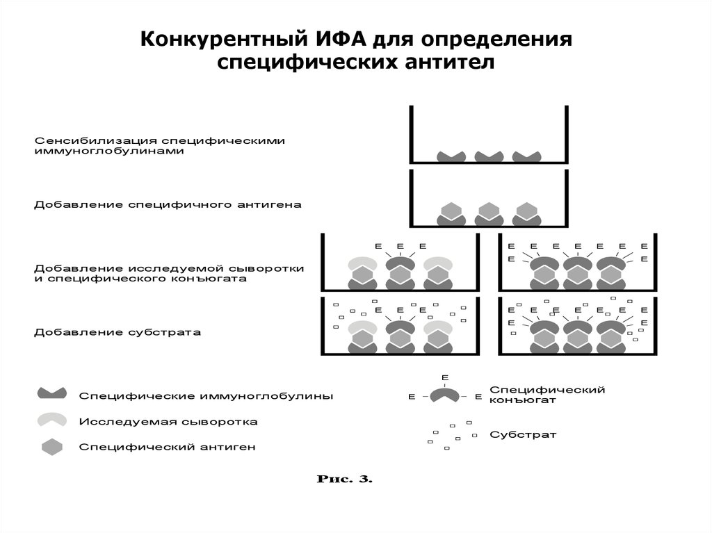
Конкурентный ИФА для определенияспецифических антител
Сенсибилизация специфическими
иммуноглобулинами
Добавление специфичного антигена
Е
Е
Е
Е
Е
Е
Е
Е
Е
Е
Е
Е
Добавление исследуемой сыворотки
и специфического конъюгата
Е
Е
Е
Е
Е
Е
Е
Е
Е
Добавление субстрата
Е
Специфические иммуноглобулины
Е
Е
Специфический
конъюгат
Исследуемая сыворотка
Субстрат
Специфический антиген
Рис. 3.
Е
Е
Е
40.
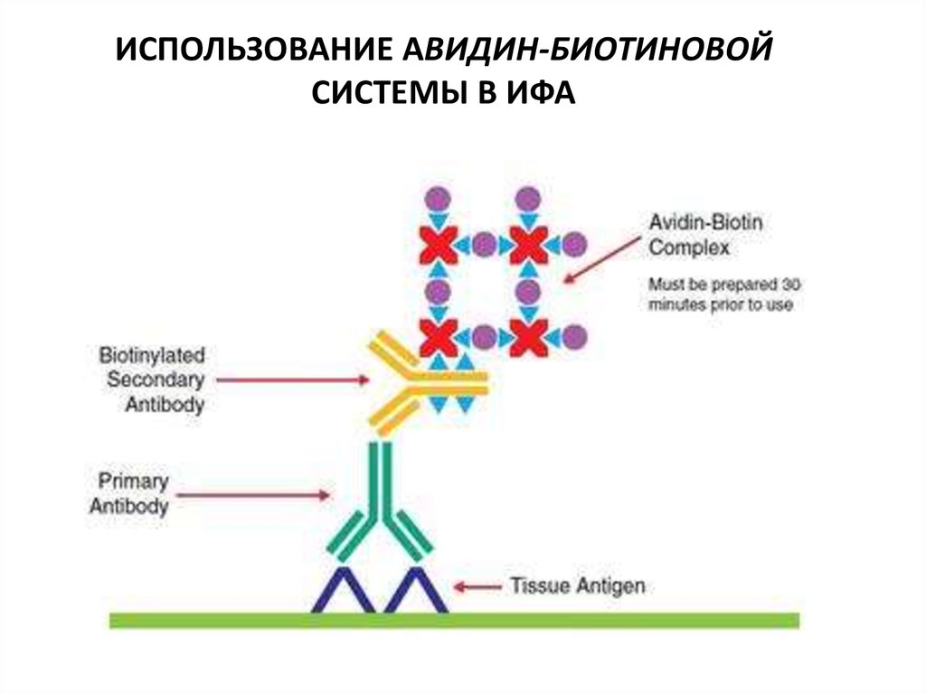
ИСПОЛЬЗОВАНИЕ АВИДИН-БИОТИНОВОЙСИСТЕМЫ В ИФА
41.
42. Dot-ИФА
43. Сравнительная характеристика dot-ИФА и ИФА.
Показатели, характеристикиdot-ИФА
ИФА
Продолжительность
2 час
3-6 часов
Число этапов постановки
3 часа
4-5 часов
3
3
Цена анализа (в долларах
США)
0,05-0,1
1-2
Спецподготовка персонала
нет
да
Применяемость в полевых
условиях
да
нет
Необходимость фотометра
нет
да
Возможность непрерывной
регистрации
да
неб
Число промывок
44. ПРИНЦИП ИММУНОХРОМАТОГРАФИЧЕСКОГО АНАЛИЗА
45. Оборудование для производства иммунохроматографической тест - системы
Оборудование для производства иммунохроматографической тест системыПрезиционный диспенсер
автоматический
Автоматический гильотинный
резак
Вакуумный сушильный шкаф
для упаковки тестов