


 biology
Similar presentations:


Protein splicing
1.
RECAP (1)In eukaryotes, large primary transcripts are
processed to smaller, mature mRNAs.
What was first evidence for this precursorproduct relationship?
2.
Observation:Nuclear RNA pool consists of very high molecular weight
species as well as lower molecular weight.
Darnell asked if there is a relationship between the high
and low molecular weight RNAs
DNA
3.
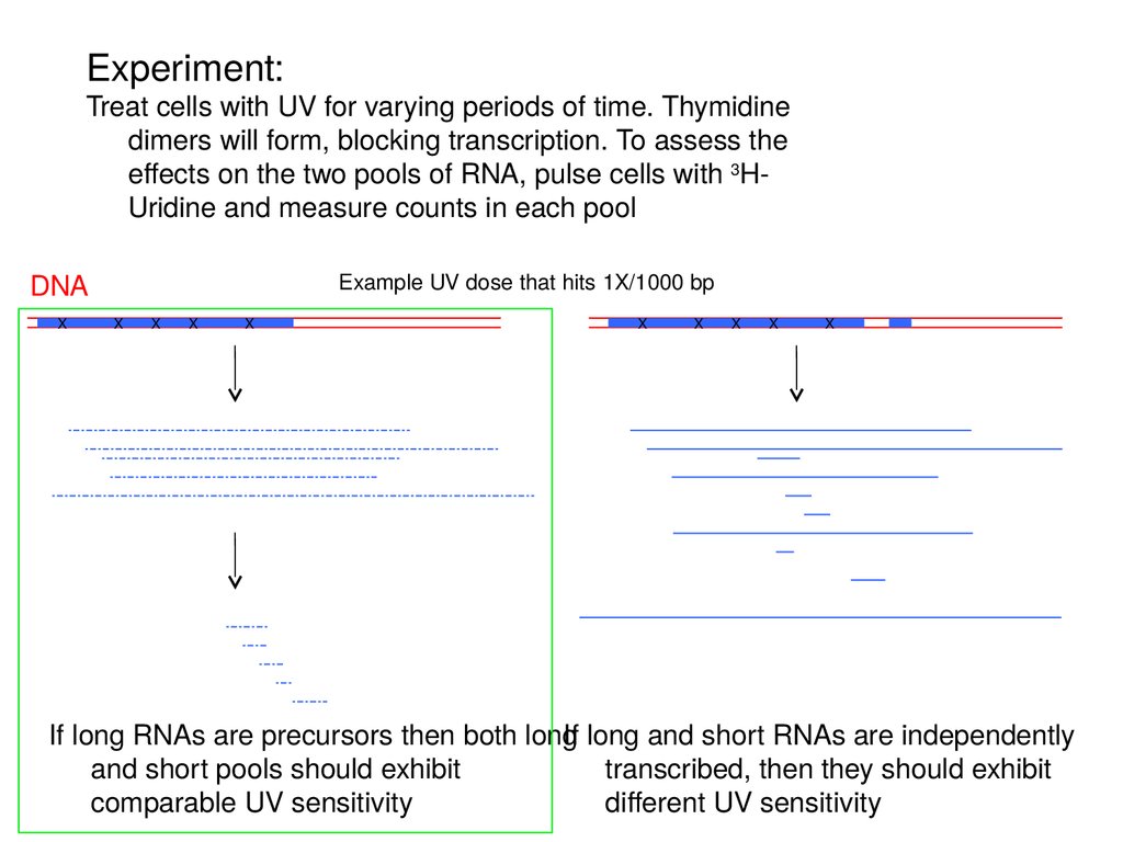
Experiment:Treat cells with UV for varying periods of time. Thymidine
dimers will form, blocking transcription. To assess the
effects on the two pools of RNA, pulse cells with 3HUridine and measure counts in each pool
DNA
X
Example UV dose that hits 1X/1000 bp
X
X
X
X
X
X
X
X
X
If long RNAs are precursors then both long
If long and short RNAs are independently
and short pools should exhibit
transcribed, then they should exhibit
comparable UV sensitivity
different UV sensitivity
4.
Experiment:Treat cells with UV for varying periods of time. Thymidine
dimers will form, blocking transcription. To assess the
effects on the two pools of RNA, pulse cells with 3HUridine and measure counts in each pool
DNA
X
Example UV dose that hits 1X/1000 bp
X
X
X
X
X
X
X
X
X
If long RNAs are precursors then both long
If long and short RNAs are independently
and short pools should exhibit
transcribed, then they should exhibit
comparable UV sensitivity
different UV sensitivity
5.
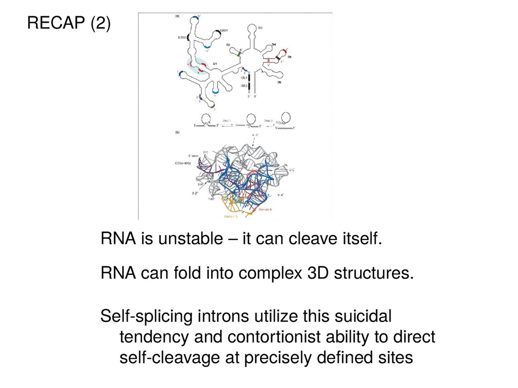
RECAP (2)RNA is unstable – it can cleave itself.
RNA can fold into complex 3D structures.
Self-splicing introns utilize this suicidal
tendency and contortionist ability to direct
self-cleavage at precisely defined sites
6.
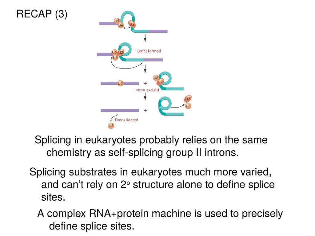
RECAP (3)Splicing in eukaryotes probably relies on the same
chemistry as self-splicing group II introns.
Splicing substrates in eukaryotes much more varied,
and can’t rely on 2o structure alone to define splice
sites.
A complex RNA+protein machine is used to precisely
define splice sites.
7.
The spliceosomeis made up of 5
small nuclear
ribonucleoprotein
subunits + > 100
proteins. These
snRNPs are
called: U1, U2, U4,
U5, U6, and
assemble in a
stepwise pathway
onto each intron.
There are also
many additional
non-snRNP
proteins in the
spliceosome.
8.
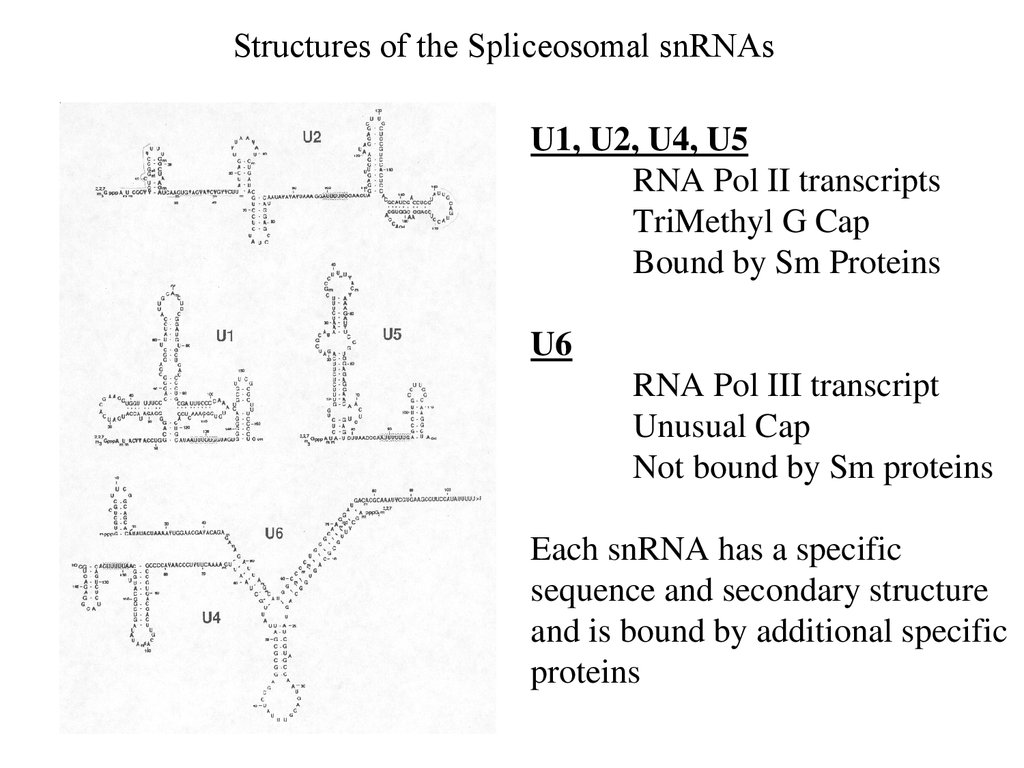
Structures of the Spliceosomal snRNAsU1, U2, U4, U5
RNA Pol II transcripts
TriMethyl G Cap
Bound by Sm Proteins
U6
RNA Pol III transcript
Unusual Cap
Not bound by Sm proteins
Each snRNA has a specific
sequence and secondary structure
and is bound by additional specific
proteins
9.
The earliest snRNP to bind to the pre-mRNA is U1,which uses its snRNA to base-pair to the 5’ splice
site.
10.
The U2 snRNP binds to the branchpoint via RNA/RNAbase-pairs to create a bulged A residue. This forms the
pre-spliceosomal “A” complex.
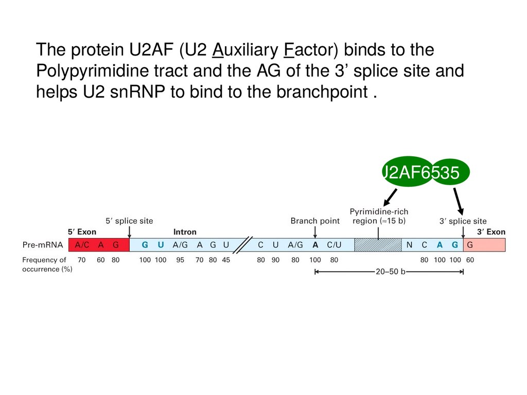
11.
The protein U2AF (U2 Auxiliary Factor) binds to thePolypyrimidine tract and the AG of the 3’ splice site and
helps U2 snRNP to bind to the branchpoint .
U2AF6535
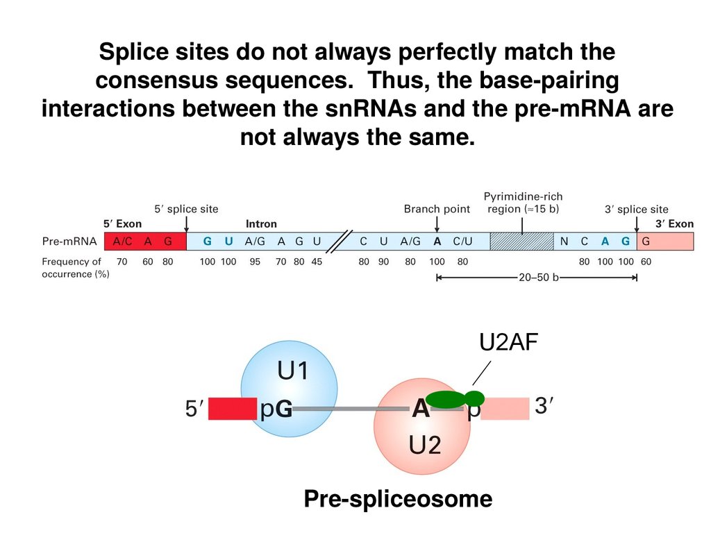
12.
Splice sites do not always perfectly match theconsensus sequences. Thus, the base-pairing
interactions between the snRNAs and the pre-mRNA are
not always the same.
U2AF
Pre-spliceosome
13.
The interactions ofU1 with the 5’ splice
site and U2 with the
branchpoint were
proven by creating
mutant splice sites
that bound the
Pre-mRNA snRNA so poorly
that splicing was
inhibited.
Compensating
mutations in the
snRNA that
restored
complementarity
(base-pairing) with
the splice site
restored splicing.
14.
The full spliceosomeis formed from the
pre-spliceosome by
the addition of the
U4/U5/U6 Tri-snRNP.
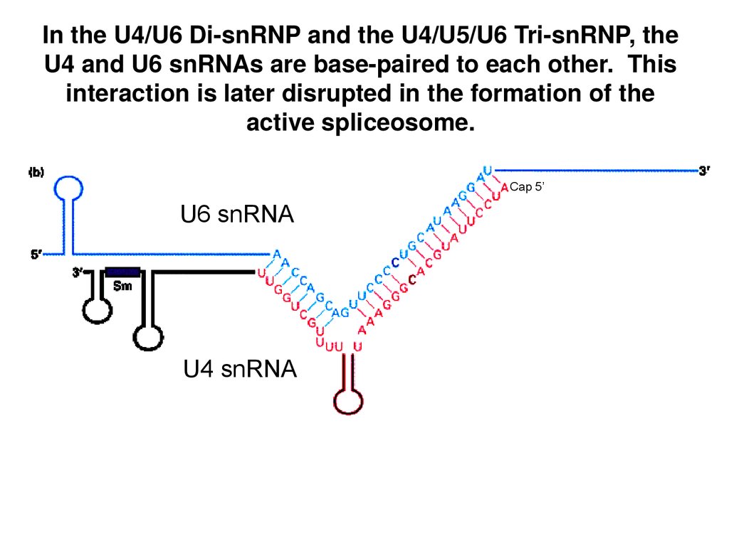
15.
In the U4/U6 Di-snRNP and the U4/U5/U6 Tri-snRNP, theU4 and U6 snRNAs are base-paired to each other. This
interaction is later disrupted in the formation of the
active spliceosome.
Cap 5’
U6 snRNA
U4 snRNA
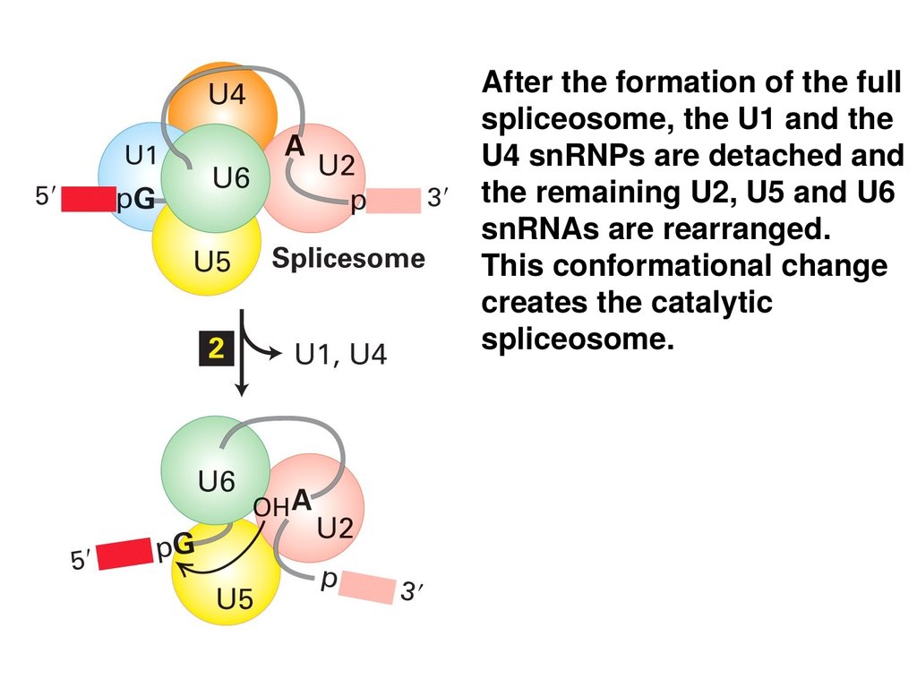
16.
After the formation of the fullspliceosome, the U1 and the
U4 snRNPs are detached and
the remaining U2, U5 and U6
snRNAs are rearranged.
This conformational change
creates the catalytic
spliceosome.
17.
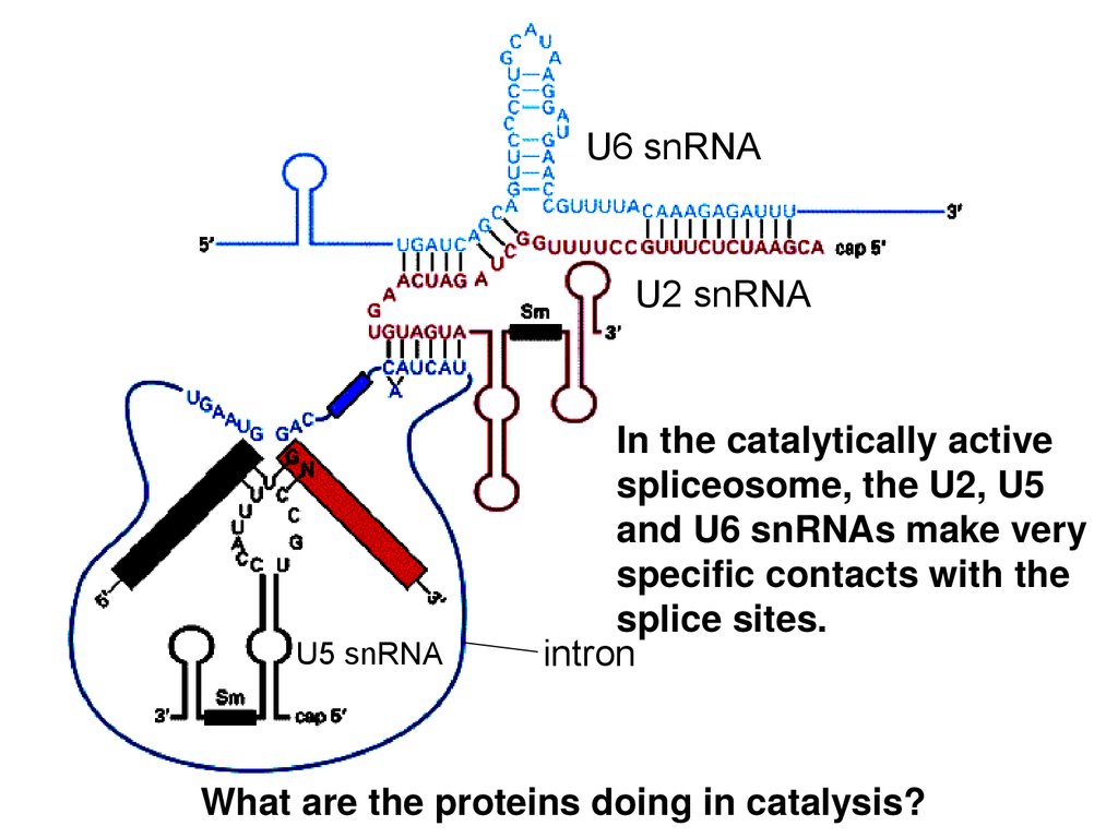
U6 snRNAU2 snRNA
U5 snRNA
In the catalytically active
spliceosome, the U2, U5
and U6 snRNAs make very
specific contacts with the
splice sites.
intron
18.
The twotransesterification
reactions of splicing
take place in the
mature spliceosome.
19.
After the secondtransesterification reaction,
the spliceosome comes
apart. The snRNPs are
recycled, and the spliced
exons and the lariat intron
are released.
20.
The lariat intron is debranched by Debranching Enzyme returning itto a typical linear state. This linear intron is quickly degraded by
ribonucleases.
21.
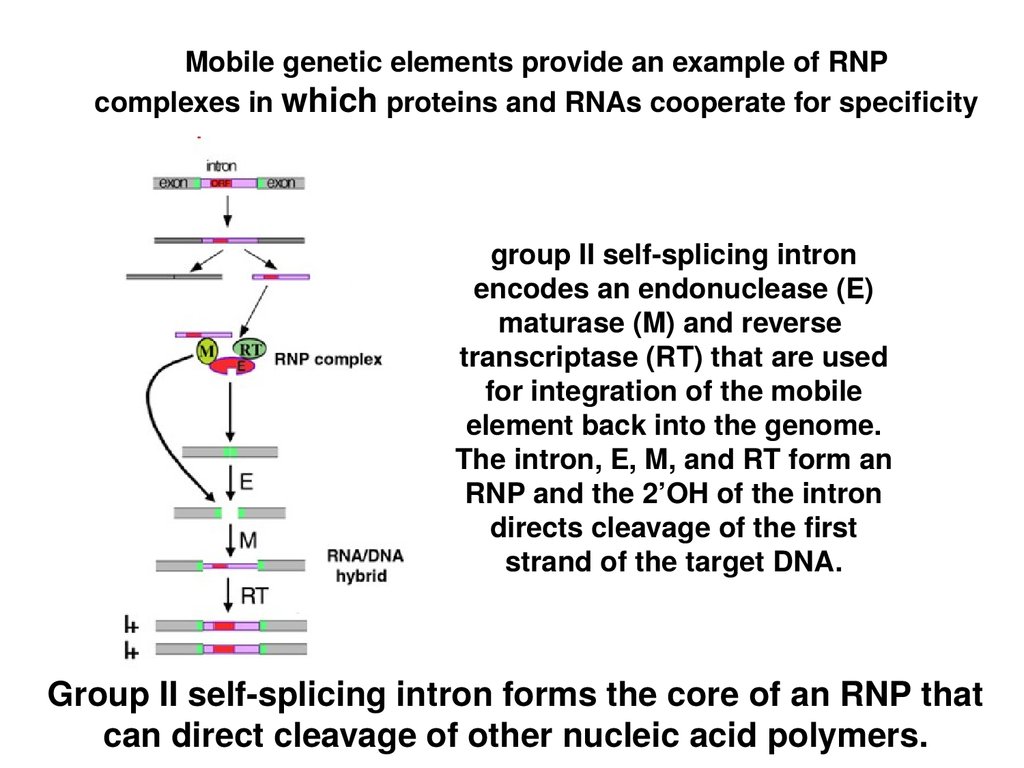
Mobile genetic elements provide an example of RNPcomplexes in which proteins and RNAs cooperate for specificity
group II self-splicing intron
encodes an endonuclease (E)
maturase (M) and reverse
transcriptase (RT) that are used
for integration of the mobile
element back into the genome.
The intron, E, M, and RT form an
RNP and the 2’OH of the intron
directs cleavage of the first
strand of the target DNA.
Group II self-splicing intron forms the core of an RNP that
can direct cleavage of other nucleic acid polymers.
22.
U6 snRNAU2 snRNA
U5 snRNA
In the catalytically active
spliceosome, the U2, U5
and U6 snRNAs make very
specific contacts with the
splice sites.
intron
What are the proteins doing in catalysis?
23.
A tale of the U5 protein, Prp8.Prp8 mutants are splicing defective.
Many Prp8 mutations suppress splicing defects
caused by 5’-SS, 3’-SS and branch point mutations.
Prp8 cross links to crucial U5, U6, 5’-SS, 3’-SS and
branch point residues.
Prp8 interacts with Brr2 and Snu114, which unwind
U4/U6 and allow U2 to pair with U6
24.
Crystal structure of Prp8 reveals a cavity of appropriatedimensions to position spliceosomal RNAs for catalysis.
Group II intron
Prp8
Structural domains of Prp8 (endonuclease, reverse
transcriptase) suggest ancient evolutionary origins as a
homing endonuclease.
25.
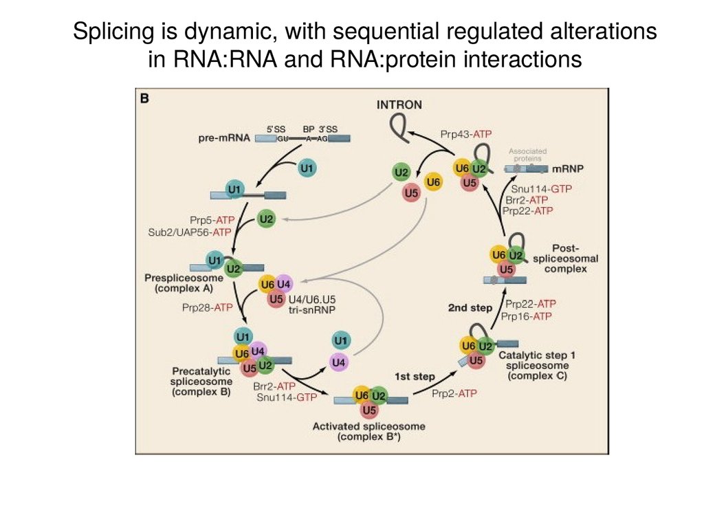
Splicing is dynamic, with sequential regulated alterationsin RNA:RNA and RNA:protein interactions
26.
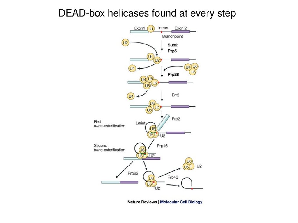
DEAD-box helicases found at every step27.
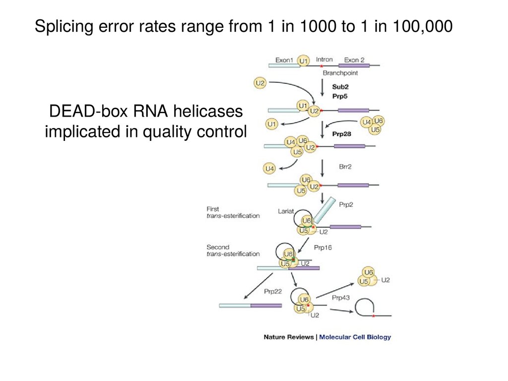
Splicing error rates range from 1 in 1000 to 1 in 100,000DEAD-box RNA helicases
implicated in quality control
28.
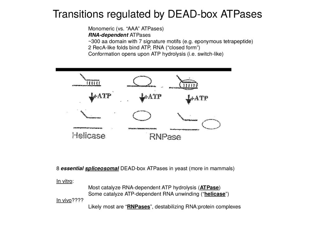
Transitions regulated by DEAD-box ATPasesMonomeric (vs. “AAA” ATPases)
RNA-dependent ATPases
~300 aa domain with 7 signature motifs (e.g. eponymous tetrapeptide)
2 RecA-like folds bind ATP, RNA (“closed form”)
Conformation opens upon ATP hydrolysis (i.e. switch-like)
8 essential spliceosomal DEAD-box ATPases in yeast (more in mammals)
In vitro:
In vivo????
Most catalyze RNA-dependent ATP hydrolysis (ATPase)
Some catalyze ATP-dependent RNA unwinding (“helicase”)
Likely most are “RNPases”, destabilizing RNA:protein complexes
29.
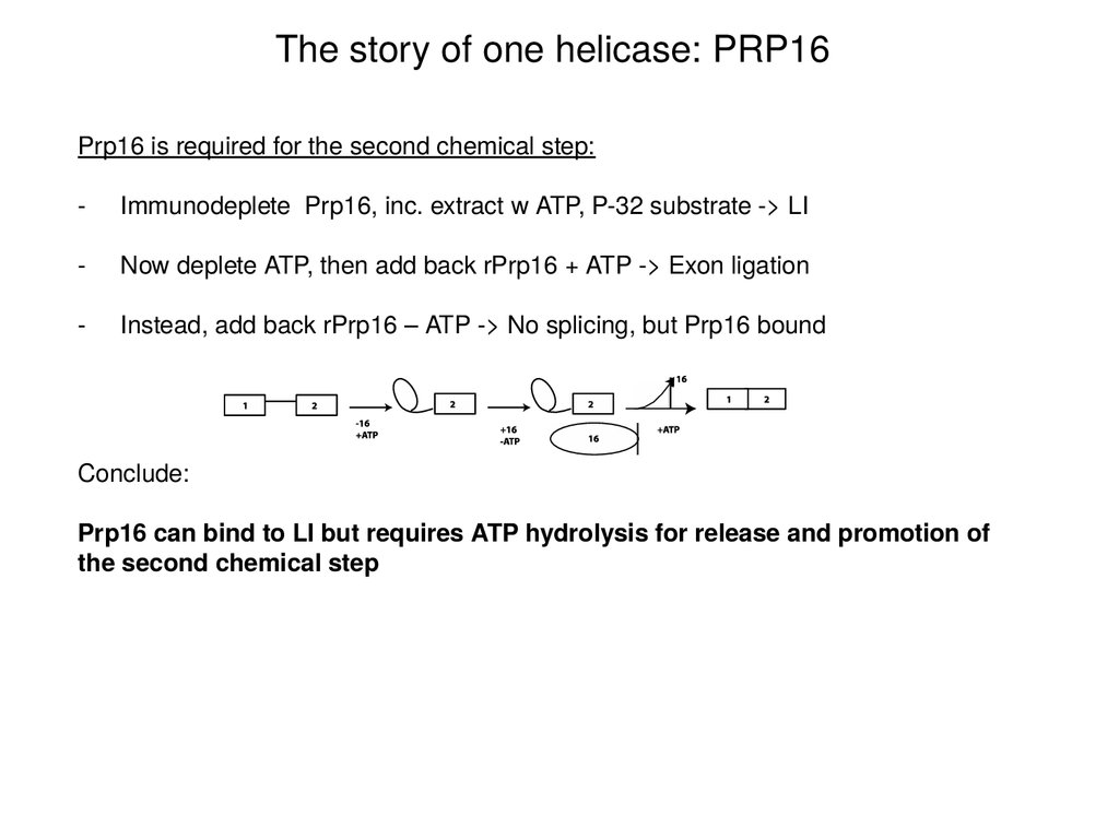
The story of one helicase: PRP16Prp16 is required for the second chemical step:
-
Immunodeplete Prp16, inc. extract w ATP, P-32 substrate -> LI
-
Now deplete ATP, then add back rPrp16 + ATP -> Exon ligation
-
Instead, add back rPrp16 – ATP -> No splicing, but Prp16 bound
Conclude:
Prp16 can bind to LI but requires ATP hydrolysis for release and promotion of
the second chemical step
30.
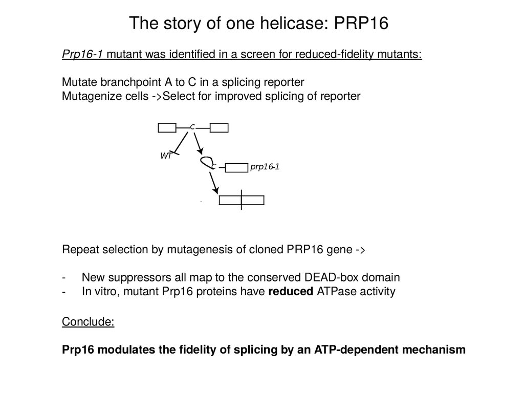
The story of one helicase: PRP16Prp16-1 mutant was identified in a screen for reduced-fidelity mutants:
Mutate branchpoint A to C in a splicing reporter
Mutagenize cells ->Select for improved splicing of reporter
C
WT
C
prp16-1
Repeat selection by mutagenesis of cloned PRP16 gene ->
-
New suppressors all map to the conserved DEAD-box domain
In vitro, mutant Prp16 proteins have reduced ATPase activity
Conclude:
Prp16 modulates the fidelity of splicing by an ATP-dependent mechanism
31.
The story of one helicase: PRP16Hypothesis: Prp16 promotes fidelity
1) branchpoint mutations -> slow conformational rearrangement -> rejection
2) suppressor mutations in Prp16 -> more time
32.
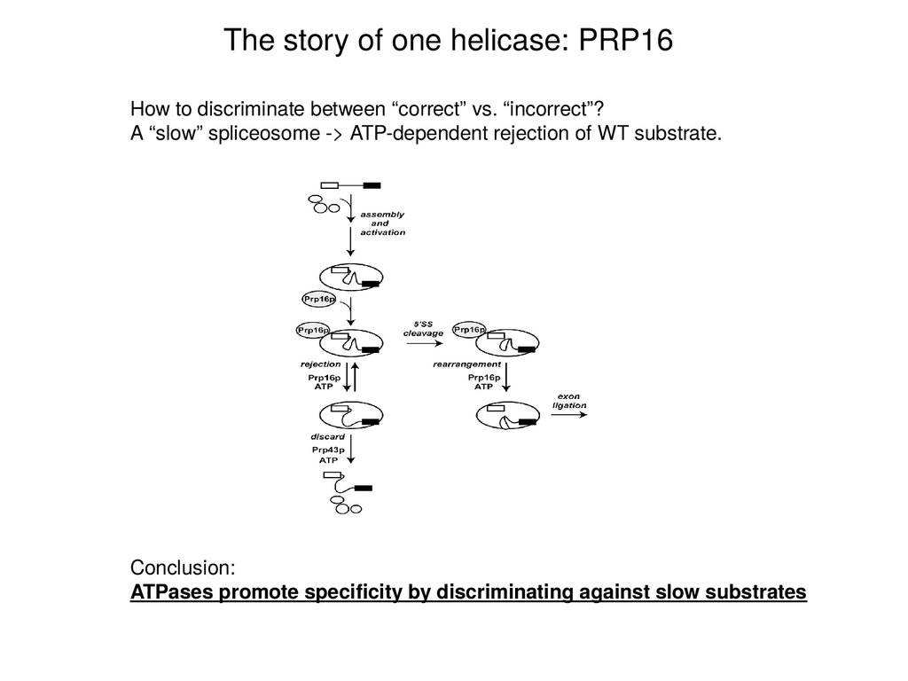
The story of one helicase: PRP16How to discriminate between “correct” vs. “incorrect”?
A “slow” spliceosome -> ATP-dependent rejection of WT substrate.
Conclusion:
ATPases promote specificity by discriminating against slow substrates
33.
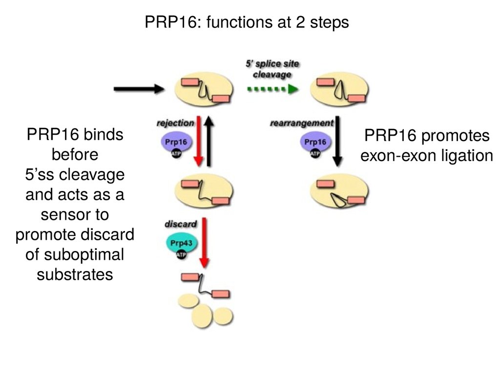
PRP16: functions at 2 stepsPRP16 binds
before
5’ss cleavage
and acts as a
sensor to
promote discard
of suboptimal
substrates
PRP16 promotes
exon-exon ligation
34.
QuestionsHow are the intervening sequences removed?
How are the splice sites identified?
35.
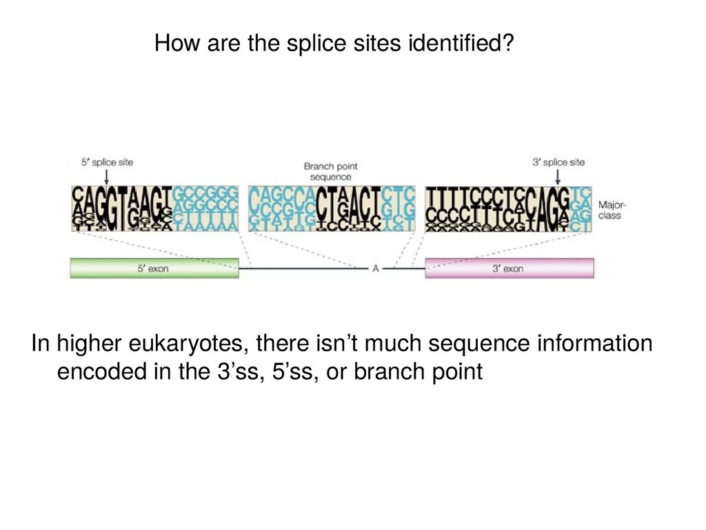
How are the splice sites identified?In higher eukaryotes, there isn’t much sequence information
encoded in the 3’ss, 5’ss, or branch point
36.
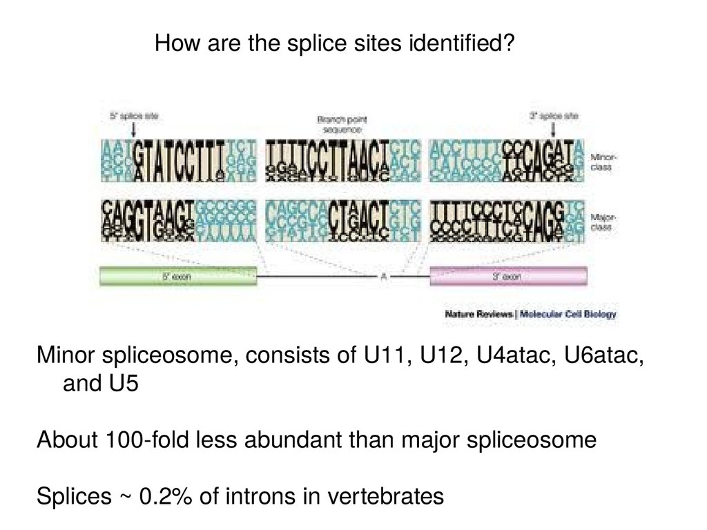
How are the splice sites identified?Minor spliceosome, consists of U11, U12, U4atac, U6atac,
and U5
About 100-fold less abundant than major spliceosome
Splices ~ 0.2% of introns in vertebrates
37.
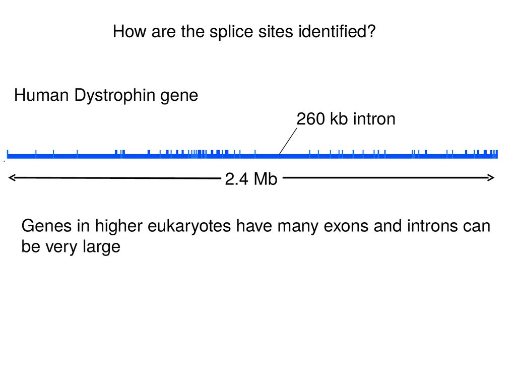
How are the splice sites identified?Human Dystrophin gene
260 kb intron
2.4 Mb
Genes in higher eukaryotes have many exons and introns can
be very large
38.
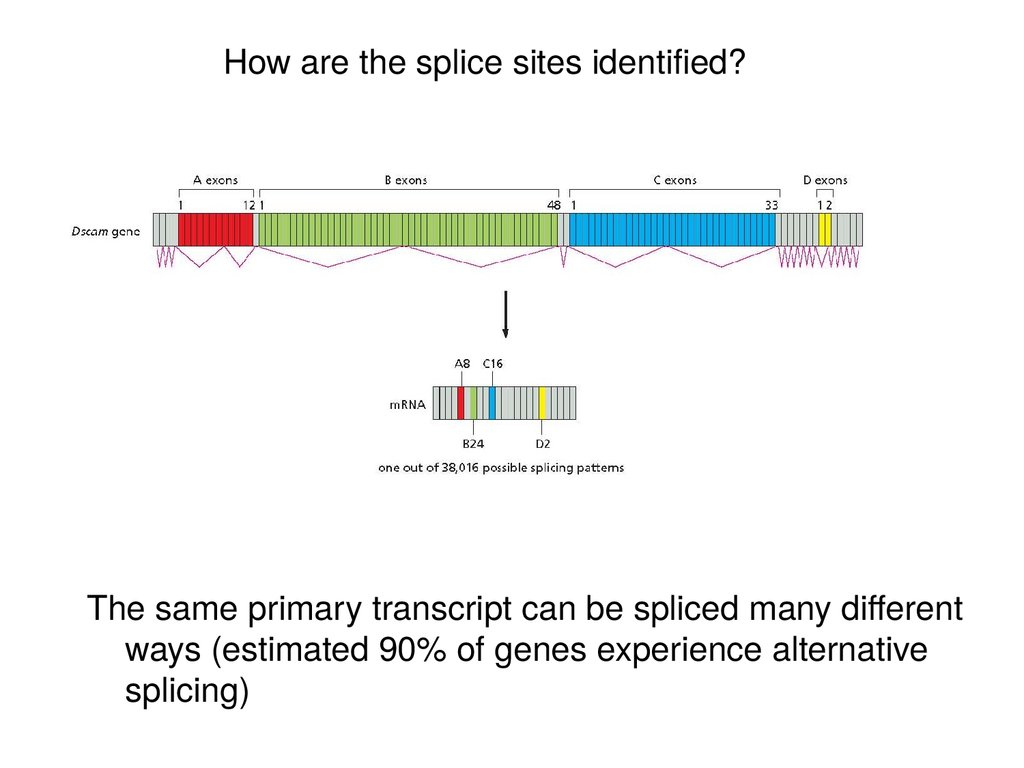
How are the splice sites identified?The same primary transcript can be spliced many different
ways (estimated 90% of genes experience alternative
splicing)
39.
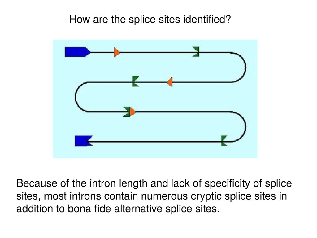
How are the splice sites identified?Because of the intron length and lack of specificity of splice
sites, most introns contain numerous cryptic splice sites in
addition to bona fide alternative splice sites.
40.
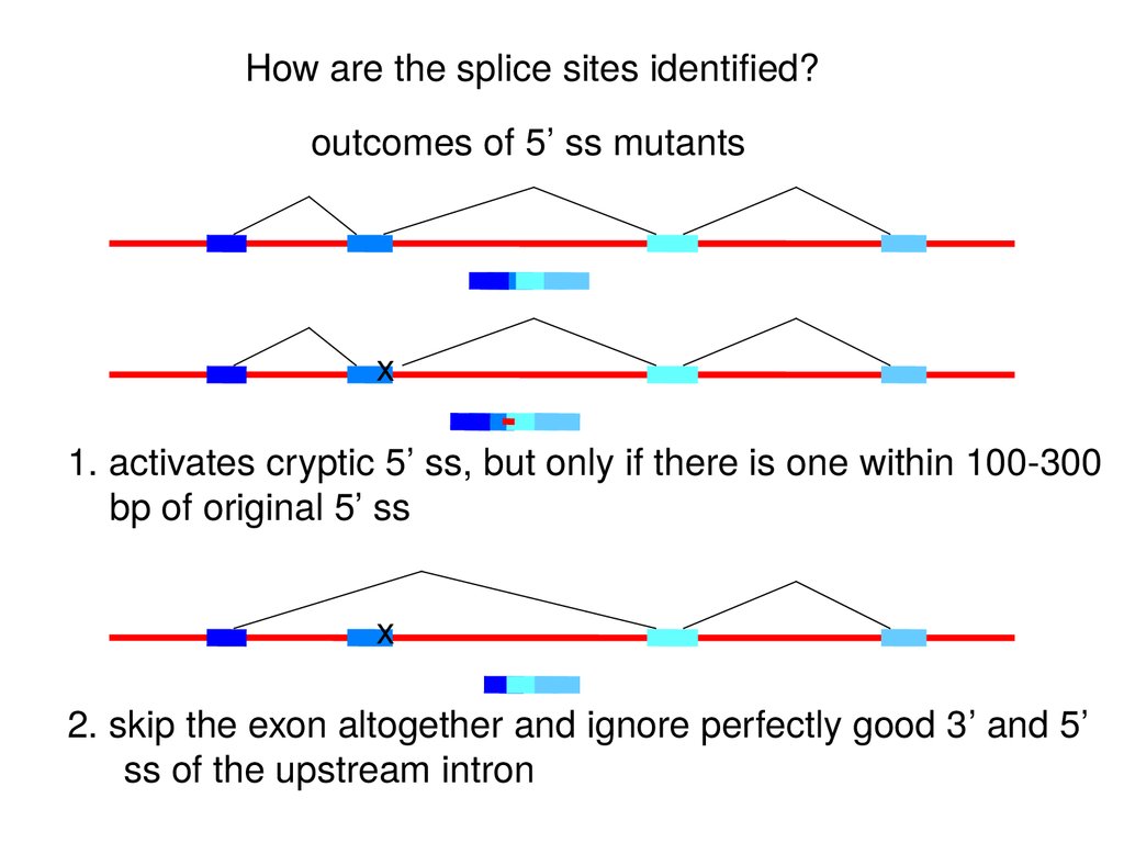
How are the splice sites identified?outcomes of 5’ ss mutants
x
1. activates cryptic 5’ ss, but only if there is one within 100-300
bp of original 5’ ss
x
2. skip the exon altogether and ignore perfectly good 3’ and 5’
ss of the upstream intron
41.
How are the splice sites identified?beta-globin mutants that create a new 3’ ss within
an intron:
x
also create a new exon???
42.
In multicellular organisms, exons are recognized as units prior to assembly ofthe spliceosome across the long introns. This “exon definition” step involves
interactions between the splice sites across the exon and special sequences in
the exon called Exonic Splicing Enhancers (ESE).
The sequences in exons are selected to not just code for particular
peptide sequences, but also for binding of regulatory proteins to ESE’s.
43.
How are the splice sites identified?Boundaries between introns & exons are recognized through its interaction with multiple
proteins either across exon or intron
Intron definition:
Uses intron as the unit of
recognition mechanism.
Complex forms through stabilized
protein interactions across the
intron
RS
70K
Exon 1
SR
RS
SF2
SR
A
U2AF
U2AF35
RS
Exon 2
SF1
U1
snRNP
Exon Definition:
Complex can easily form stabilized
protein interactions across the exon.
Excises out the flanking introns
SR
Intron Definition
SR
A
U2AF
U2AF35
RS
SR
SR
RS
70K
RS
SF2
Exon
SF1
(Cote, Univ. of Ottawa)
Exon Definition
U1
snRNP
Stable interaction confirms accuracy of splice site choice
44.
Why are exons preferentially recognized?Differential size distributions of exons (~50 to 300 nt) vs. introns (<100-100,000 nt)
• SR protein - preferentially binds to exon sequences
- mark the 5’ & 3’ splicing sites in conjunction w/ U1 & U2 during transcription
• hnRNP - heterogenous nuclear ribonucleoproteins (twice the diameter of nucleosome)
- consists at least eight different proteins
- compacts introns, thereby masking cryptic splicing sites
- preferentially binds to introns, but also bind to exons, although less frequently
45.
Cross-exon bridging interactions involve SR domains of U2AF, U170KAnd 1 or more SR-family proteins
~12 in mammals (and # AS isoforms!)
Tissue-specific differences in concentration
RRMs vary in degree of sequence preferences
Outstanding question:
What triggers the switch from Exon- to Intron-Defined interactions?
46.
Vertebrate external exons47.
Splicing is co-transcriptional and all introns assayed are splicedwithin 5-10 minutes of transcription of the downstream exon
and 3’ splice site, regardless of intron size (1 kb or 240 kb)
48.
Defining an exon involves thespecific stabilization or
destabilization of splice site
recognition
Stabilization: exon inclusion
Destabilization: exon skipping
49.
Regulation of alternativesplicing involves the specific
stabilization or destabilization of
splice site recognition
Stabilization: exon inclusion
Destabilization: exon skipping
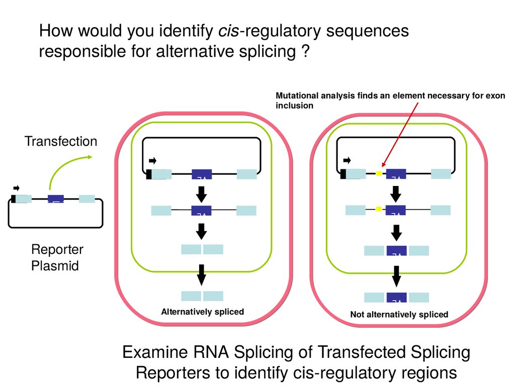
50.
How would you identify cis-regulatory sequencesresponsible for alternative splicing ?
Mutational analysis finds an element necessary for exon
inclusion
Transfection
Reporter
Plasmid
Alternatively spliced
Not alternatively spliced
Examine RNA Splicing of Transfected Splicing
Reporters to identify cis-regulatory regions
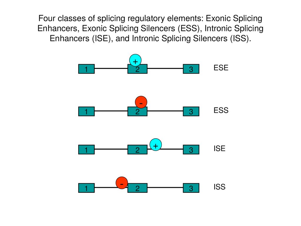
51.
Four classes of splicing regulatory elements: Exonic SplicingEnhancers, Exonic Splicing Silencers (ESS), Intronic Splicing
Enhancers (ISE), and Intronic Splicing Silencers (ISS).
+
1
2
-
1
2
1
2
1
-
2
+
3
ESE
3
ESS
3
ISE
3
ISS
52.
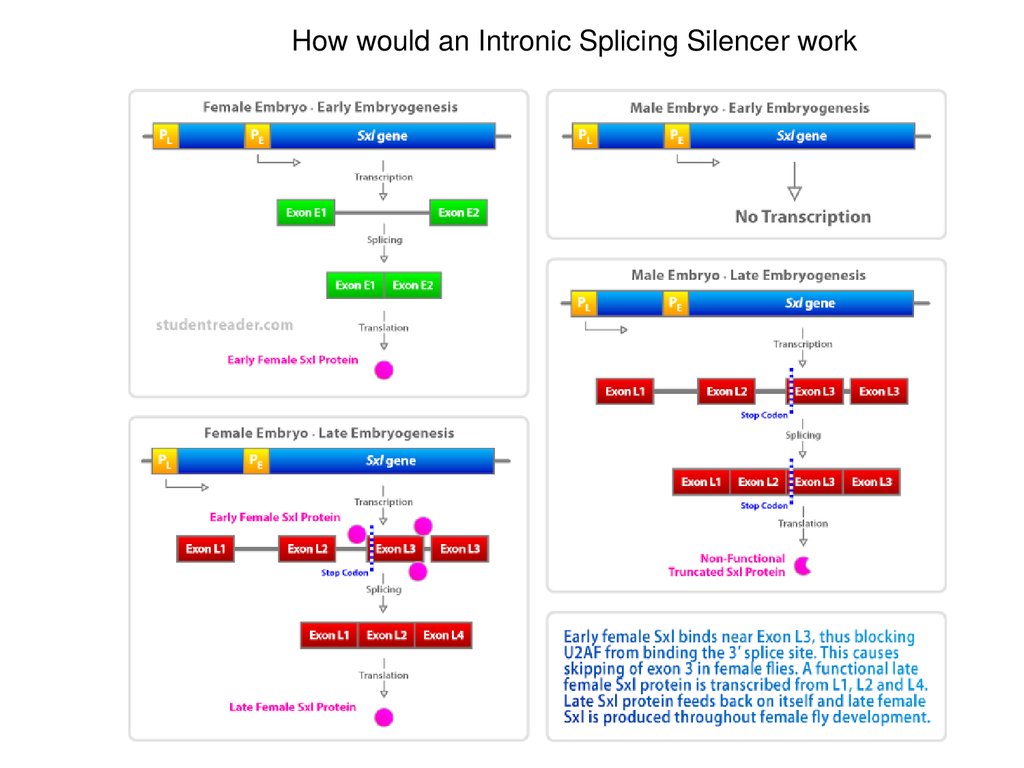
How would an Intronic Splicing Silencer work53.
SR proteins generally bind ESE, ESS, ISE, and ISSs54.
The SR Proteins are a family ofproteins with a common
domain structure of 1 or 2 RNP
RNA binding domains (also
called RRMs) and a C-terminal
domain rich in SR dipeptides.
These proteins are involved in
many aspects of splicing, but
most significantly they bind to
Exonic Splicing Enhancers
(ESEs) and stimulate
spliceosome assembly at the
adjacent sights.
It is thought that most exons
carry ESE’s and require SR
proteins for exon recognition.
55.
SR Proteins bind to specific RNA elements using their RNA binding domainssimilar to those in the Sex-Lethal protein.
56.
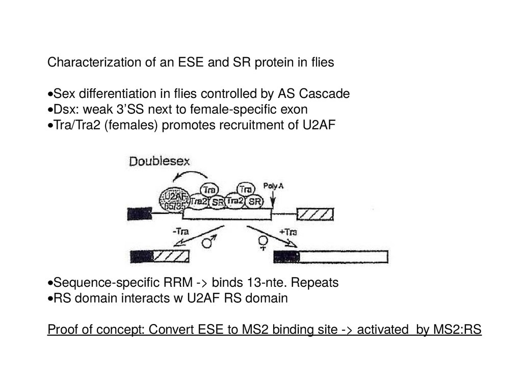
Characterization of an ESE and SR protein in fliesSex differentiation in flies controlled by AS Cascade
Dsx: weak 3’SS next to female-specific exon
Tra/Tra2 (females) promotes recruitment of U2AF
Sequence-specific RRM -> binds 13-nte. Repeats
RS domain interacts w U2AF RS domain
Proof of concept: Convert ESE to MS2 binding site -> activated by MS2:RS
57.
hnRNP function at ISSshnRNP contain RRMs but not SR domain
Can block sterically, tighter binding affinity than U2AF
58.
SR Proteins bind to CTD of polII: promote co-transcriptional splicing?59.
CTD of RNA pol II plays important role in pre-mRNA splicing(Kornblihtt et al, 2004)
60.
Does splice site strength affect alternative splicing?61.
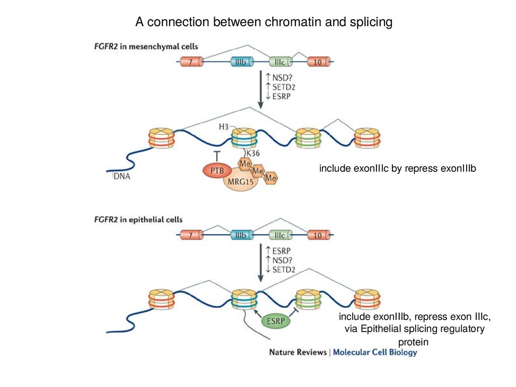
A connection between chromatin and splicinginclude exonIIIc by repress exonIIIb
include exonIIIb, repress exon IIIc,
via Epithelial splicing regulatory
protein
62.
mRNA export - formation of an export competent mRNPBalbiani Rings (Chironomus tentans)
Sees formation of mRNP as
transcription commences
Follow mRNP through NPC
• Why export as a protein/DNA complex? RNAs are too big
and lack the signals to interact w/ nuclear export receptors
• Specific “adaptor” proteins must first bind to the RNA and chaperone this
molecule to the export receptor, which, in turn, guides the RNA across the
NPC
63.
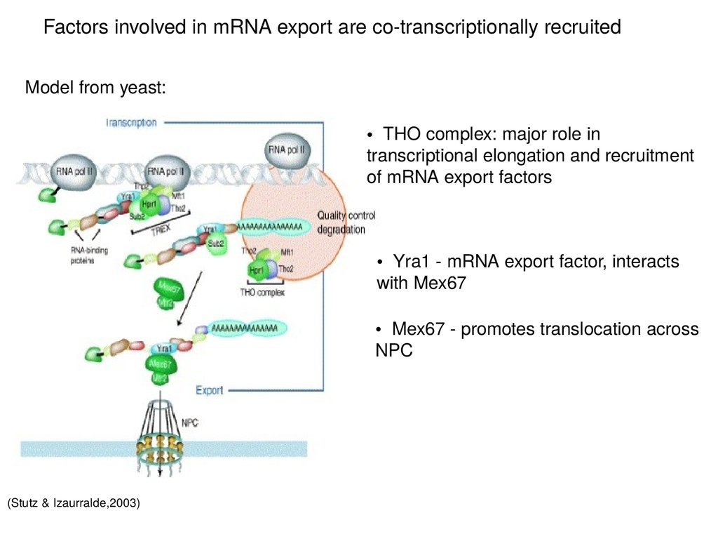
Factors involved in mRNA export are co-transcriptionally recruitedModel from yeast:
• THO complex: major role in
transcriptional elongation and recruitment
of mRNA export factors
• Yra1 - mRNA export factor, interacts
with Mex67
• Mex67 - promotes translocation across
NPC
(Stutz & Izaurralde,2003)
64.
Proteins involved in the nuclear export of mRNAs(Sub2p)
(Mex67p)
(Yra1p)
(Cullen, 2003)
(yeast homolog is indicated in parentheses)
(Mtr2p)
65.
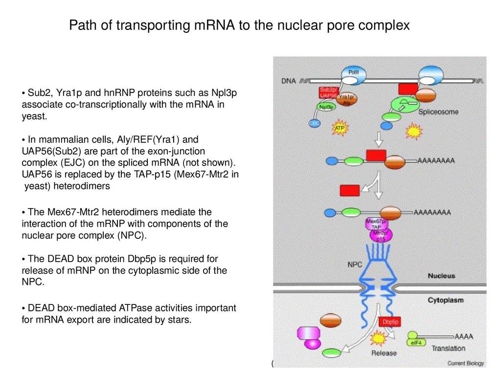
Path of transporting mRNA to the nuclear pore complex• Sub2, Yra1p and hnRNP proteins such as Npl3p
associate co-transcriptionally with the mRNA in
yeast.
• In mammalian cells, Aly/REF(Yra1) and
UAP56(Sub2) are part of the exon-junction
complex (EJC) on the spliced mRNA (not shown).
UAP56 is replaced by the TAP-p15 (Mex67-Mtr2 in
yeast) heterodimers
• The Mex67-Mtr2 heterodimers mediate the
interaction of the mRNP with components of the
nuclear pore complex (NPC).
• The DEAD box protein Dbp5p is required for
release of mRNP on the cytoplasmic side of the
NPC.
• DEAD box-mediated ATPase activities important
for mRNA export are indicated by stars.
(Linder & Stutz, 2001)
66.
Genetic approach to identify genes involved in mRNA export processNon-essential genes
RNA FISH w/
oligo dT
Mutagenized cells or
collection of nonessential gene KOs
(Lei et al, 2003)
essential genes
Growth at permissive
temperature
RNA FISH w/ oligo dT
Shift to non-permissive
temperature
67.
Mex67(yeast) and NXF1(Drosophila) are essential genes involved inmRNA export
Nuclear mRNA accumulation is observed after shifting
mex67 TS mutant to the restrictive temperature (37°C)
(Stutz & Izaurralde, 2003)
Visualization of poly(A) mRNA is accomplished by in situ using
fluorescently-labeled oligo-dT probe
68.
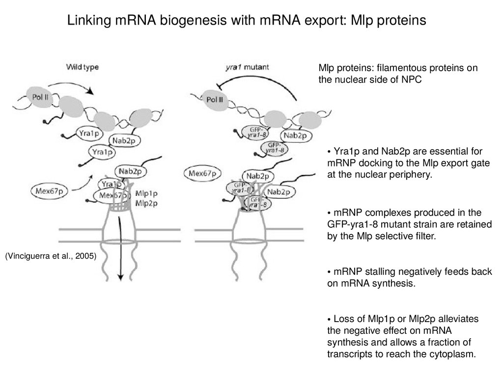
Linking mRNA biogenesis with mRNA export: Mlp proteinsMlp proteins: filamentous proteins on
the nuclear side of NPC
• Yra1p and Nab2p are essential for
mRNP docking to the Mlp export gate
at the nuclear periphery.
• mRNP complexes produced in the
GFP-yra1-8 mutant strain are retained
by the Mlp selective filter.
(Vinciguerra et al., 2005)
• mRNP stalling negatively feeds back
on mRNA synthesis.
• Loss of Mlp1p or Mlp2p alleviates
the negative effect on mRNA
synthesis and allows a fraction of
transcripts to reach the cytoplasm.
69.
Mlp proteins act as selective filters at NPC entrance• The perinuclear Mlp1p protein contributes to
mRNP surveillance by retaining unspliced
transcripts within the nucleus
• This is achieved possibly via recognition of a
component associated with the 5´ splice site.
(Vinciguerra & Stutz, 2004)
70.
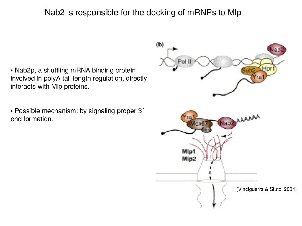
Nab2 is responsible for the docking of mRNPs to Mlp• Nab2p, a shuttling mRNA binding protein
involved in polyA tail length regulation, directly
interacts with Mlp proteins.
• Possible mechanism: by signaling proper 3´
end formation.
(Vinciguerra & Stutz, 2004)