


 medicine
Similar presentations:


 – Digestion and Organs of digestive system")


")

Histology of gallbladder and biliary tract
1.
Semey state medical universityDepartment of anatomy and histology
SIW
Theme: Histology of gallbladder and
biliary tract
Prepared by: Erlanova N.E,
343 group
Checked by: Uzbekova S.E
Semey 2016
2.
Plan:1. Biliary tract
2. Histology of biliary tract
3. Histology of gallbladder
4. Age changes
5. Regeneration
6. Conclusion
3.
Biliary tract— a channel of a liver for a bileconclusion, part of a gastrointestinal tract of the
highest vertebral and the person. It is formed at
merge of a hepatic channel and channel of a gall
bladder. At the highest vertebrata conducts in a
duodenum gleam (at the lowest goes to the top
department of an average gut). As a rule, together
with an output channel of a pancreas. In walls of a
bilious channel there is Oddi's sphincter regulating
frequency of intake of bile in intestines.
4.
5.
The wall of biliary tract consistsof 3 layer:
Mucous-single-layer (high) prismatic
epithelium
Muscular –bundles of smooth
myocytes
External- LFCT
6.
Epitelial cells are rich with lysosomes andmitochondria which concentrate mainly in their
apical part. The epithelium of tract in the
functional relation can carry out as secretion
(mucous glands of bilious channels), and a
resorption. Also goblet cells which quantity
sharply increases at an inflammation of tract
meet. A surface of a mucous membrane of
channels on a big extent smooth, but in some sites
it forms folds: a spiral fold (plica spiralis) — in a
vesical duct, a row the pocket figurative of folds —
in distal part of the general bilious tract. (these
folds extremely complicate or do impossible
sounding of a channel from a duodenum).
7.
Generally the muscular layer is expressed better and presentedto the item by two layers — external and internal; between
them the vegetative (autonomous) intermuscular nervous
texture containing nervous cells lies. In a place of merge of
hepatic channels in the general hepatic concentric congestions of
muscle fibers form similarity of a sphincter — a physiological
sphincter of Mirissi. Thickenings of a muscular layer are noted
and in other places: in a vesical duct — at an release from a
bubble neck, generally. Тhe item — in its intramural part. The
muscular device of intramural part of the general is most
difficult arranged. Тhe item where distinguish two circular
sphincters — a sphincter of the general. Тhe item (m. sphincter
ductus choledochi, PNA) located in a channel wall before an
ampoule, and a sphincter of a hepatopancreatic ampoule (m.
sphincter ampullae hepatopancreaticae, PNA). The specified
sphincters in total with a sphincter of a pancreatic channel
make the combined sphincter, the described Oddi (R. Oddi). The
external cover (tunica externa) of channels is formed by friable
not properly executed connecting tissue. In it vessels and
8.
Gallbladder- thinwalled body, thebile containing 4070 ml. Walls of a
gall bladder
consists of three
layers:
1. Mucous
2. Muscular
3. Serous
9.
The mucous membrane forms numerousfolds. It is covered by the high prismatic epithelial
cells having a border.
Under an epithelium lamina propria of a
mucous membrane containing a large number the
elastic fibers settles down. In a vesicle neck in it
there are alveolar and tubular glands , which
secreted mucus.
The epithelium of a mucous membrane
possesses ability to soak up water and some other
substances from the bile filling a bubble cavity.
Therefore vesical bile always more dense
consistence and more dark color, than the bile
streaming directly from a liver.
10.
The muscular layer of a gall bladderconsists of bunches of the smooth myocytes
located a type of a network in which their
circular direction prevails. In a bubble neck
circular bunches of muscular cells are especially
strongly developed. Together with muscular a
layer of a biliary duct they form a sphincter.
Between bunches of cells always there
are well expressed layers of loose connective
tissue. The adventitial layer of a gall bladder
consists of dense fibrous connective tissue
which contains a lot of thick the elastic of the
fibers forming networks.
11.
Age changes• In
hepatocytes the quantity of a lipofusin
which paints cells in brown color increases.
• The number of the sharing cells sharply
decreases.
• Hypertrophy of nucleus of hepatocytes
• Increasing of DNA in hepatocytes
• growth of connective tissue between liver
lobes
12.
RegenerationThe liver possesses high ability to
physiological and reparative regeneration. At
animals during removal from 50 to 70% of
tissue of liver its initial weight is restored for
10-14 day. Processes of regeneration happen by
compensatory increase in the sizes of cells and
reproduction of hepatocytes. The food rich with
carbohydrates and proteins stimulates
regeneration of a liver.
13.
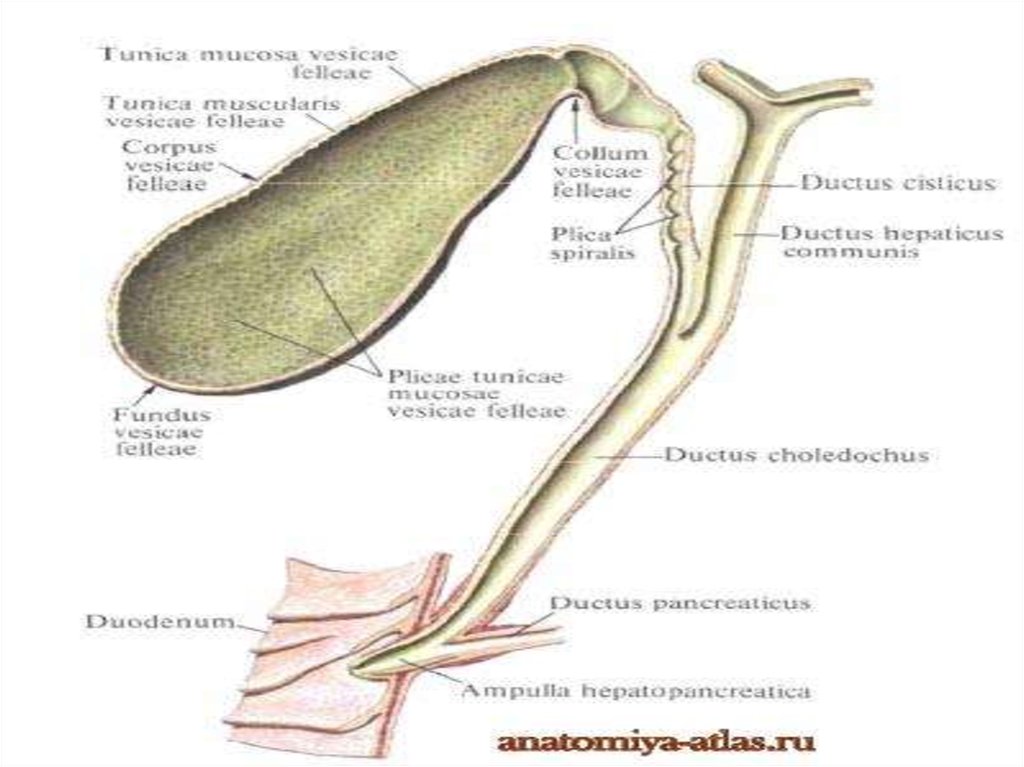
ConclusionThe gall bladder represents the sacciform tank
for the bile developed in a liver; it has the extended form
with one wide, other narrow end, and bubble width from
a bottom to a neck decreases gradually. Length of a gall
bladder fluctuates from 8 to 14 cm, width — from 3 to 5
cm, it reaches capacity 40 — 70 cm ³. It has dark green
coloring and rather thin wall. In a gall bladder
distinguish a bottom (Latin of fundus vesicae fellae), the
most distal and wide part, a body (Latin of corpus
vesicae fellae) — a middle part, and a neck (Latin of
collum vesicae fellae) — peripheral narrow part from
which the biliary duct (Latin of ductus cysticus), the
reporting bubble with the general bilious channel (Latin
of ductus choledochus departs).