


. The long glands begin with the")


 medicine
Similar presentations:

")

")


Lab 2 Esophagus & Stomach
1. Lab 2 Esophagus & Stomach
Lab 2Esophagus & Stomach
Georgia Duker, PhD
2. K34 Esophagus - Upper As with all the tubular portion of the GI tract, the esophagus is built of four layers: 1. mucosa =
stratified squamous mucous type epithelium, lamina propria, and a smooth muscle muscularis mucosa (slightly darker red instaining,just below the lymphocyte nodules)
2. submucosa =loose connective tissue
3. muscularis externa = inner circular and outer longitudinal muscle; here in the upper third of the esophagus, both are made of skeletal muscle
4. adventitia = the outer most layer blends into surrounding connective tissue of the mediastinum.
3. K35 Esophagus - Upper Esophagus - Upper The mucosa itself is composed of three layers. 1. The epithelium is stratified
squamous, mucous type, therefore nuclei persist out to the surface. Rete pegs interdigitate to attach theepithelium to the underlying lamina propria.
2. The lamina propria contains scattered lymphocytes, normal for the GI tract
3. The muscularis mucosa is smooth muscle, longitudinally oriented (therefore cut here in cross section).
4. K36 Esophagus - Upper The esophageal submucosa is a wide layer of varying densities. Larger blood vessels and the submucosal or
esophageal glands (mucous type) areprominent here. A scattered number of automonic fibers are present as the submucosal or Meisner's plexus (not seen on this slide).
Skeletal muscle fibers are easily discerned in the muscularis externa.
5. K37 Esophagus - Upper The ducts from the submucosal or esophageal glands course through the layers of the mucosa on their way
to the esophageal lumen. Theseglands provide lubrication for swallowing.
Can you differentiate the fine smooth muscle bundles of the muscularis mucosa from the surrounding dense connective tissue?
6. K38 Esophagus - Upper Both layers of the muscularis externa are composed of skeletal muscle.
7. K39 Esophagus - Upper Skeletal muscle of the muscularis externa
8. K40 Esophagus - Upper Skeletal muscle of the muscularis externa, longitudinal section.
9. K41 Esophagus - Upper Skeletal muscle of the muscularis externa, cross section.
10. K42 Esophagus - Middle Distinguish the four major layers: 1. Mucosa = epithelium, stratified squamous, mucous type; lamina
propria & muscularis mucosa.2. Submucosa = note the esophageal submucosal glands.
3. Muscularis externa = inner circular & outer longitudinal
4. Adventitia
11. K43 Esophagus - Middle The mucosa is composed of a stratified squamous epithelium, lamina propria with lymphocyte nodule, and
the musclaris mucosa, smoothmuscle oriented longitudinally, therefore cut in cross section here.
12. K44 Esophagus - Middle Nuclei are clearly visible to the surface of the stratified squamous epithelium. The lamina propria
contains lymphocytes expressing the integrin "addressin", a4b7, that recognize a homing ligand on themucosal endothelial cells, MadCAM-1. (see the Mucosal Immunology Lecture)
13. K45 Esophagus - Middle Small bundles of smooth muscle of the muscularis mucosa can be distinguished from the surrounding
connective tissue. Rememberthat since smooth muscle cells are not individually innervated, they communicate via gap junctions. Thus the clusters of smooth
muscle cells.
14. K46 Esophagus - Middle Deep to the three strata of the mucosa, esophageal glands are found in the submucosa.
15. K47 Esophagus - Middle Ducts from the submucosal esophageal glands penetrate the strata of the mucosa to access the esophageal
lumen.16. K48 Esophagus - Middle The muscularis externa is a blend of both skeletal and smooth muscle. This is the only unique feature
that distinguishes the middlethird from the upper and lower esophagus.
17. K49 Esophagus - Middle Muscle fibers of the inner circular layer are cut in longitudinal orientation. Both smooth and skeletal
muscle are present.18. K50 Esophagus - Middle Muscularis externa - longitudinal section through both smooth and skeletal muscle.
19. K51 Esophagus - Middle Muscularis externa - cross section through both smooth and skeletal muscle.
20. K52 Esophagus - Lower Distinguish the four major layers: 1. Mucosa = epithelium, lamina propria, muscularis mucosa 2. Submucosa
= here, rather a dense, slightly orange tissue layer3. Muscularis externa = all smooth muscle; note the thickened circular layer that forms the lower esophageal sphincter
4. Adventitia or serosa = as the esophagus penetrates below the diaphragm, it is covered by a serosa
21. K53 Esophagus Lower
22. K54 Esophagus Lower The lower esophagus is unique for the muscularis externa built of only smoth muscle.
23. K55 Esophagus Lower Higher power view of smooth muscle cut in longitudinal section.
24. K56 Esophagus Lower Both the circular and longitudinal layers of the muscularis externa are smooth muscle in the lower third of
the esophagus.25. K57 Esophagus Lower Smooth muscle cut in cross section.
26. K58 Espohageal-Stomach Junction There is an abrupt transition from the histology of the esophagus to the cardiac stomach. The
stomach epithelium is characterized by pits andglands that project into the lamina propria. The muscularis mucosa becomes circularly oriented and lies immediately at the base of the gastric
glands. Due to the unusual shape of the stomach, the muscularis externa becomes distorted into three layers: inner oblique, middle circular and
outer longitudinal.
27. K59 Esophagus-Stomach Junction The transition between esophagus and stomach is abrupt and reflects the different functions of
the two organs from conduit todigestion and storage.
28. K60 Esophagus-Stomach Junction Occasionally, at the junction of the cardiac stomach and the esophagus there is a back growth of
the cardiac glands of the stomachinto the lamina propria of the esophagus. These are still called cardiac glands and help to protect the esophagus from acid reflux.
29. K61 Stomach - Cardiac The cardiac stomach displays pits and glands with a 1:1 ratio in depth. While all gastric pits are all
lined by surface mucous cells, the glands of thecardiac region contain predominantly mucous cells with scattered parietal cells. The large, round bright pink cells of the glands are the acid
secreting parietal cells. The bottom of the glands rest on the muscularis mucosa.
30. K62 Stomach - Cardiac The cardiac stomach is characterized by surface pits into which drain 2-4 cardiac glands. The ratio of
the lengths of pits:glands is 1:1 in thecardiac stomach. The pits are lined by surface mucous cells. These secrete an alkaline, bicarbonate rich mucous to coat and protect the stomach
surface. Within the glands are the round, bright pink parietal cells that secrete HCl and Vit B12 intrinsic factor. Mucous cells within the glands
release a neutral or slightly acid mucous, so as to not dilute the acid from the parietal cells.
31. K63 Stomach - Cardiac The round morphology of the parietal cells is due to the massive intracellular cannaliculus (requires EM
to visualize). The extrasurface area is needed to insert the transporters for H+K+ATPase and Cl-. The nuclei are therefore pushed up to the center of
these plump cells. Large numbers of mitochondria (for ion pumping) renders the cytoplasm eosinophilic with H&E staining.
32. K64 Stomach - Body This is the histological appearance of the stomach in the fundus, body and antrum. Note the broad, loose
submucosa with large blood vessels toassimilate absorbed water. The thick muscularis externa churns and mixes the stomach contents. The mucosa is expand by very deep glands. The
ratio of the lengths of pits to glands is approximately 1:4.
33. K65 Stomach - Body The pits are lined by surface mucous cells (note pale pink cytoplasm). The long glands begin with the
isthmus (source of stem cells for both thepits and glands); the neck (mucous neck cells and parietal cells); and the base (parietal cells and chief cells - pink and blue respectively). The
muscularis mucosa is the pale pink band at the bottom of the glands (top of the slide), bordering the watery submucosa.
34. K66 Stomach - Body The base of the glands are slightly basophilic due to the stored granules of enzymes in the chief cells.
Chief cells secrete pepsinogen, rennin andlipase.
35. K67 Stomach - Body Surface mucous cells of the gastric glands. These secrete mucous in response to the friction of food in the
lumen, or chemical stimuli such asethanol. The HCO3- concentration in the mucous is increased by Ca2+, prostaglandins E & F, cholinergic agents and dibutyryl cGMP. Drugs that
decrease the HCO3- include aspirin and NSAIDs.
36. K68 Stomach - Body The surface pits penetrate the lamina propria. Lymphocytes can be seen throughout the lamina of most
sections of the GI tract.37. K69 Stomach - Body Cross section from the base of the gastric glands displaying chief cells. The round nuclei are pushed to the
base of the cells, while the gritty,basophilic apical end is fill with stored granules of secretions. The few parietal cells display eosinophilic cytoplasm and a central nucleus
38.
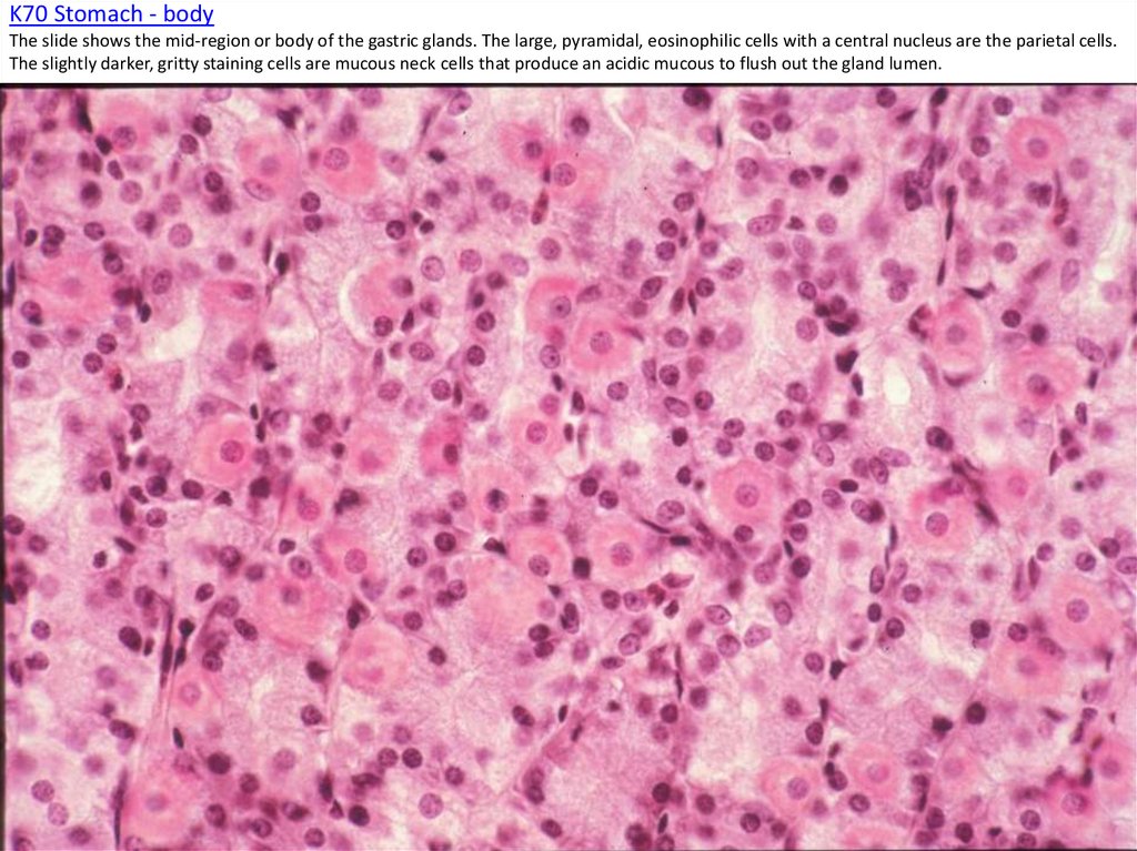
K70 Stomach - bodyThe slide shows the mid-region or body of the gastric glands. The large, pyramidal, eosinophilic cells with a central nucleus are the parietal cells.
The slightly darker, gritty staining cells are mucous neck cells that produce an acidic mucous to flush out the gland lumen.
39. K71 Stomach - Body The rugae of the stomach are large folds that contain a core of the submucosal layer. The entire mucosa,
limited by the muscularis mucosaporject into the rugae.
40. K72 Stomach - Pylorus In the pyloric stomach the ratio of the lengths of pits to glands is again 1:1. The pits are deeper and
more regular than in thecardiac stomach. The pyloric glands contain mucous cells only; parietal cells are notably absent.
41. K73 Stomach - Plyorus Pits with surface mucous cells and glands with neutral mucous cells form the pylorus.
42. K74 Stoamch - Pylorus The glands of the pylorus contain mainly neutral mucous secreting cells. The pale staining “fried egg”
looking cells are G cells that secretegastrin.
43. K75 Stomach - Muscularis Externa The smooth muscle of the muscularis externa is innervated by the autonomic nervous system.
Sandwiched between the circular & longitudinallayers is an Auerbach's (Myenteric) plexus. The large cell are the postganglionic parasympathetic neurons. Axonal fibers that pass through the
plexus are postganglionic sympathetic and both the pre- and postganglionic parasympathetic.
44. K76 Stomach, Pylorus - Duodenum Junction The lumen of the pylorus is in the upper left; the lumen of the duodenum is in the
lower right. The thick expanse of the circular layer of themuscularis externa (center)is the pyloric sphincter. Shifting around the right side of the slide, one transitions from the stomach into the
duodenum. It is difficult to see the transition of the mucosa, but a distinct difference in the submucosa is apparent. The most notable feature of
the duodenum are the extensive glands that pack the submucosa. These Brunner's glands secrete HCO3- enriched mucus to neutralize the acidic
chyme being delivered from the stomach.