")


 medicine
Similar presentations:


")


Gallstones (Cholelithiasis)
1. JSC “Astana Medical University” Department of Internal Diseases №1 IWS Gallstones (Cholelithiasis)
Checked by: Baydurin S.A.Prepared by: Issabayeva A.
463 GM
Astana, 2018
2.
Gallstone (Cholelithiasis)is a chronic recurrent hepatobiliary
disease, the basis for which is the
impaired metabolism of cholesterol,
bilirubin and bile acids, which is
characterized by the formation of
gallstones in the hepatic bile duct,
common bile duct, or gallbladder.
3. Epidemiology
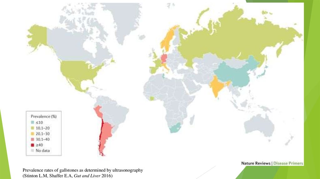
GD is a common disorder all over the world. Theprevalence of GD varies widely by region.
In Western countries, the prevalence of gallstone disease
reportedly ranges from approximately 7.9% in men to
16.6% in women. In Asians, it ranges from approximately
3% to 15%, is nearly non-existent (less than 5%) in
Africans, and ranges from 4.21% to 11% in China.
4.
Prevalence rates of gallstones as determined by ultrasonography(Stinton L.M, Shaffer E.A, Gut and Liver 2016)
5. Etiology
There are three main pathways in the formation of gallstones:Cholesterol
supersaturation
Excess bilirubin:
Gallbladder
hypomotility or
impaired contractility
• Normally, bile can dissolve the amount of cholesterol excreted by the liver.
But if the liver produces more cholesterol than bile can dissolve, the excess
cholesterol may precipitate as crystals. Crystals are trapped in gallbladder
mucus, producing gallbladder sludge. With time, the crystals may grow to
form stones and occlude the ducts which ultimately produce the gallstone
disease.
• Bilirubin, a yellow pigment derived from the breakdown of red blood cells,
is secreted into bile by liver cells. Certain hematologic conditions cause the
liver to make too much bilirubin through the processing of breakdown of
hemoglobin. This excess bilirubin may also cause gallstone formation.
• If the gallbladder does not empty effectively, bile may become
concentrated and form gallstones.
6. Etiology
Depending on the etiology, gallstones have differentcompositions. The three most common types are cholesterol
gallstones, black pigment gallstones, and brown pigment
gallstones. Ninety percent of gallstones are cholesterol
gallstones.
Each stone has a unique set of risk factors. Some risk factors
for the development of cholesterol gallstones are obesity, age,
female gender, pregnancy, genetics, diet, liver and
pancreatic diseases, rapid weight loss, and certain
medications.
7.
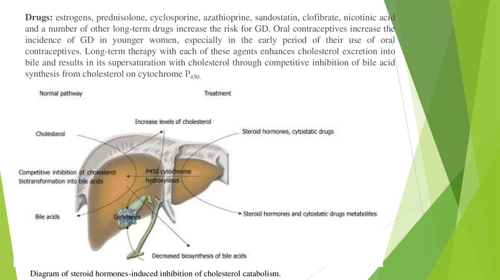
Drugs: estrogens, prednisolone, cyclosporine, azathioprine, sandostatin, clofibrate, nicotinic acidand a number of other long-term drugs increase the risk for GD. Oral contraceptives increase the
incidence of GD in younger women, especially in the early period of their use of oral
contraceptives. Long-term therapy with each of these agents enhances cholesterol excretion into
bile and results in its supersaturation with cholesterol through competitive inhibition of bile acid
synthesis from cholesterol on cytochrome Р450.
Diagram of steroid hormones-induced inhibition of cholesterol catabolism.
8. Composition of gallstones
Stones in the gallbladder and/or bile ducts are a morphological substrateof GD.
The major components of virtually all types of GS are free unesterified
cholesterol, unconjugated bilirubin, bilirubin calcium salts, fatty
acids, calcium carbonates and phosphates, and mucin
glycoproteins.
Three main categories of gallstones can be identified according to their
predominant chemical composition, cholesterol and pigment stones:
cholesterol stones, constituting as high as 75% of all gallstones in GD;
pigment stones;
mixed stones.
9. Composition of gallstones
they are round or ovalin shape, light (they do
not sink in water) and,
when ignited, burn with
a bright flame. When
sectioned, they are
radial in structure due
to the radial alignment
of cholesterol crystals.
These are black (compact and
small) and brown (softer and
large) pigment stones.They are
composed
predominantly
of
calcium bilirubinate, phosphate
and
carbonate
without
a
cholesterol impurity. They have
different shapes, are more
commonly very small and
numerous, greenish black in color,
compact, but fragile.
they sink in water and
burn poorly; when cut,
they have a lamellar
pattern.
10. Pathogenesis
The pathogenesis of GD is suggested to be multifactorial andprobably develops from complex interactions between many
genetic and environmental factors.
Unphysiological biliary supersaturation from hypersecretion of
cholesterol, gallbladder hypomotility and the accumulation of
mucin gel contribute to the formation of cholesterol GS, while
black pigment stones derive from the precipitation of calcium
hydrogen bilirubinate where pigment supersaturation and
deposition of inorganic salts, phosphate and calcium bicarbonate
accelerate the nucleation.
11.
Cholesterol stones are formed in the gallbladder due to impaired relationshipsbetween the major bile components, cholesterol, phospholipids and bile acids. The
pathophysiology of GS formation involves three steps: saturation, crystallization
and growth. Bile cholesterol supersaturation is an obligatory, but not the only,
factor that contributes to GS formation. An important role in this is played by the
state of pronucleating and antinucleating factors and the functional state of the
gallbladder.
In parallel with this, there are morphological changes in the gallbladder mucosa.
The surface epithelium passes into goblet, mucus cells that secret much mucus, the
columnar epithelium flattens and microvilli are lost. This results in impaired water
and electrolyte absorption processes. Mucin and mucus hypersecretion gives rise to
a parietal colloid solution that is turned to viscoelastic glycoprotein-mucin gel. The
latter promotes the aggregation of phospholipid vesicles and the nucleation and
precipitation of cholesterol monohydrate crystals and/or bilirubin.
The bulk of intrahepatic stones are formed due to biliary tract infection. The neck
of the gallbladder hosts the biggest bacterial load in comparison with the body and
the fundus.
Foreign bodies, such as suture materials, clips, swallowed metal or plastic
fragments, or parasites, may become foci of nucleation.
12.
It shows a diagram of gallstone formation by taking into accountthe above impaired bile production and excretion processes.
13. Symptoms and Signs
About 80% of people with gallstones are asymptomatic. The remainder have symptoms ranging from acharacteristic type of pain (biliary colic) to cholecystitis to life-threatening cholangitis. Biliary colic is the
most common symptom.
Stones occasionally traverse the cystic duct without causing symptoms. However, most gallstone migration
leads to cystic duct obstruction, which, even if transient, causes biliary colic. Biliary colic characteristically
begins in the right upper quadrant but may occur elsewhere in the abdomen. The pain may radiate into
the back or down the arm.
Episodes begin suddenly, become intense within 15 min to 1 h, remain at a steady intensity (not colicky) for
up to 12 h (usually < 6 h), and then gradually disappear over 30 to 90 min, leaving a dull ache. The pain is
usually severe enough to send patients to the emergency department for relief. Nausea and some vomiting
are common, but fever and chills do not occur unless cholecystitis has developed. Mild right upper quadrant
or epigastric tenderness may be present; peritoneal findings are absent. Between episodes, patients feel
well.
Although biliary colic can follow a heavy meal, fatty food is not a specific precipitating factor. Nonspecific
GI symptoms, such as gas, bloating, and nausea, have been inaccurately ascribed to gallbladder disease.
These symptoms are common, having about equal prevalence in cholelithiasis, peptic ulcer disease, and
functional GI disorders.
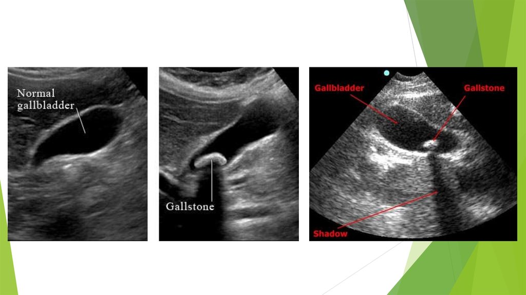
14. Diagnosis Ultrasonography
Gallstones are suspected in patients with biliary colic. Abdominalultrasonography is the imaging test of choice for detecting gallbladder stones;
sensitivity and specificity are 95%. Ultrasonography also accurately detects
sludge. CT, MRI , and oral cholecystography are alternatives. Endoscopic
ultrasonography accurately detects small gallstones (< 3 mm) and may be
needed if other tests are equivocal.
Laboratory tests usually are not helpful; typically, results are normal unless
complications develop.
Asymptomatic gallstones and biliary sludge are often detected incidentally
when imaging, usually ultrasonography, is done for other reasons. About 10 to
15% of gallstones are calcified and visible on plain x-rays.
15.
16.
CONDITIONSSIGNS / SYMPTOMS
INVESTIGATIONS
Peptic
ulcer May have hx of Helicobacter pylori infection, NSAID use, EGD: peptic ulcer.
disease (PUD)
smoking, increased age, or positive family history of H pylori breath/stool antigen test: may be positive if H
PUD. Presents with burning or gnawing pain in the upper pyloricausative.
abdomen, particularly with food consumption and often
improved with antacids.
Gallbladder
cancer
Can present with painless jaundice and/or weight loss, CT abdomen: may reveal intrahepatic mass lesion, dilated
although often presents late with upper abdominal pain.
intrahepatic ducts, and/or localised lymphadenopathy.
Gallbladder
polyps
Often found incidentally on imaging for other conditions.
Acalculous
cholecystitis
Positive Murphy's sign (tenderness suddenly becomes Abdominal ultrasound: no gallstones; may produce Murphy's sign.
worse during deep inspiration, and produces inspiratory Hepatobiliary iminodiacetic acid (HIDA) scan: gallbladder nonarrest). In ICU setting, findings are often subtle.
visualisation.
Sphincter
Oddi
dysfunction
(SOD)
Non-biliary
acute
pancreatitis
of Postcholecystectomy biliary pain.
Abdominal ultrasound: polypoidal lesion.
ERCP with biliary manometry: lack of sludge or retained stones;
should only be undertaken in those with abdominal pain after
cholecystectomy who have significant laboratory or imaging
abnormalities
History is helpful in identifying alcohol use, possible Triglycerides: elevated; usually >11.3 mmol/L (>1000 mg/dL)
offending medications, or recent biliary tract (can be lower in fasting patients).
endoscopy/surgery.
Calcium: elevated; checking ionised calcium is useful.
IgG4: for autoimmune pancreatitis.
MRCP/abdominal ultrasound: normal bile ducts.
17. Treatment
For symptomatic stones: Laparoscopic cholecystectomyor sometimes stone dissolution using ursodeoxycholic acid.
For asymptomatic stones: Expectant management.
Most asymptomatic patients decide that the discomfort,
expense, and risk of elective surgery are not worth removing
an organ that may never cause clinical illness. However, if
symptoms occur, gallbladder removal (cholecystectomy) is
indicated because pain is likely to recur and serious
complications can develop.
18. Surgery
Surgery can be done with an open or a laparoscopictechnique.
Open cholecystectomy, which involves a large
abdominal incision and direct exploration, is safe and
effective. Its overall mortality rate is about 0.1%
when done electively during a period free of
complications.
Laparoscopic cholecystectomy is the treatment of
choice. Using video endoscopy and instrumentation
through small abdominal incisions, the procedure is
less invasive than open cholecystectomy. The result
is a much shorter convalescence, decreased
postoperative discomfort, improved cosmetic results,
yet no increase in morbidity or mortality.
19. Stone dissolution
For patients who decline surgery or who are at high surgical risk, gallbladder stonescan sometimes be dissolved by ingesting bile acids orally for many months. The best
candidates for this treatment are those with small, radiolucent stones (more likely to
be composed of cholesterol) in a functioning nonobstructed gallbladder.
Ursodeoxycholic acid 4 to 5 mg/kg po bid or 3 mg/kg po tid (8 to 10 mg/kg/day)
dissolves 80% of tiny stones < 0.5 cm in diameter within 6 mo. For larger stones (the
majority), the success rate is much lower, even with higher doses of ursodeoxycholic
acid. Further, after successful dissolution, stones recur in 50% within 5 yr. Most
patients are thus not candidates and prefer laparoscopic cholecystectomy. However,
ursodeoxycholic acid 300 mg po bid can help prevent stone formation in morbidly
obese patients who are losing weight rapidly after bariatric surgery or while on a
very low calorie diet.
Stone fragmentation (extracorporeal shock wave lithotripsy) to assist stone
dissolution and clearance is now unavailable (rarely used).
20. Prognosis
The outlook for patients with symptomatic cholelithiasismanaged by cholecystectomy is favourable. The same holds for
patients with choledocholithiasis who undergo endoscopic
retrograde cholangiopancreatography (ERCP) with biliary
sphincterotomy and stone extraction, followed later by
cholecystectomy.
Recurrent choledochal problems. Risk factors for recurrent
choledochal problems are common with: bile duct dilatation to
>15mm; a periampullary diverticulum; brown pigment stones;
or the gallbladder being left intact.
21. References
https://www.nlm.nih.gov/https://www.ncbi.nlm.nih.gov/
https://www.ncbi.nlm.nih.gov/pubmed/
Clinical Protocol of Diagnosis and Treatment No. 10 of the Ministry of Health of the
Republic of Kazakhstan from September 30, 2015