


")


 medicine
Similar presentations:


")

Регенерация костной ткани
1. Регенерация костной ткани
Выполнил: Капанадзе ГеоргийГеоргиевич
2. Основные функции:
1. Опорно-механическая2. Защитная
3. Гемопоэтическая функция
4. Депонирующая функция
3. Определение
Регенерация – способность живых организмоввосстанавливать поврежденные ткани, а иногда
и поврежденные органы.
4. Виды
Физиологическая регенерация – регенерация впроцессе нормальной жизнедеятельности
организма, обычно не связанная с
повреждением или утратой части органа.
Репаративная регенерация – регенерация
происходящая в случаях повреждения или
утраты какого либо органа или части
организма.
5. Bone Composition
Extracellular MatrixOrganic (35%)
Collagen (type I) 90%
Osteocalcin, osteonectin, proteoglycans,
glycosaminoglycans, lipids (ground substance)
Inorganic (65%)
Primarily hydroxyapatite Ca5(PO4)3(OH)2
Cells
Osteocytes
Osteoblasts
Osteoclasts
6. Остеогенные клетки
Остеогенный дифферон: остеогенная клетка –остеобласт – преостеоцит – остеоцит.
7.
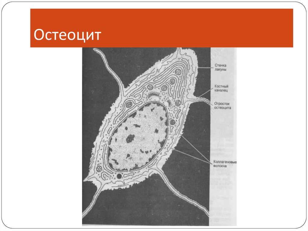
ОстеобластСинтезирует межклеточное
вещество.
Участвует в синтезе
коллагена и минерализации
кости
8.
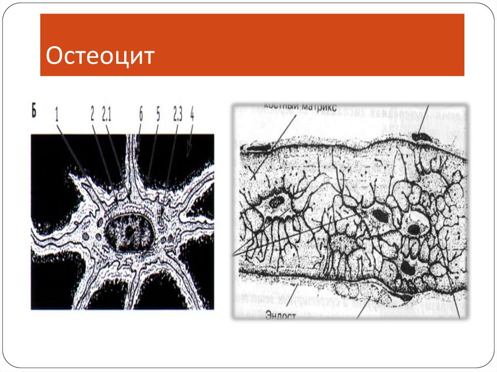
Остеоцит9.
Остеоцит10. Остеокласт, его развитие и функции
Остеокластразвитие и
функции
11. Цикл ремоделирования кости
12.
Типы костейПластинчатая кость
коллагеновых фибрилл, идущих в одном
направлении, но под углом к другой пластинке.
Нормальная взрослая кость.
Грубоволокнистная
состоит из пучков костных волокон, которые
идут в разных направлениях.
У взрослых наблюдается в местах заживления
переломов, прикрепления сухожилий или связок и
при патологических состояниях.
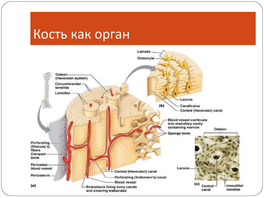
13.
Кость как орган14.
Компоненты костеобразованияCortex
Periosteum
Bone marrow
Soft tissue
15.
Требования для регенерацииМеханическая стабильность
Кровоснабжение
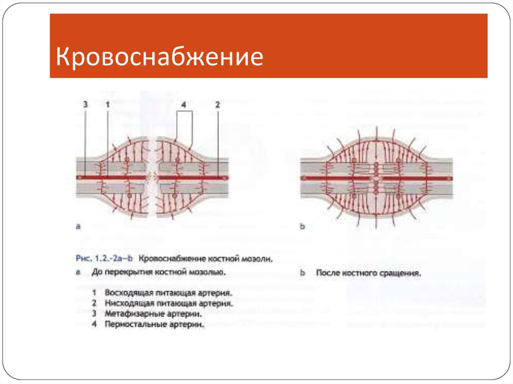
16.
КровоснабжениеОбычно основное кровоснабжение
представлено диафизом(80 to 85%)
В надкостнице нижней части кости имеется
маленькое отверстие, через которое внутрь
кости проходит питающая артерия
В костном мозге эта артерия разделяется на
верхнюю и нижнюю ветви
17.
Кровоснабжение18. 1.
Сращениеперелома
1.
Первичное, сращение путем внутренней
перестройки
Вторичное, сращение путем формирования
костной мозоли
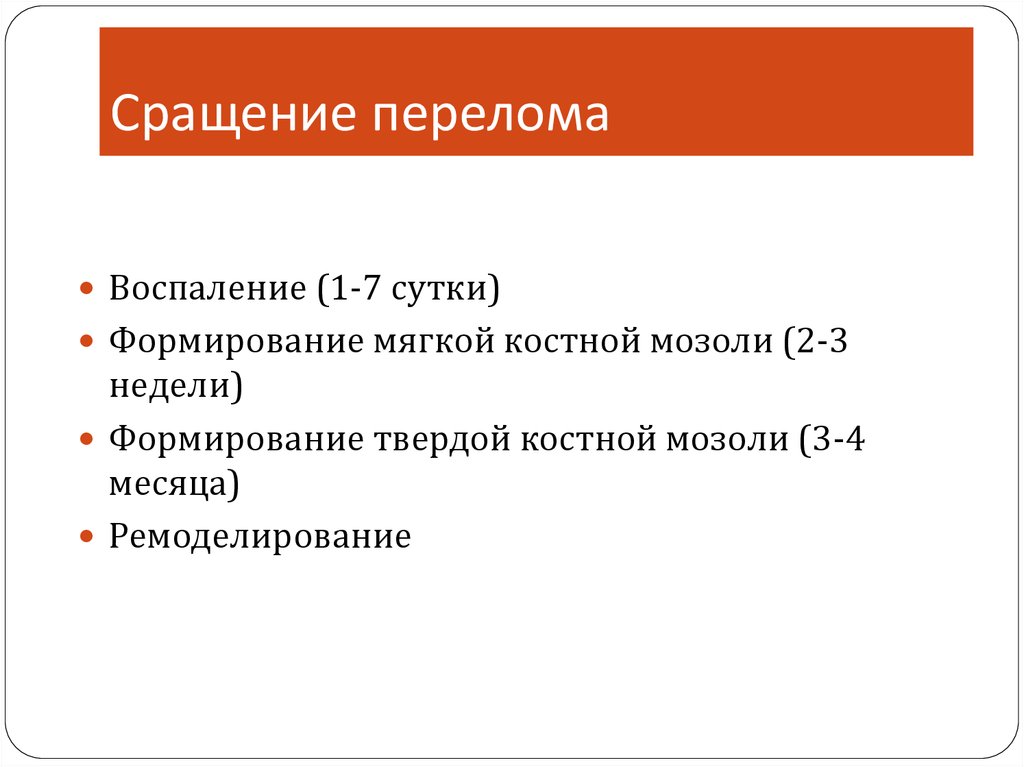
19.
Сращение переломаВоспаление (1-7 сутки)
Формирование мягкой костной мозоли (2-3
недели)
Формирование твердой костной мозоли (3-4
месяца)
Ремоделирование
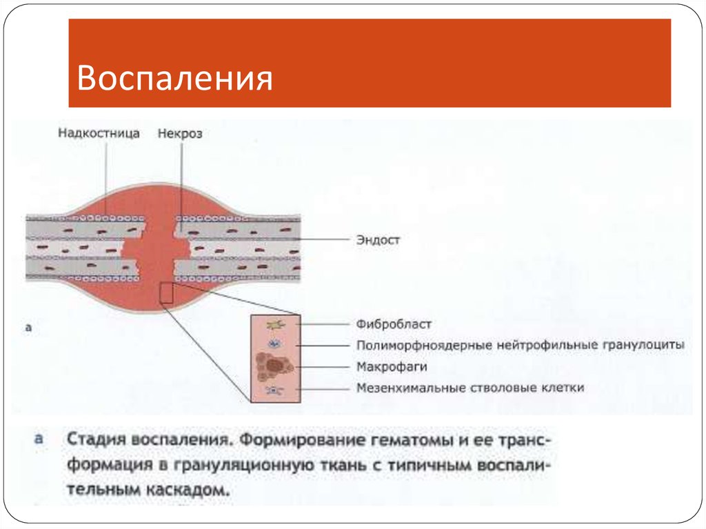
20.
Воспаления21.
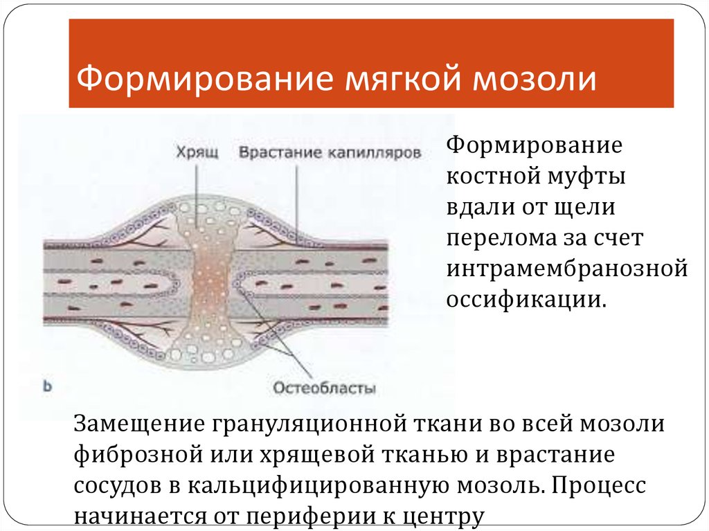
Формирование мягкой мозолиФормирование
костной муфты
вдали от щели
перелома за счет
интрамембранозной
оссификации.
Замещение грануляционной ткани во всей мозоли
фиброзной или хрящевой тканью и врастание
сосудов в кальцифицированную мозоль. Процесс
начинается от периферии к центру
22. Формирование твердой мозоли
Полная замена мозоликальцифицированной
тканью путем
интрамембранозной и
энхондральной
оссификации
23. Ремоделирование
24. Local Regulation of Bone Healing
Growth factorsCytokines
Prostaglandins/Leukotrienes
Hormones
Growth factor antagonists
25. Growth Factors
Transforming growth factorBone morphogenetic proteins
Fibroblast growth factors
Platelet-derived growth factors
Insulin-like growth factors
26. Transforming Growth Factor
Superfamily of growth factors (~34 members)Act on serine/threonine kinase cell wall receptors
Promotes proliferation and differentiation of
mesenchymal precursors for osteoblasts,
osteoclasts and chondrocytes
Stimulates both endochondral and
intramembranous bone formation
Induces synthesis of cartilage-specific
proteoglycans and type II collagen
Stimulates collagen synthesis by osteoblasts
27. Bone Morphogenetic Proteins
Osteoinductive proteins initially isolated fromdemineralized bone matrix
Proven by bone formation in heterotopic muscle
pouch
Induce cell differentiation
BMP-3 (osteogenin) is an extremely potent inducer of
mesenchymal tissue differentiation into bone
Promote endochondral ossification
BMP-2 and BMP-7 induce endochondral bone
formation in segmental defects
Regulate extracellular matrix production
BMP-1 is an enzyme that cleaves the carboxy termini
of procollagens I, II and III
28. Bone Morphogenetic Proteins
These are included in the TGF-β familyExcept BMP-1
BMP2-7,9 are osteoinductive
BMP2,6, & 9 may be the most potent in osteoblastic
differentiation
Work through the intracellular Smad pathway
Follow a dose/response ratio
29. BMP Antagonists
May have important role in bone formationNoggin
Extra-cellular inhibitor
Competes with BMP-2 for receptors
30. Fibroblast Growth Factors
Both acidic (FGF-1) and basic (FGF-2) formsIncrease proliferation of chondrocytes and
osteoblasts
Enhance callus formation
FGF-2 stimulates angiogenesis
31. Platelet-Derived Growth Factor
A dimer of the products of two genes, PDGF-A andPDGF-B
PDGF-BB and PDGF-AB are the predominant
forms found in the circulation
Stimulates bone cell growth
Mitogen for cells of mesenchymal origin
Increases type I collagen synthesis by increasing
the number of osteoblasts
PDGF-BB stimulates bone resorption by increasing
the number of osteoclasts
32. Insulin-like Growth Factor
Two types: IGF-I and IGF-IISynthesized by multiple tissues
IGF-I production in the liver is stimulated by
Growth Hormone
Stimulates bone collagen and matrix synthesis
Stimulates replication of osteoblasts
Inhibits bone collagen degradation
33. Cytokines
Interleukin-1,-4,-6,-11, macrophage andgranulocyte/macrophage (GM) colony-stimulating
factors (CSFs) and Tumor Necrosis Factor
Stimulate bone resorption
IL-1 is the most potent
IL-1 and IL-6 synthesis is decreased by estrogen
May be mechanism for post-menopausal bone
resorption
Peak during 1st 24 hours then again during
remodeling
Regulate endochondral bone formation
34. Prostaglandins / Leukotrienes
Effect on bone resorption is species dependent andtheir overall effects in humans unknown
Prostaglandins of the E series
Stimulate osteoblastic bone formation
Inhibit activity of isolated osteoclasts
Leukotrienes
Stimulate osteoblastic bone formation
Enhance the capacity of isolated osteoclasts to
form resorption pits
35. Hormones
EstrogenStimulates fracture healing through receptor
mediated mechanism
Modulates release of a specific inhibitor of IL-1
Thyroid hormones
Thyroxine and triiodothyronine stimulate
osteoclastic bone resorption
Glucocorticoids
Inhibit calcium absorption from the gut causing
increased PTH and therefore increased
osteoclastic bone resorption
36. Hormones (cont.)
Parathyroid HormoneIntermittent exposure stimulates
Osteoblasts
Increased bone formation
Growth Hormone
Mediated through IGF-1 (Somatomedin-C)
Increases callus formation and fracture strength
37. Vascular Factors
MetalloproteinasesDegrade cartilage and bones to allow invasion of
vessels
Angiogenic factors
Vascular-endothelial growth factors
Mediate neo-angiogenesis & endothelial-cell
specific mitogens
Angiopoietin (1&2)
Regulate formation of larger vessels and
branches
38. Local Anatomic Factors That Influence Fracture Healing
Soft tissue injuryInterruption of local blood supply
Interposition of soft tissue at fracture site
Bone death caused by radiation, thermal or
chemical burns or infection
39. Systemic Factors That Decrease Fracture Healing
MalnutritionCauses reduced activity and proliferation of
osteochondral cells
Decreased callus formation
Smoking
Cigarette smoke inhibits osteoblasts
Nicotine causes vasoconstriction diminishing
blood flow at fracture site
Diabetes Mellitus
Associated with collagen defects including
decreased collagen content, defective crosslinking and alterations in collagen sub-type ratios
40. PEMF
Перменноеэлектромагнитное поле
PEMF
Одобрено FDA для лечения несращения переломов
Эффективность костной стимуляции зависит от
частоты.
Наиболее эффективны низкочастотные
синусоидальные электрические поля в
физиологическом (диапазон от 15 до 30 Гц)
41. Ultrasound
УльтразвукUltrasound
Ультразвук низкой интенсивности одобрен FDA
для стимулирования заживления свежих
переломов
Модулирует передачу сигнала, увеличивает
экспрессию генов, увеличивает кровоток,
усиливает ремоделирование кости и
увеличивает скорость образования мозоли
42. Ultrasound
УльтразвукUltrasound
Клинические испытания на людях показывают
снижение времени регенерации при свежих
переломах