


 medicine
Similar presentations:


. Область предплечья и кисти. Практическое занятие №3")


Апоневрозы региона верхней конечности
1. Апоневрозы региона верхней конечности
2. Подмышечная область
3. Клетчатка подмышечной области и фасции передней поверхности плеча
Fasciae of the Upper LimbКлетчатка подмышечной области и
фасции передней поверхности плеча
Coracobrachialis muscle
covered by its fascia.
It is in continuity with
the clavipectoral facia.
Pectoralis minor
muscle
Clavipectoral
fascia
Deltoid
muscle
Insertion of the deltoid fascia
into the brachial fascia
FIGURE 7.31 Anterior view of the shoulder region. The pectoralis major muscle was removed to show the clavipectoral fascia. This fascia continues
4. Подмышечный апоневроз
Fasciae of the Upper LimbПодмышечный апоневроз
Superficial fascia
of the axilla
Connections between
the superficial fascia and
the brachial fascia in the axilla
Deltoid muscle
covered by its
deep fascia
Fat lobules
inside the
superficial fascia
5. Фасции передней поверхности плеча
Fasciae of the Upper LimbMyofascial
expansion of the
pectoralis major
muscle into the
brachial fascia
Biceps brachii
muscle enveloped
by its epimysium
Brachial fascia
Brachial fascia
Medial
intermuscular
septum
6. Фасции задней поверхности плеча
Deep fascia ofthe latissimus
dorsi muscle
Brachial fascia
Reinforcement of the brachial fascia
due to the myofascial expansion of
the latissimus dorsi muscle
Fasciae of the Upper Limb
Deltoid fascia
7. Фасции лопатки, задней поверхности плеча и дельтовидная
he Upper LimbФасции лопатки, задней поверхности
плеча и дельтовидная
The subscapular fascia is a thin aponeurotic fascia attached to the entire circumference of the subscapular fossa (Fig. 7.23). Some bres of the subscapularis
muscle originate from its deep surface. Singer (1935)
describes the subscapular fascia as being the thinnest
Insertion of the
deltoid fascia into
Infraspinatus the infraspinatus
fascia
fascia
Latissimus dorsi muscle
covered by its fascia
Teres major
muscle
of the bursa is also linked to the coracoid process by
a brous attachment called the suspensory ligament.
During movement of the glenohumeral joint, the subscapularis muscle sustains huge changes of orientation, particularly in the upper part of the muscle that
Deltoid muscle
enveloped
by its fascia
Insertion of the deltoid fascia
into the brachial fascia
8. Межмышечные перегородки и фасциальные ложа плеча
Biceps brachiimuscle
Pectoralis minor muscle
Clavipectoral fascia
Coracobrachialis
muscle
Medial
intermuscular
septum
Brachial fascia
Fasciae of the Upper Limb
Межмышечные
перегородки и
фасциальные ложа плеча
9. Расположение сосудисто-нервного пучка плеча
10. Апоневрозы предплечья
Fasciae of the Upper LimbХод волокон поверхностного листка
апоневроза предплечья
Longitudinal fibrous
bundles inside the
brachial fascia
Oblique fibrous
bundles inside
the
antebrachial
fascia
11. Костно-фасциальные мышечные ложа предплечья
Содержимое переднегокостно-фасциального
ложа
Содержимое заднего
и наружного костнофасциального ложа
4
5
12. Костно-фасциальные мышечные ложа предплечья
1- лучевая кость; 2- поверхностный апоневроз; 3- локтевая кость4- передняя межмышечная перегородка; 5- задняя межмышечная перегородка
1
2
III
3
I
II
4
5
6
6- межкостная мембрана; I- переднее
ложе; II – заднее ложе; III- наружное
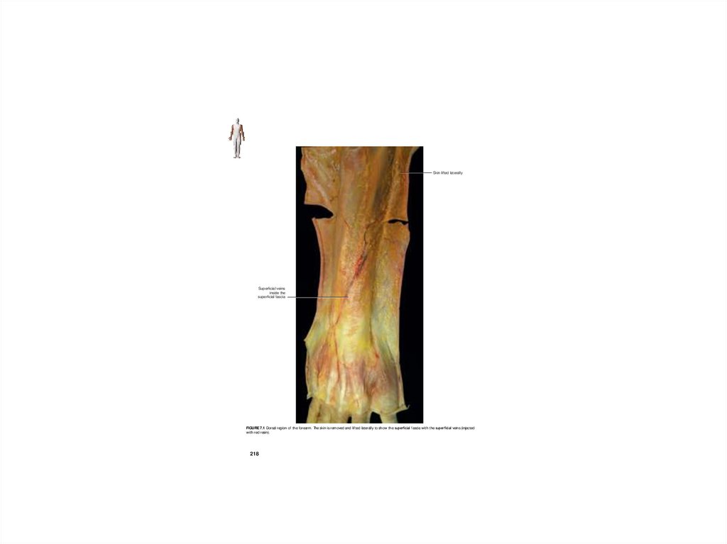
13. Прохождение артерий и поверхностных вен в районе предплечья
14. Ладонные апоневрозы кисти
Palmarislongus
tendon
Flexor carpi
retinaculum
Expansion
for the
thenar
fascia
Expansio
for the
palmar
aponeuro
Palmar
aponeurosis
FIGURE 7.57 Anterior view of the wrist. In this subject the palmaris longus tendon opens as a fan, having a very thin insertion into t
aponeurosis, while the larger portion inserts into the exor carpi r etinaculum.
15. Тыльные апоневрозы кисти
Myofascialexpansion of the
extensor
carpi radialis
longus tendon
Fasciae of the Upper Limb
Dorsal
retinaculum
carpi
Fasciae of the Upper Limb
Antebrachial
fascia
Dorsal fascia
of the hand
Extensor pollicis
longus tendon
FIGURE 7.62 Posterior view of the hand. The extensor digitorum tendons are removed. The extensor carpi radialis longus tendon is stretched t
show its myofascial expansion into the deep lamina of the dorsal fascia of the hand.
FIGURE 7.61 Posterior view of the hand. The extensor pollicis longus tendon is lifted to show that it is enveloped by the dorsal fascia of the hand.
16. Костно-фасциальные ложа кисти и прохождение сосудов
17.
Fasciae of the Upper LimbSkin lifted laterally
Superficial veins
inside the
superficial fascia
FIGURE 7.1 Dorsal region of the forearm. The skin is removed and lifted laterally to show the super cial fascia with the super cial veins (injected
with red resin).
218