


 english
Similar presentations:


")
Appendicular skeleton the scull
1.
Lecture number 2.Topic:
APPENDICULAR SKELETON
THE SCULL
2.
Clavicle | Collar BoneIt is a modified long bone having
two curves.
Medial 2/3 is convex and lateral
1/3 is concave as seen from front.
Like all long bones, it has two ends:
the acromial end and the sternal and,
superior and inferior surfaces with
3.
It is a flat triangularScapula
|
bone. It has three
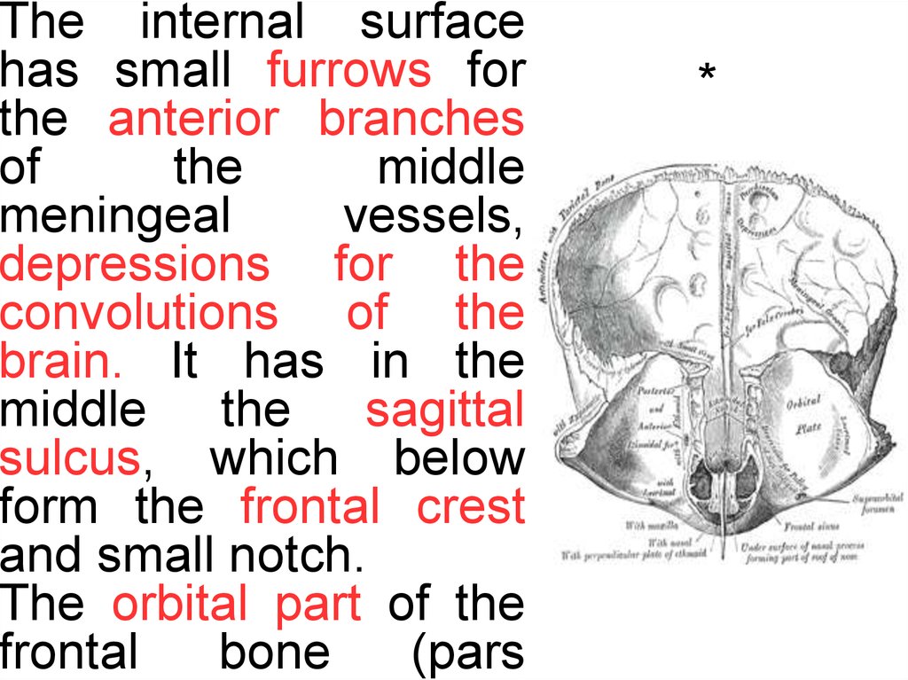
borders;
Vertebral Shoulder Blade
(medial)
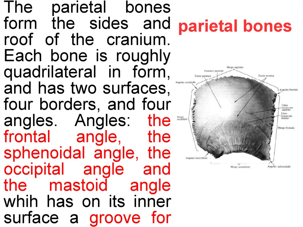
border,
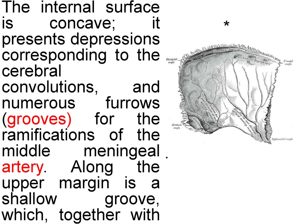
Superior border, and
Axillary
(lateral)
border. Also it has
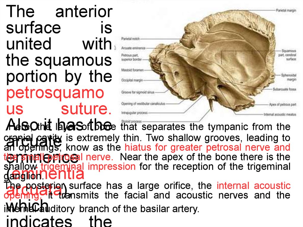
three angles; Medial
angle, Lateral angle
and Inferior angle
Glenoid fossa is a
pear shaped fossa
that articulates with
humerus to form
4.
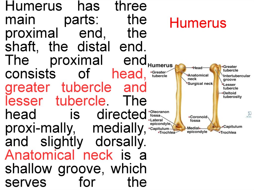
Humerus has threemain
parts:
the
proximal end, the
shaft, the distal end.
The proximal end
consists
of
head,
greater tubercle and
lesser tubercle. The
head
is directed
proxi-mally, medially,
and slightly dorsally.
Anatomical neck is a
shallow groove, which
serves
for
the
Humerus
5.
The distal end ofthe humerus is
furnished with two
articular surfaces.
The lateral of these
is the capitulum for
the head of the
radius.
In
front
there is a shallow
depression (radial
fossa). The medial
articular surfaces is
called trochlea for
ulna.
On
the
6.
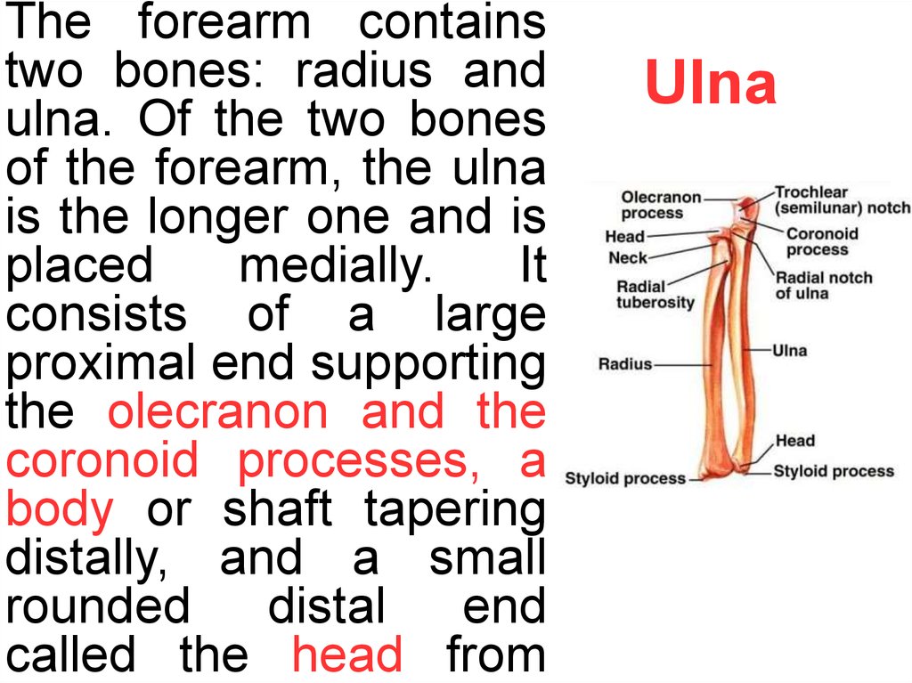
The forearm containstwo bones: radius and
ulna. Of the two bones
of the forearm, the ulna
is the longer one and is
placed
medially.
It
consists of a large
proximal end supporting
the olecranon and the
coronoid processes, a
body or shaft tapering
distally, and a small
rounded
distal
end
called the head from
Ulna
7.
Proximal end ofradius consists of
head, neck and
tuberosity.
The
head
of
radius
provided with a
shallow
concave
surface proximally
for articulation with
the capitulum of the
humerus.
The
circumference
of
the head is smooth.
radius
8.
The skeleton of theThe
skeleton
hand is subdivided
into three segments: of the hand
the carpus or wrist
bones;
the
metacarpus or bones
of the palm; and the
phalanges or bones of
the digits. Bones of
the wrist are small
bones,
and
are
arranged in two rows.
The first or proximal
row is made
of
9.
Hip boneHip bone consists of three parts:
Ilium, ischium and pubis. These
three bones meet one another at the
acetabulum. Acetabulum articulates
with the head of femur to form hip
joint. Inferiorly, the margin of
acetabulum is deficient and is
10.
iliumThe ilium possesses a iliac crest.The
crest ends in the front at the anterior
superior iliac spine below which lies
the anterior inferior iliac spine.
Posteriorly, the crest ends at the
11.
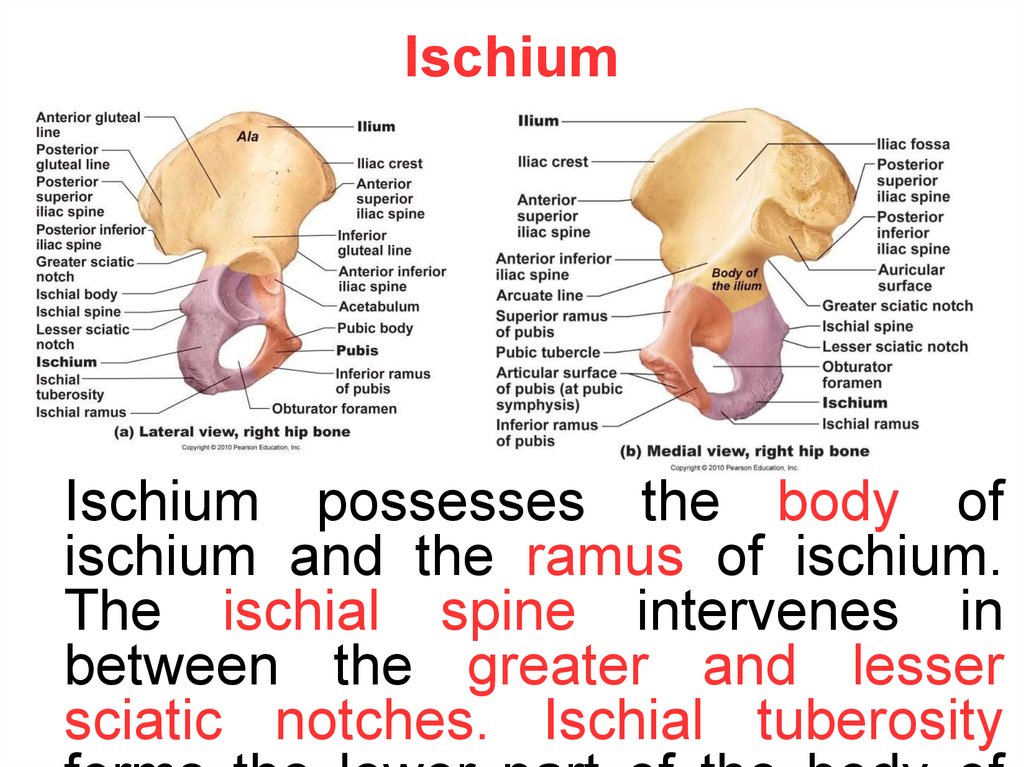
IschiumIschium possesses the body of
ischium and the ramus of ischium.
The ischial spine intervenes in
between the greater and lesser
sciatic notches. Ischial tuberosity
12.
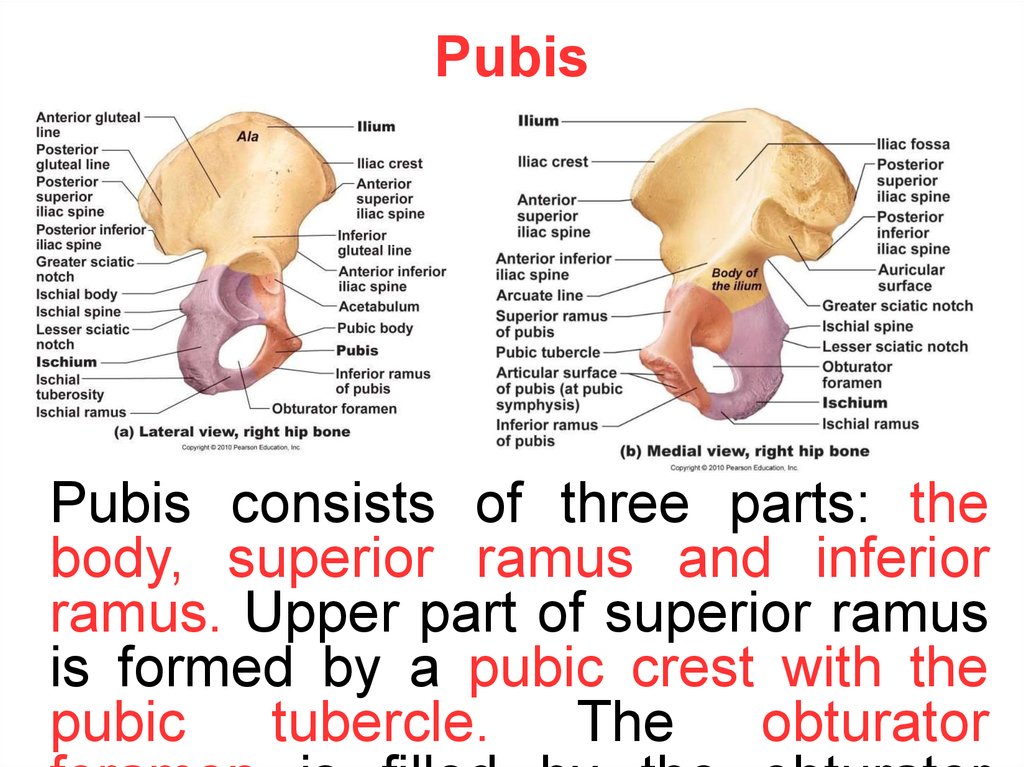
PubisPubis consists of three parts: the
body, superior ramus and inferior
ramus. Upper part of superior ramus
is formed by a pubic crest with the
pubic
tubercle.
The
obturator
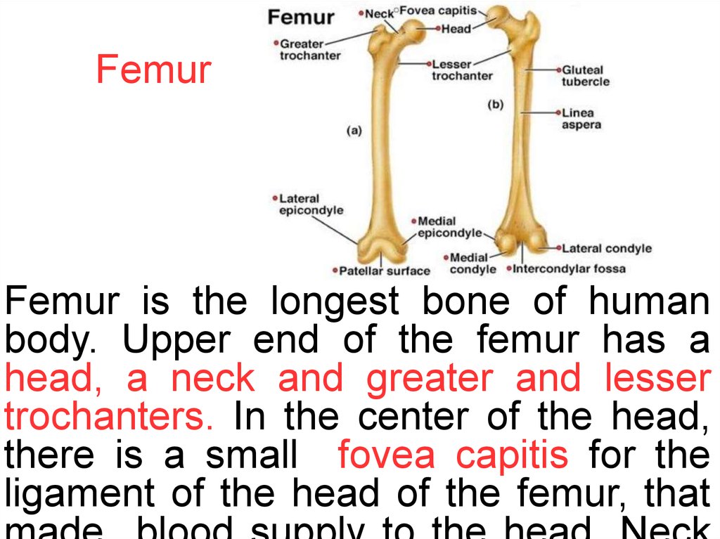
13.
FemurFemur is the longest bone of human
body. Upper end of the femur has a
head, a neck and greater and lesser
trochanters. In the center of the head,
there is a small fovea capitis for the
ligament of the head of the femur, that
14.
The shaft of femur has a ridge for manymuscles of thigh known as linea
aspera; also it has triangular area on
the posterior surface known as popliteal
surface. Lower end of femur consists of
lateral and medial condyles, which are
15.
Tibia | ShinboneThe proximal end of tibia has massive
medial and lateral condyles and an
intercondylar area intervening between
the condyles. There is also a prominent
tibial tuberosity for the patellar tendon,
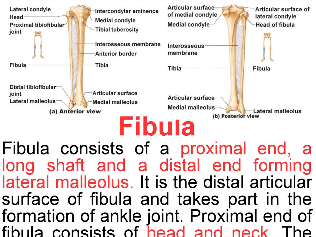
16.
FibulaFibula consists of a proximal end, a
long shaft and a distal end forming
lateral malleolus. It is the distal articular
surface of fibula and takes part in the
formation of ankle joint. Proximal end of
17.
The human foot isa complex structure
containing
26
bones.
The foot can be
subdivided into the
tarsus (7): talus,
calcaneus, cuneiformes (3), cuboid,
and
navicular;
metatarsus
(5):
first, second, third,
fourth, and fifth
metatarsal
bone;
foot
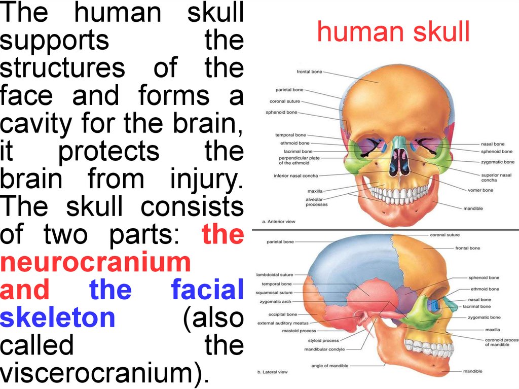
18.
The human skullsupports
the
structures of the
face and forms a
cavity for the brain,
it
protects
the
brain from injury.
The skull consists
of two parts: the
neurocranium
and the facial
skeleton
(also
called
the
viscerocranium).
human skull
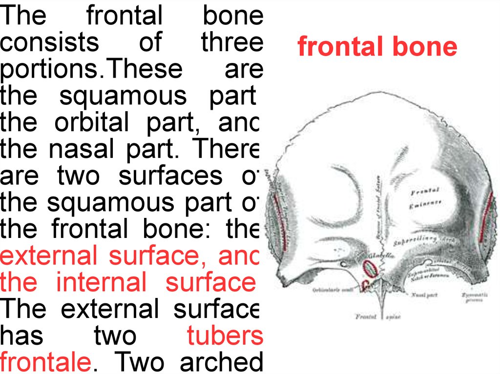
19.
The frontal boneconsists of three
portions.These are
the squamous part,
the orbital part, and
the nasal part. There
are two surfaces of
the squamous part of
the frontal bone: the
external surface, and
the internal surface.
The external surface
has
two
tubers
frontale. Two arched
frontal bone
20.
The internal surfacehas small furrows for
the anterior branches
of
the
middle
meningeal
vessels,
depressions for the
convolutions of the
brain. It has in the
middle the sagittal
sulcus, which below
form the frontal crest
and small notch.
The orbital part of the
frontal
bone
(pars
*
21.
The occipital bone isthe main bone of the occipital bone
occiput. The occipital
bone, like the other
cranial bones, has
outer and inner plates
of cortical bone tissue
between which is the
cancellous
bone
tissue
known
as
diploë. The occipital
bone has the basilar
part, at the sides of
the foramen magnum
22.
*Near the middle of
the outer surface of
the squamous part of
the occipital (the
largest part) there is
a prominence – the
external
occipital
protuber-ance. Along
the midline of the
squamous part runs
a ridge – the external
23.
The parietal bonesform the sides and parietal bones
roof of the cranium.
Each bone is roughly
quadrilateral in form,
and has two surfaces,
four borders, and four
angles. Angles: the
frontal
angle,
the
sphenoidal angle, the
occipital angle and
the mastoid angle
whih has on its inner
surface a groove for
24.
The internal surfaceis
concave;
it
presents depressions
corresponding to the
cerebral
convolutions,
and
numerous
furrows
(grooves) for the
ramifications of the
middle
meningeal .
artery. Along the
upper margin is a
shallow
groove,
which, together with
*
25.
The ethmoid bone(from Greek ethmos,
"sieve") is an unpaired
bone in the skull that
separates the nasal
cavity from the brain.
It is located between
the two orbits. The
ethmoid has three
parts: cribriform plate,
ethmoidal
labyrinth,
and
perpendicular
plate. The cribriform
ethmoid
bone
26.
The sphenoid bone sphenoid boneconsists of a body,
paired greater wings
and lesser wings,
and two pterygoid
processes. The body
lies at the centre,
lesser wing forms the
and it contains the The
the optic canal for optic
sphenoidal sinuses. nerve and ophthalmic
and the superior
Anteriorly it is the artery,
orbital fissure there is for 7
sinuses open up. numerous and vessels
The superior surface structures. The pterygoid
process consists of two
contains:
parts: medial and lateral
1. Sella turcica with pterygoid plates.
27.
The temporal boneconsists of 3 parts— temporal bone
the
squamous,
petrous and tympanic
parts. The squamous
part has the zygomatic
and
mastoid
processes.The
tympanic
part
is
relatively small. The
petrous part is shaped
like
a
pyramid.
Directed
medially,
forward, and a little
28.
The anterior*
surface
is
united
with
the squamous
portion by the
petrosquamo
us
suture.
Here, the
bone that separates the tympanic from the
Also
it layer
hasofthe
cranial cavity is extremely thin. Two shallow grooves, leading to
arcuate
an
openings, know as the hiatus for greater petrosal nerve and
the
small petrosal nerve. Near the apex of the bone there is the
eminence
shallow trigeminal impression for the reception of the trigeminal
(eminentia
ganglion.
The posterior surface has a large orifice, the internal acoustic
arcuata),
opening,
it transmits the facial and acoustic nerves and the
which
internal
auditory branch of the basilar artery.
indicates the
29.
The maxilla consistsof the body of the
maxilla
and
four
processes:
1. The body of the
maxilla.
In
the
midline
of
the
anterior surface is
found the anterior
nasal spine, and the
nasal notch, that
forms the piriform
aperture.
The
superior surface of
maxilla
5. The palatine process. It
articulate with each other in the
midline and with the horizontal
plate of the palatine bone
posteriorly. There is the incisive
canal, which transmits the
nasopalatine
nerve
and
branches of the
palatine vessels.
greater
30.
The mandible is theonly mobile bone of
mandible
the facial skeleton. It
is composed of a
body and the ramus.
On
the
anterior
region of the body
are
the
mental
protuberance,
2
inferior alveolar nerve
mental
tubercles, The
and blood vessels run
and
2
mental through this aperture and
mandibular
canal
The
foramines
that mandible houses the lower
Interdental septi
transmit the mental dentition.
run between the dental
nerves and vessels. alveoles.