


")
")


 medicine
Similar presentations:


Colon diseases
1. Colon diseases professor Youry Vladimirovitch Plotnicov
2. anatomy
3. anatomy
4. Arterial blood supply
5. Venouse outflow
6. Intraparietal lymphatic vessels
7. Lymphatic drainage
8. Differences of the right and left half
• Anatomy: on the right the lumen iswider, than at the left (except for the
ileocecal valve)
• Contention on the right is liquid, at the
left dense
• Tumours on the right is more often
exophytic, at the left endophytic
• Exophytic tumours destroyed with a
bleeding more often
9. Special investigation methods
1. Physical investigation2. A proctosigmoidoscopy
3. Fibrocolonoscopy
10. Colonoscopy - an initial cancer
11. Modern colonoscopy
12. Special investigation methods
4. irrigoscopy (including virtual)5. abdominal cavity US
6. radial methods (CТ, PET,
etc.)
7. laparoscopy
8. intravenous urography
9. reactions to an occult blood
10. cancer markers
13. Virtual colonoscopy
14. At what a cancer localization more often
anemy?15. At what a cancer localization more often
Visiblebleeding?
16. AT WHAT A CANCER LOCALIZATION MORE OFTEN
Disturbanceof passability
17. AT WHAT A CANCER LOCALIZATION MORE OFTEN
Perforation ismore
possible?
18. AT WHAT A CANCER LOCALIZATION MORE OFTEN
Fistulas,phlegmons
are possible?
19. Colon cancer localisation
20. Cancer clinical signs
1. Functional signs without intestinaldisorders (a pain, etc.)
2. Intestinal disorders (diarrheas, constipations, alternating)
3. Disturbances of intestinal passability
4. Pathological discharge
5. Disturbance of the general condition of patients
6. Palpating detection of a tumour
21. Cancer clinical forms
1) toxico-anemic2) enterocolitic
3) dyspeptic
4) obturational
5) pseudo-inflammatory
6) tumoral
22. Colon cancer diagnosis
23. Colon cancer diagnosis
24.
25.
26. TNM
27. TNM - T
•Tx - the estimation of aprimary tumour is impossible
•T0 - the primary tumour is
not found out
•Tis - a cancer in situ: cancer
cells find out within the limits
of a basal membrane of glands
or in own plate of a mucous
membrane
28. T1 – The tumour amazes a submucouse layer
29. T2 - the tumour spreads into a muscular layer
30. Т3 - the tumour gets into a subserous layer or not covered by a paracolitis and pararectal peritoneum fat
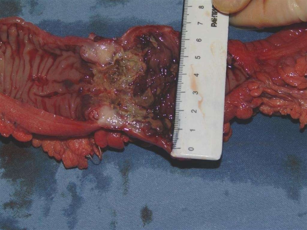
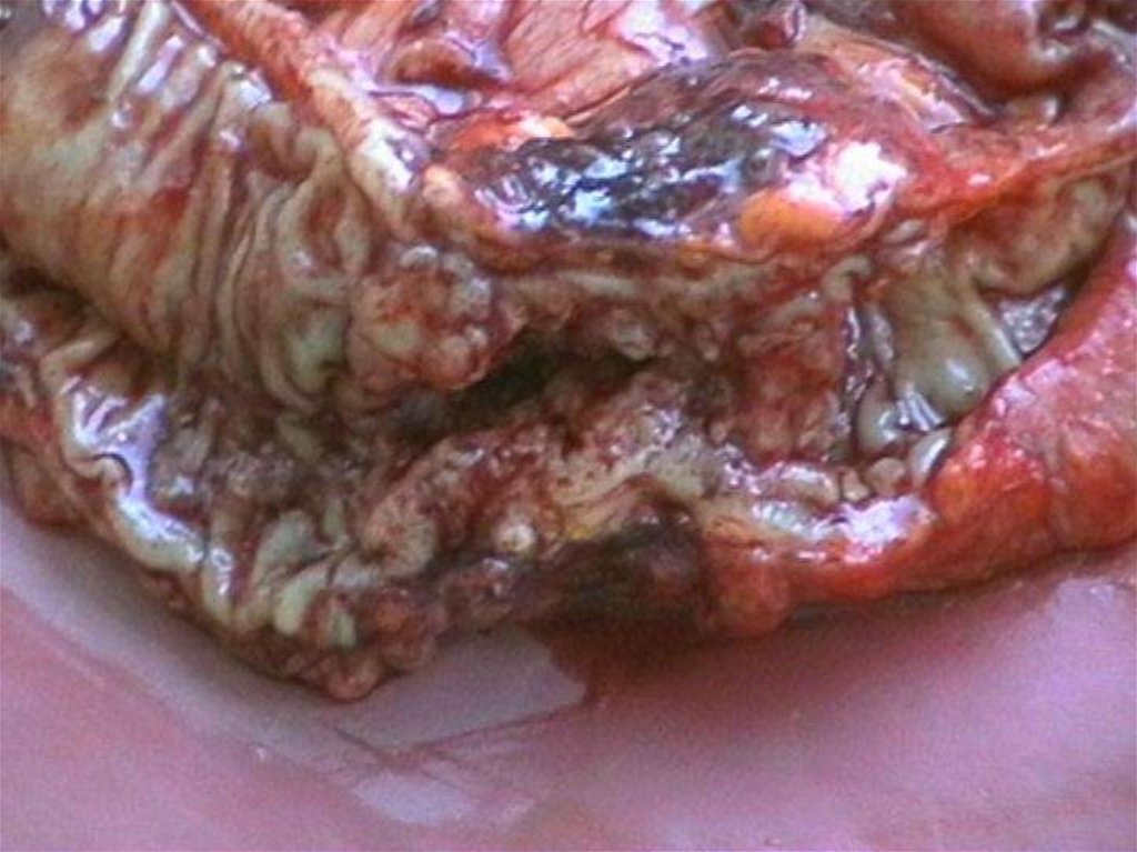
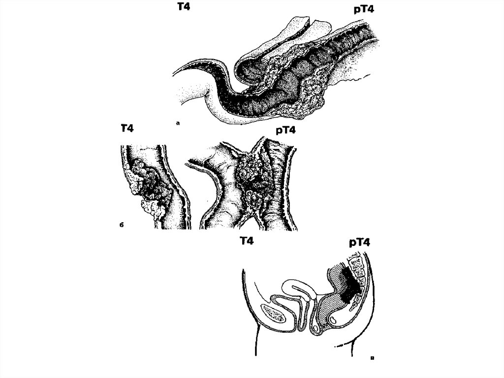
31. Т4 - the tumour amazes the neighboring organs and tissues and/or spread through a visceral peritoneum
32.
33. N1 - it is amazed from 1 up to 3 regional lymphonoduses
34. N2 - it is amazed 4 and more regional lymphonoduses
35. Manual suturing of an intestine
36. Staplers
37. Hardware seam
38. Hardware seam
39. Left half resection (hemicolectomy)
40. Right half resection (hemicolectomy)
41. Transversum resection
42. Type Hartmann resection
43. Terminal flat colostomy on E.G.Topuzov
44. Terminal flat colostomy on E.G.Topuzov
45. Terminal flat colostomy on E.G.Topuzov
46. Terminal flat colostomy on E.G.Topuzov
47. E.G.Topuzov's updating of Hartmann type operation
E.G.Topuzov's updating of Hartmann type operation48. Double-barrelled colostomy
49. Colostomy formation places
50. stenting
51. stenting
52. complications
The intestinal obstruction is most typical for a tumor localization in the colon left half or in a sigmoid intestine (here is more often marked endophytictumour growth, fecal masses more dense, diameter of an intestine is less). The principal cause of an
obstruction - narrowing of an intestine lumen, but
sometimes it causes an invagination of an intestine
at exophytically growing tumour or volvulus of the
intestine amazed by a tumour. Harbingers of development of an obstruction are the constipations,
replaced diarrheas, rumbling in an abdomen, a periodic abdominal distention.
53. complications
The inflammation in tissuessurrounding a tumour (up
to phlegmon or abscess development) is marked at 810% of patients. It is more
often marked at tumours of
caecum and ascending
colon.
54. Question
Pain in the right ilealregion, a tumour and
a heat.
With what diseases
you should
differentiate?
55. complications
Perforation of an intestine can be asin a zone of the tumour, at its disintegration or a ulceration, and in adducent loop (more often in a caecum) at
the phenomena of an obstruction
(overdistension). Perforation in a free
abdominal cavity conducts to development of a fecal peritonitis. At perforation phlegmons develop in a fat
behind of an intestine and abscesses
of a retroperitoneal fat.
56. Question
At what colon cancer complicationSchetkin-Blumberg
sign more often is
defined?
57. complications
Formation of fistulas atspreading at the nearest hollow organs (colo-small intestinal, colo-gastric, colo-vesical)
carry to rare complications
58. Cancer complication - fistula
59. Cancer complication - fistula
60. Cancer complication - fistula
61. complications
The intestinal bleedinghappens, as a rule, insignificant. Sometimes it is
shown in the form of an
impurity of not changed
blood in a feces. Is hidden (occult) is more often.
62. Colon diseases
63. Cancer on a background a polyposis
64. Poliposis
65. Nonspecific colitises
1. Ulcerouse2. Granulomatous
(Crohn's disease)
3. Ischemic
66. «Drainpipe» sign
67. colitis
68. Cystous colitis
69. Extraintestinal displays
vesselsvasculitis
thromboembolism
liver
fatty steatosis
chronic active hepatitis
primary sclerosing cholangitis
joints
peripheral arthropathy
sacroiliac disease
spondylitis
eyes
episcleritis
uveitis
conjunctivitis
heart
plevroperikardit
myocarditis
kidneys
oxalate stones
renal tubular damage
skin
pyoderma gangrenosum
erythema nodosum
70. complications
Toxic megacolonPerforation
Peritonitis
Intestinal obstruction
Bleedings
Abscesses
Fistulas
Infiltrates
71. Indications to operation at ulcerouse colitis
• Intestinal bleeding.•1. The frequency of bowel movements
12 or more per day with a
macroscopically severe admixture of
blood against the background of the
introduction of combined therapy with
steroid hormones for 7 days;
•2. The volume of the stool with the
intense bloody 1000 ml per day or
more;
•3. The volume of blood loss, confirmed
by scintigraphy, 150 ml per day or more.
• Toxic dilatation of the colon
• Perforation.
72. Pseudomembranous colitis
73. Polips
HyperplasticTubular adenoma
Tubulary-villiferous
adenoma
Villiferous adenoma
74. polips
75. poliposis
76. poliposis
77. Congenital diseases
1. Hirshsprungdisease
2. Megacolon
3. Dolichocolon
78. Hirshsprung disease
79. Differential diagnostics
1. Myxedema2. Medicinal influences
(morphinum and so forth)
5. Depressions
6. Schizophrenia
7. Scleroderma
8. Chagas disease
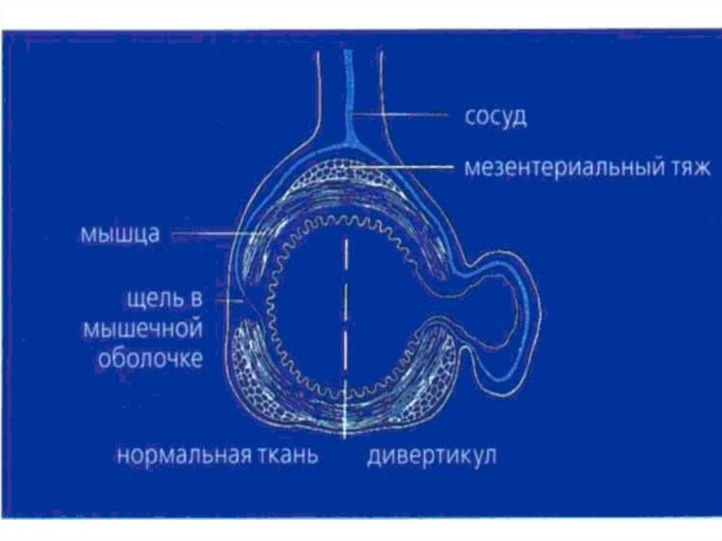
80. diverticuls
DiverticulDiverticulosis
Diverticulitis
81. diverticul
82.
83. diverticulosis
84. diverticulosis
85. diverticuls
86. Multiple diverticuls
87. Diverticul - obturation
88. diverticulosis
89. Fecal stone in a diverticulum
90. diverticulitis
91. Clinical features
• Acute diverticulitis is well nicknamed 'leftsided appendicitis'; an acute onset of centralabdominal pain which shifts to the left iliac
fossa accompanied by fever, vomiting and local
tenderness and guarding. A vague mass may
be felt in the left ileal fossa and also on rectal
examination. Perforation into the general
peritoneal cavity produces the signs of general
peritonitis. A pericolic abscess is comparable
to an appendix abscess but on the left side; a
tender mass accompanied by a swinging fever
and leucocytosis.
92. Clinical features
•Chronic divertlcular disease exactly mimics the localclinical features of carcinoma of the colon; there may
be diarrhoea alternating with constipation which
progresses to a large bowel obstruction with
vomiting, distension, colicky abdominal pain and
constipation: (note that small bowel obstruction
from adhesion of a loop of small Intestine to the
inflammatory mass is not uncommon). There may be
episodes of pain in the left ileal fossa, passage of
mucus or bright red blood per rectum or of melaena,
or there may be anaemia due to chronic occult
bleeding. Examination reveals tenderness in the left
ileal fossa and there is often a thickened mass in the
region of the sigmoid colon, which may also be felt
per rectum.
93. Diverticulitis
•This results from infection of one or moredivertlcula. An inflamed diverticulum may.
•1. Perforate:
•a) into the general peritoneal cavity;
•b) with formation of pericolic abscess;
•c) into adjacent structures; bladder, small
bowel and vagina;
•2. Produce chronic infection with inflammatory fibrosis resulting in strictures and
obstructive symptoms — acute or chronic.
•3. Haemorrhage, as a result of erosion of
a vessel in the bowel wall. The bleeding
varies from acute to a chronic occult loss.
94. Diverticulitis
The Hinchey classification - proposed by Hinchey etal. in 1978[1] classifies a colonic perforation due to
diverticular disease. The classification is I-IV:
•Hinchey I - localised abscess (paracolonic)
•Hinchey II - pelvic abscess
•Hinchey III - purulent peritonitis (the presence of
pus in the abdominal cavity)
•Hinchey IV - faeculent peritonitis.
The Hinchey classification is useful as it guides
surgeons as to how conservative they can be in
emergency surgery. Recent studies have shown with
anything up to a Hinchey III, a laparoscopic washout
is a safe procedure[2], avoiding the need for a
laparotomy and stoma formation.