


")


")


 medicine
Similar presentations:


")

Inflammatory Bowel Diseases
1. Inflammatory Bowel Diseases
2. INFLAMMATORY BOWEL DISEASES
3. ULCERATIVE COLITIS AND CROHN’S DISEASE
4.
5. Etiology and Pathogenesis
• Genetically predisposed individuals• Chronic activation of the mucosal immune system may represent an
appropriate response to an unidentified infectious agent
• Inappropriate response to the endogenous microbial flora within the
intestine, with or without some component of autoimmunity
6. Genetic Considerations
• CARD15– senses bacterial muramyl dipeptide and regulates intracellular signaling
– expressed by intestinal epithelial cells, including Paneth cells,
monocytes, macrophages, and dendritic cells
– Loss-of-function mutations in CARD15 are highly associated with CD
– decreased intestinal antimicrobial activity by diminishing defensin
production by Paneth cells
– excess NF-kB activation
7. VARIETIES OF COLITIS
8. DIFFERENTIAL DIAGNOSIS OF INFECTIOUS AND ULCERATIVE COLITIS
9. DIFFERENTIAL DIAGNOSIS OF IBD AND IBS
10. CHARACTERISTIC FEATURES OF ULCERATIVE COLITIS
11. Pathology
• Ulcerative Colitis: Macroscopic Features– mucosal disease that usually involves the rectum and extends proximally to
involve all or part of the colon
– 40–50%-rectum and rectosigmoid, 30–40%- extending beyond the sigmoid,
20%- total colitis
– Proximal spread occurs in continuity without areas of uninvolved mucosa
– terminal ileum (1-2 cm) in 10–20% of patients- backwash ileitis
– biopsies from normal-appearing mucosa are usually abnormal
– mucosa is erythematous, hemorrhagic, edematous, and ulcerated
– inflammatory polyps (pseudopolyps) may be present as a result of epithelial
regeneration
– mucosa may appear normal in remission
– In prolonged disease mucosa is atrophic and featureless and the entire colon
becomes narrowed and shortened
12. UC Physical findings
• Abdomen: tenderness and distension, but can benormal
• Extra colonic: arthritis, skin changes liver disease
• Usually normal perineum
13. UC Laboratory findings
• No specific findings• ESR , CRP , anemia (chronic disease,
Fe ), WBC
K , Albumin (protein loosing)
Disturbed LFT
14. UC Clinical Features
• Relapsing disease (~ 80% 1yr)• Symptoms usually parallel disease extent
(More disease more systemic signs & need for
operation)
• Proctitis may be hard to treat and cause blood loss and
disturbing tenesmus
• Disease may extent more proximally with follow up
(~40% in proctitis, ~ 10% in left sided)
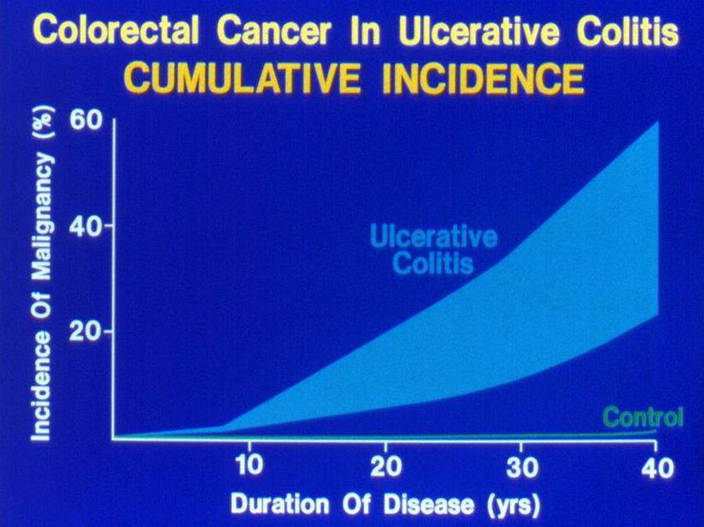
15. UC- Complications
• Bleeding• Perforation
• Toxicity
• Cancer
16.
17. Crohn’s disease (CD)
• Transmural disease, symptoms depend on siteof involvement and complications
• Abdominal pain, diarrhea (usually not bloody),
weight loss, fever
• Mouth to anus
18. ANATOMIC DISTRIBUTION
Terminal ileum is involved in 75%19. CD Small bowel
• Abdominal pain (mainly RLQ), may be constantand dull, may be colicky (obstruction)
• Diarrhea
• Vomiting (obstruction)
• Weight loss, fatigue, fever
• Acute presentation may resemble appendicitis
• May present as FUO or chronic subtle disease
20. CD Colon
• Colon: diarrhea, less rectal bleeding(less colon & rectum involved),
characteristic rectal sparing.
• Perianal involvement: fissures, fistulas,
perirectal abscess
21. CD Perianal Disease
• Fissures• Fistulas
• Perirectal abscess
22. CD Pathology Macroscopic Features
– terminal ileum is involved in 75%– the rectum is often spared in CD
– CD is segmental with skip areas
– Perirectal fistulas, fissures, abscesses, and anal stenosis are
present in one-third of patients with CD, particularly those
with colonic involvement
– serosal and mesenteric inflammation promotes adhesions and
fistula formation
– "creeping fat"
23. VIENNA CLASSIFICATION
24. CLINICAL PATTERNS
25. FISTULIZATION
26. CONFINED PERFORATION
27. Natural history of CD accumulation of disease complications
100Cumulative probability %
90
1
80
70
0.75
60
penetrating
50
0.5
40
30
Stricturing
0.25
Inflammatory
20
10
0
0
0
12 24 36
48 60 72 84
96 108 120 132 144 156 168 180 192 204 216 228 240
months
Patients at risk:
2002
552
229
95
Kaplan-Meier estimates of remaining free of
complications in 2,002 patients with Crohn’s disease
since onset of the disease.
37
Upper GI
T.ileum
inflammatory
Ileocolon
stricturing
Colon
penetrating
Kaplan-Meier 20-year cumula
cidence of stricturing and penetrating
complication
2065 pts
Follow up 1974-2000
Cosnes J. et al, Inflammatory Bowel Diseases 2002;8:244-250
28. APPROACH TO DIFFERENTIAL DIAGNOSIS OF ULCERATIVE VERSUS CROHN’S COLITITS
29. Extraintestinal Manifestations
• Arthritis- Peripheral -dependent on disease activity
- Axial-independent of disease activity
• Ocular
- episcleritis, uveitis
• Skin
- erythema nodosum
- pyoderma gangrenosum
• Liver
- PSC
30. Extra-intestinal manifestations, co-morbidities and complications of CD
Extra-intestinal manifestations, comorbidities and complications of CDUveitis1
Pyoderma gangrenosum2,3
Psoriasis4
Spondyloarthropathy5
31. Extraintestinal Manifestations Rheumatologic
Peripheral arthritis- 15–20% of IBD patients• more common in CD
• worsens with exacerbations of bowel activity
• asymmetric, polyarticular, and migratory and most often
affects large joints of the upper and lower extremities
• In severe UC, colectomy frequently cures the arthritis
Ankylosing spondylitis
• more common in CD than UC
• HLA-B27 antigen
• AS activity is not related to bowel activity
32. Extraintestinal Manifestations Rheumatologic
Sacroilitis– Symmetric
– equally in UC and CD
– often asymptomatic
– does not correlate with bowel activity
– does not always progress to AS
33. Extraintestinal manifestations - Skin
Pyoderma gangrenosum- more in UC patients• may occur years before the onset of bowel symptoms
• independent of the bowel disease
• respond poorly to colectomy
• very difficult to treat and often require intravenous
antibiotics, intravenous glucocorticoids, dapsone,
azathioprine, thalidomide, intravenous cyclosporine, or
infliximab
34.
35. Extraintestinal Manifestations - Skin
-Erythema nodosum (15% of CD patients and 10% of
UC patients)
• correlate with bowel activity
• concomitant active peripheral arthritis
– Perianal skin tags are found in 75–80% of patients with CD
– Aphthous stomatitis and "cobblestone" lesions of the buccal
mucosa
– Metastatic CD- cutaneous granuloma formation
36. Erythema nodosum
37. Extraintestinal Manifestations
• Ocular:– The most common are conjunctivitis, anterior uveitis/iritis, and
episcleritis
– Uveitis is associated with both UC and Crohn's colitis
– Prompt intervention, sometimes with systemic glucocorticoids, is
required to prevent scarring and visual impairment
• Hepatobiliary
– Fatty liver
– Cholelithiasis is more common in CD than UC
– PSC- 1–5% of patients with IBD have PSC, but 50–75% of patients
with PSC have IBD
• fatigue, jaundice, abdominal pain, fever, anorexia, and malaise
• Ds: ERCP or MRCP
• cholangiocarcinoma
• increased risk of colon cancer
• ursodeoxycholic acid (ursodiol)
38. Extraintestinal Manifestations
• Urologic– calculi, ureteral obstruction, and fistulas
– nephrolithiasis (10–20%) occurs in patients with CD
• hyperoxaluria
• Metabolic Bone Disorders
– Low bone mass
• risk is increased by glucocorticoids, cyclosporine, methotrexate and total
parenteral nutrition (TPN)
• Malabsorption and inflammation mediated by IL-1, IL-6, and TNF
–
Osteonecrosis
• bone scan or MRI
• within 6 months of starting glucocorticoids
39. Extraintestinal Manifestations
• Thromboembolic Disorders– increased risk of both venous and arterial thrombosis
• Other Disorders
– cardiopulmonary manifestations: endocarditis, myocarditis,
pleuropericarditis
– interstitial lung disease
– amyloidosis
40. Diagnosis
• History- How long?
- How bad: no. of stools? Blood?
• Signs of rectal involvement (urgency, frequency
incomplete evacuation)
• Pain (nature, awakes at night, location, relation to
defecation)
• Additional inflammatory signs: fever, weight loss
(anorexia, diarrhea, sitophobia)
• Additional signs of complications:
arthritis, rashes, ulcers, perineal diseases
41. Diagnosis
• Laboratory tests- non specific and reflectdisease severity & involvement
• Anemia- normocytic normochromic (chronic
disease), Iron , B12 (CD of TI, BOG), FA
(malabsorption due to disease involvement)
• Electrolytes- K , Ca , Mg , Zn
• Albumin (malabsorption, protein losing)
42. Diagnosis
• Stool: Steatorrhea (mild), WBC in stool,Increased calprotectin
• Disturbed Liver function tests
(Alk. P- PSC, TA- inflammation)
43. Diagnosis
• Determine anatomic involvement• Determine nature of involvement
(UC Vs CD Vs others)
44. Diagnosis
• Endoscopic examinations:Rectosigmoidoscopy- rectum? Mucosal morphology?
(ulcer type, skip areas)
Colonoscopy- Same + disease extent + terminal ileoscopy
• Pathologic examination: biopsies
(granulomas in 10-25 % of cases), other features less
specific
45. ENDOSCOPIC SPECTRUM OF SEVERITY
46.
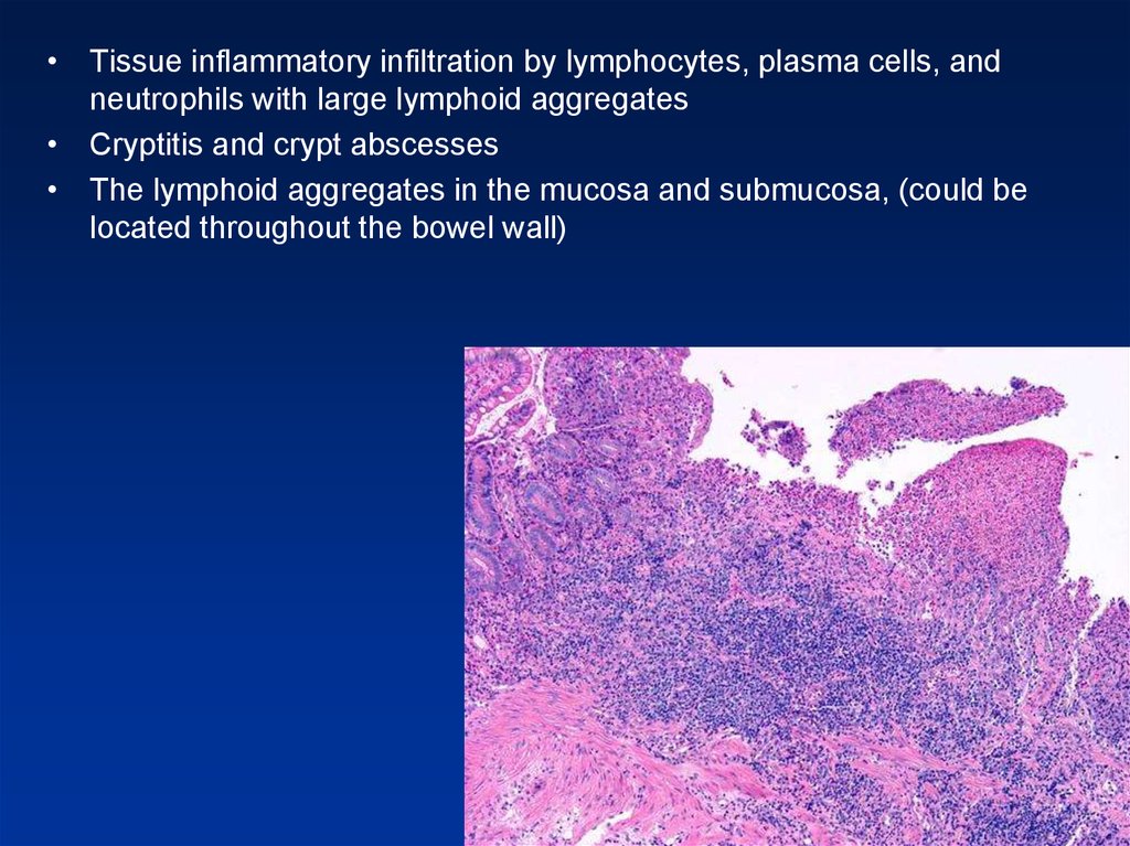
• Tissue inflammatory infiltration by lymphocytes, plasma cells, andneutrophils with large lymphoid aggregates
• Cryptitis and crypt abscesses
• The lymphoid aggregates in the mucosa and submucosa, (could be
located throughout the bowel wall)
47. ENDOSCOPIC APPEARANCES
stellate ulcerCD
aphthae APPEARANCES
ENDOSCOPIC
longitudinal ulcer
Macroulcerations and pseudoplyps
48. Diagnosis Radiology
• Barium enema:fistula, sinus tract, stricturing (not used today)
• Small bowel follow through- small bowel
anatomy and involvement, strictures, fistula (rarely
used today)
49. TRANSVERSE COLON STRICTURE
50. SPECTRUM OF ILEITIS
CDSPECTRUM OF ILEITIS
marked edema and
nodularity in addition
to ulceration
narrowing and spasm
deeper ulceration+
mesenteric sinus
tract formation
51. Diagnosis
• CT – replaced SBFT, allows for detection ofextramural complications
( abscess, fistula, retroperitoneal disease)
• MRI: MRE – replaces CT?
- MR for pelvic CD
• EUS- pelvic CD, biliary disease
52.
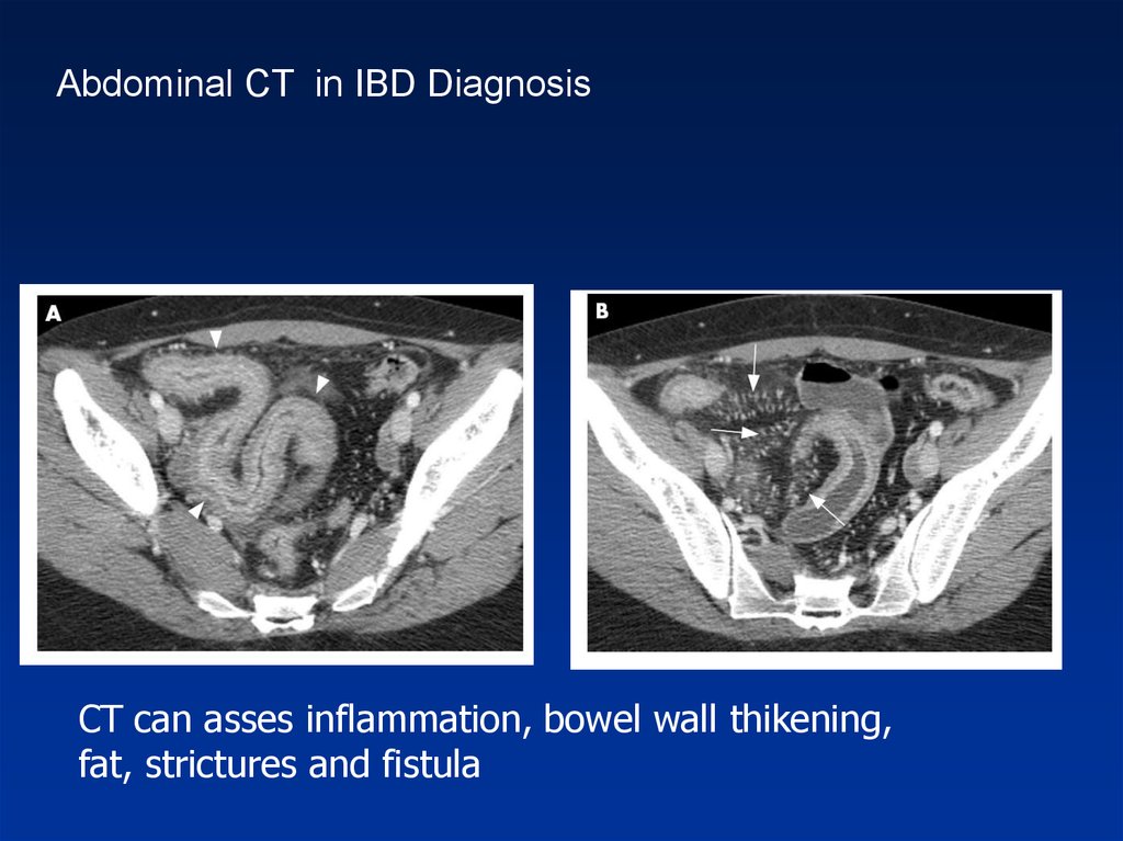
Abdominal CT in IBD DiagnosisCT can asses inflammation, bowel wall thikening,
fat, strictures and fistula
53. DISTINGUISHING FEATURES OF CROHN’S DISEASE
54. GOALS OF THERAPY
55. CONVENTIONAL DRUG THERAPIES
BiologicsAnti- TNF
Anti-cytokine
Anti Migration
56. SULFASALAZINE
57. AMINOSALICYLATES
58. AMINOSALICYLATE DISTRIBUTION
59. STEROID PREPARATIONS
Systemic / TopicalSTEROID PREPARATIONS
60.
61. Immuno-suppressors in IBD
• Azathioprine, 6-Mercaptopurine• Methotrexate
• Cyclosporin
• Tacrolimus
62.
63. Side effects thiopurines (cont.)
• Small increased risk of developing lymphoma• Increased risk of non- melanoma skin cancer
64. TOXICITY OF CYCLOSPORINE
65. Chronic Inflammation: Imbalance Between Mediators
66. Migration of Cells into Tissues
ROLLINGSelectin ligands,
L-Selectin
E, P Selectins
Mucosa
ACTIVATION
ARREST
Integrins
TRANSMIGRATION
Adhesion
Molecule
67. Biologicals
• Anti TNF agents:- Infliximab (Remicade), Adalimumab (Humera),
Golimumab (Simponi)
• Anti migration:
- Natalizumab
- Vedolizumab
Binds α4β7-integrin heterodimer, inhibits the pathologic effects of
CD4 T-cell
68. Chimerized and Humanized Antibodies
69. Infliximab Mechanism of Action
70. Integrin Structure
b 1,7a4
Plasma
membrane
71. ADVERSE EFFECTS OF INFLIXIMAB
72. Biologicals: Pre-therapy preparations
• TB exposure: Skin test/quatiferon + Rx• HBV, HIV, Varicella exposure
• Immunize: Pneumovax, Influenza
(HBV, varicella)
73.
DiagnosisCD
UC
?
?
74.
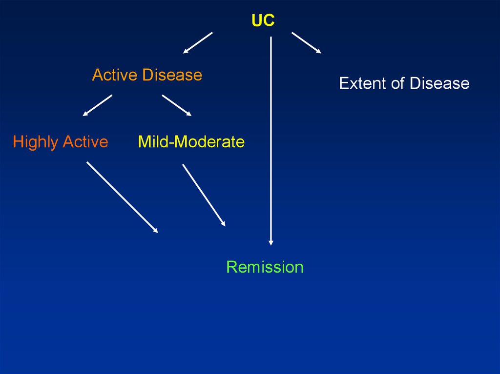
UCActive Disease
Highly Active
Extent of Disease
Mild-Moderate
Remission
75. Main clinical points to address
Factors that affect treatment choice:- Disease distribution (proctitis, left sided,
extensive)
- Disease behavior (frequent relapse?)
- Response to previous medications
- Side effects
- Extraintestinal manifestations
76. Patient assessment
• Exclusion of infectious agents:STD in proctitis
Bacterial (including C. Diff) and parasitic
infections
CMV- in the context of immune suppression
(biopsy)
• Endoscopic evaluation:
Infectious?
Crohn’s?
Mucosal prolapse?
IBS & haemorrhoidal bleeding ?
77. Outpatient assessment of the severity of active UC: T&W- Important not to miss severe progressive disease
Outpatient assessment of the severity of active UC:T&W- Important not to miss severe progressive disease
Mild
Bloody
stools/day
Moderate
Severe
<4
>6
Pulse
<90bpm
>90bpm
Temperature
<37.5oC
In between
>37.8oC
Hb
>11.5g/dL
<10.5g/dL
ESR
<20mm/hr
>30mm/hr
o
r
or
or
• Easy to remember, easy to apply, defines
severe attacks
78. UC - Mild to moderate activity
• 5-ASA/SZP:Both induction of remission and maintenance
Dose – dependent
Combine topical & systemic
If Failure:
• Steroids:
Induction of remission only
Combine topical & systemic
Start high does and taper
79. UC - Left sided & Pan colitis Mild to moderate activity
UC - Left sided & Pan colitisMild to moderate activity
If steroid dependent:
• Azathioprine/ 6-MP
If non responsive:
• Infliximab
Can be used to induce & maintain remission
Note: Role of Adalimumab & Methotrexate not
formally established for UC
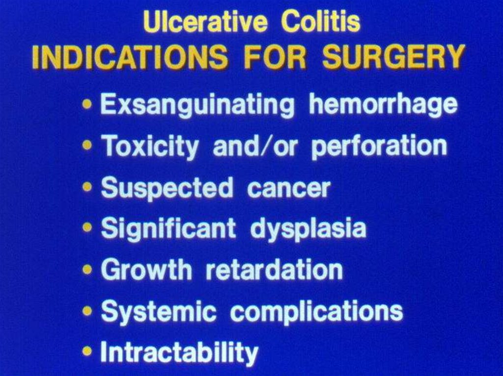
80. Severe UC
• Prevalence ~ 20% for first and recurrent attacks• Severe active UC with systemic toxicity →hospitalize
• Usually IV, hydrocortison 100 mg X 3 for 5 days
• Lower doses – less effective, > 7-10 days – no benefit
• Systematic review 32 trials (1991 pts) 2:
Response 67%
Colectomy 29%
Death 1%
81. Severe UC
• Correct:Hypokalemia, hypomagnesemia (toxic dilatation )
Hemoglobin
Nutritional support
(complications enteral Vs parenteral 9% Vs 35%)1
Withdraw anticholinergics, antidiarrheals, NSAID, opiod
Abx – only if infection suspected or preoperative
• Cyclosporin monotherapy = 40 mg Methylpredinsolone
use in steroid intolerant
82.
83.
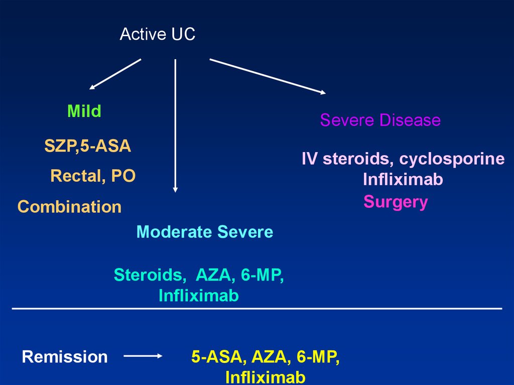
Active UCMild
Severe Disease
SZP,5-ASA
IV steroids, cyclosporine
Infliximab
Surgery
Rectal, PO
Combination
Moderate Severe
Steroids, AZA, 6-MP,
Infliximab
Remission
5-ASA, AZA, 6-MP,
Infliximab
84.
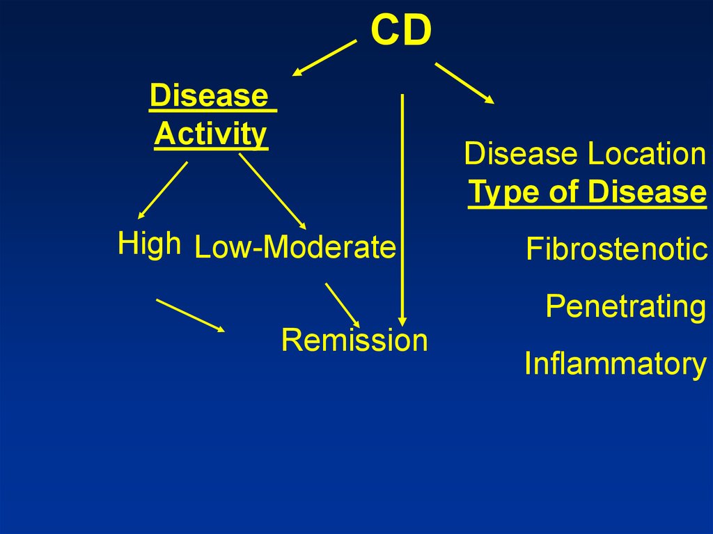
CDDisease
Activity
Disease Location
Type of Disease
High Low-Moderate
Fibrostenotic
Penetrating
Remission
Inflammatory
85. CD- Colon Mild -Moderate
• SZP-/5-ASA for colonic disease only• Side effects: paradoxical diarrhea, nausea,
vomiting, headache, hypersensitivity
• Need to check renal function
Allowed in pregnancy
86. CD-Small Bowel
• Steroids:Generally try to avoid due to side effects
– Controlled trials show definite efficacy
– Use steroids with less side effects
• Budesonide: 90% first pass effect
– TI & RT colon
– Similar effect to prednisone less SE
– Need to FU: Bone density, glucose levels allowed during pregnancy
87. CD – Moderate Activity
• Immunosuppressive agents• Azathioprine, 6 MP
• Steroid dependent or resistant disease
Steroid sparing
• 30-60% response
• Up to 6 mo to initial effect, most start earlier
• FU: CBC, LFT, Pregnancy OK
88. CD-Moderate Disease
• MethotrexateIM - 40% efficiency for 16 wks
Reduced Steroid use
Max efficiency - 6 wks
SE: leukopenia, nausea, vomiting, diarrhea
Possible liver fibrosis
• FU: CBC LFT
• Contraindicated in pregnancy
89. INFLIXIMAB IN ACTIVE CROHN’S DISEASE
Anti TNF therapy in Crohn’s diseaseINFLIXIMAB IN ACTIVE
CROHN’S DISEASE
90. Biologicals
• No difference between Infliximab and Adalimumab forefficacy
• Different modes of administration
• Loading, scheduled therapy
• Loss of response:
Dose escalation/switch
• Antibodies formation
91. CD- Severe Disease
• Hospitalization• IV steroids
• If abscess, fistula- drain, consider TPN
• Anti TNF Abs
92. CD- Effect of Disease Type
• Perianal & fistula:Antibiotics
Azathioprine/6 MP
Infliximab
• Surgery
• Treatment sequence: Image, classify, drain
sepsis – medical treatment
93. CD- Effect of Disease Type
• Fibrostenotic disease- Need to differentiate inflammation/scare
• If scare: surgery
• Medical therapy as inflammatory
94. CD- Maintenance of Remission
• Not Steroids !5-ASA: low efficiency (1:13), SE
May benefit post surgical
Not good for remission post medical Tx
Chemopreventive?
95. CD- Maintenance of Remission
• Immunomodulatory drugs• Azathioprine/6MP: efficient regardless
of therapy mode
• MTX: Good for pts that entered
remission with MTX
• Anti TNF agents
96.
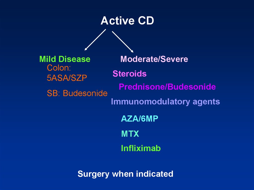
Active CDMild Disease
Colon:
5ASA/SZP
SB: Budesonide
Moderate/Severe
Steroids
Prednisone/Budesonide
Immunomodulatory agents
AZA/6MP
MTX
Infliximab
Surgery when indicated
97.
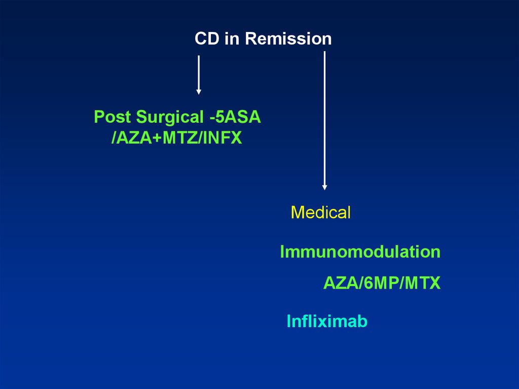
CD in RemissionPost Surgical -5ASA
/AZA+MTZ/INFX
Medical
Immunomodulation
AZA/6MP/MTX
Infliximab
98. The evolution of therapy: Should we invert the pyramid?
SurgeryWhat is the optimal use
of infliximab?
TNF inhibitors
*
Which patients should be
treated with anti-TNF?
Immunosuppressives
(AZA/6-MP, MTX)
Steroids
99. Future evolution
• Should we aim for mucosal healing?• Should we perform early surgery?
• Risk / benefit analysis of treatments and
outcomes
100. Case Study
30-year-old woman was admitted with a 4-week history of increasing bloodydiarrhea and abdominal pain; she had lost 3kg in weight. She smoked 1 pack of
cigarettes a day. On examination, she was not clinically anaemic and, apart from a
temperature of 37.8°C and some tenderness over the right iliac fossa, there were no
abnormal physical signs.
The perineum was normal but sigmoidoscopy to 15cm showed a red, granular
mucosa with aphtous lesions and contact bleeding. Laboratory investigations showed
a low haemoglobin (10.8g/l) with a raised CRP (67 mg/l) but a normal white-cell
count. Urea and electrolytes, serum vitamin B12, folate, iron, ferritin and iron-binding
capacity were normal. Her total serum proteins were 5.4g/l (NR 6.2-8.2) with a
serum albumin of 2.9g/l (NR 3.5-5.0). Faecal examination and culture revealed no
ova or Campylobacter. Clostridium difficile toxin was negative
101. The rectal biopsy : many crypt abscesses were present. The lamina propria contained a heavy infiltrate of lymphocytes, plasma
Case StudyThe rectal biopsy : many crypt abscesses were present. The lamina propria
contained a heavy infiltrate of lymphocytes, plasma cells and macrophages. Two
non-caseating granulomas were present.
A CT and a colonoscopy were performed to assmall-bowel barium infusion s the
extent of disease. Inflammatory strictures were seen at a number of separate sites
(skip lesions) in the ascending and transverse colons. She was treated with
corticosteroids and a 3-month course of metronidazole with symptomatic
improvement. She was strongly advised to stop smoking.
102. י.ע. 9/2011
י.ע9/2011 .• בת ,54מזה כחודש וחצי סובלת משלשולים רבים ,יציאות דמיות
וריריות לסירוגין ,ירידה במשקל של כ 5-ק"ג בתקופה זו .אירועים
מעירים משינה ,מלווים בכאבי בטן.
• לפני כשבועיים בוצעה קולונוסקופיה :פאן קוליטיס.
• טופלה בפנטסה ופלג'יל ללא שיפור משמעותי.
103. י.ע. 9/2011
י.ע9/2011 .•
•
•
•
•
אושפזה בפנימית להמשך בירור וטיפול.
בקבלתה הוחל טיפול בסטרואידים ורפסל.במהלך אשפוזה שיפור
ניכר בתלונות.
לאחר 3ימי טיפול ללא כאבי בטן 3-4 ,יציאות ליממה ללא דם,
CRPירד לנורמה.
בתשובת פתולוגיה ממצאים מתאימים ל IBDמסוג .Active UC
בהמשך הועברה לטיפול פומי בסטרואידים.
104. י.ע. 18/10/2011
י.ע18/10/2011 .• באשפוז הקודם הותחל גם טיפול גם ב .MP-6שוחחתי ארוכות עם
החולה ובעלה אודות הסיכונים שבטיפול זה והצורך ההדוק במעקב.
• החולה תמשיך חפיפה עם סטרואידים ותגיע בעוד כחודש לביקורת.
105. י.ע. 18/10/2011
י.ע18/10/2011 .•
•
•
•
הגיעה לביקורת ,טופלה עד כה בפרדניזון עם ירידה הדרגתית
וסיימה לפני שבועיים.
בנוסף הותחל טיפול גם ב ( MP-6פורינטול) אך הפסיקה לפני
שבועיים .למרות ההמלצות בשחרור לא נוטלת כרגע פורינטול או
רפסאל!!! מקבלת פוליקס .אסימפטומטית לחלוטין.
לויקופניה ,4350נויטרופניה של .8.8 Hb ,670עקב הירידה
בלויקוציטים ,במיוחד בנויטרופילים ,ובהמוגלובין – לא מחדש בשלב
זה טיפול בפורינטול.
ממליץ :לתת רפסאל 2גראם פעמיים ביום ,לחזור על .CBC
106. י.ע. 26/12/2011
י.ע26/12/2011 .•
•
•
•
•
•
•
שני אשפוזים בפנימית :פעם אחת בשל החמרה שטופלה
בסטרואידים ,פעם שניה בשל מחלת ריאות משנית לטיפול ברפסל.
כאשר הפחיתה לפרדניזון 10מ"ג השלשולים נשנו.
בתחילת דצמבר אנמיה ,10 Hbלויקופניה גבולית 4920
ותרומבוציטופניה.
תלוייה בסטרואידים ASA-5 ,אינן באות בחשבון בשל התפתחות
פנאומוניטיס מסכנת חיים ,ולכן האופציה הבאה היא התחלת טיפול
בפורינטול או אימוראן (אם Pltו WBCיהיו תקינות) במינון הדרגתי.
במקביל פרדניזון 30מ"ג ולרדת בהדרגה.
יהיה צורך במעקב CBCואנזימי כבד ולבלב.
דיברנו על סיכון קטן ללימפומה.
107. י.ע. 23/7/2012
י.ע23/7/2012 .•
•
•
•
מזה 4ימים עלייה בתדירות היציאות 6-7 ,ליום,
חלקן עם דם .כאבי בטן מטרימים.
התלקחות של UCבדרגה בינונית ,לאחר טיפול
במינון מספק של פורינטול ולמשך זמן מספק.
ננסה טיפול בחוקני בטנזול
לשמירה על רמיסיה ננסה אם כך טיפול ברמיקייד.
לפני כן יש לשלול שחפת ישנה.
108. י.ע. 17/06/2013
••
•
•
•
י.ע17/06/2013 .
אושפזה עקב החמרת UCוהוחל שוב טיפול
בסטרואידים.
כעת רמיקייד כל 6שבועות ,הפסיקה ליטול
פרדניזון לפני שבוע.
עושה רושם שכעת ע .ברמיסיה
:8/9/13כעת על פרדניזון 25מ"ג ליום ,העלינו
מינון רמיקייד לאחת ל 4-שבועות (מינון כפול)
:8/10/13כעת ברמיסיה ,עדיין ב"זנב" הטיפול
בפרדניזון .תמשיך טיפול ברמיקייד כל 4שבועות.