


")
")


. The presence of symptoms damage of stomach, small and large intesti")


 medicine
Similar presentations:

")


")
")

Shigella infection
1. SHIGELLA INFECTION
2. Dysentery is a common infectious disease of man, caused by bacterium of genus Shigella.
Dysentery is characterized by principaldamage of the mucous membrane of the
distal section of the large intestine.
3.
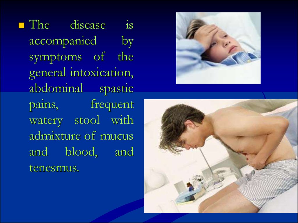
Thedisease
is
accompanied
by
symptoms of the
general intoxication,
abdominal spastic
pains,
frequent
watery stool with
admixture of mucus
and blood, and
tenesmus.
4.
INTRODUCTIONShigella organisms cause bacillary dysentery, a
disease that has been recognized since the time of
Hippocrates.
Shigellosis occurs world-wide. The incidence in
developing countries is 20 times greater than that in
industrialized countries.
>95% of shigella infections are asymptomatic hence
the actual incidence may be 20 times higher than is
reported.
5.
THE SHIGELLA BACILLUSShigella species are aerobic, non-motile,
glucose-fermenting, gram-negative rods.
It is highly contagious, causing diarrhea after
ingestion of as a few as 180 organisms.
Shigella spreads by fecal-oral contact, via
contaminated water or food.
Epidemics may occur during disasters,
in day-care centers & nursing homes.
6.
THE SHIGELLA BACILLUS4 species of shigella are identified, namely:
Shigella dysenteriae
Shigella Flexneri
Shigella Sonnei
Shigella Boydii
Every group is divided
into serologic types and subtypes.
Shigella dysenteriae is the most virulent, but sonnei is the
most common.
7.
VIRULENCEVirulence in shigella species is determined by
chromosomal & plasmid-coded genes.
Chromosomal genes control cell wall antigens that
are resistant to host defense mechanisms.
Plasmid genes control production of cytotoxin and
siderophores. The cytotoxins are both enetrotoxic and
neurotoxic.
Shigella invades colonic mucosa & causes cell
necrosis using both virulent agents.
8. Epidemiology
The sources of the infection are the patients withacute or chronic forms of dysentery, persons in the
period of convalescence and carries.
The persons with mild, chronic forms and
carries of the disease are most dangerous
9.
The mechanism and factors of the transmissionof the infection is fecal-oral.
The transmission of the infection is realized
through contaminated food-stuffs and water.
Infection of food-stuffs, water, different objects
happens due to direct contamination by infected
excrements, through dirty hands and also with
participation of flies.
Dysentery is characterized by seasonal spread like
other intestinal infections. It is registered more
frequently in summer and autumn.
10. PATHOHENESIS
Shigella adheres to intestinal epithelial cells and Mcells. After adhering to the host cells, the bacteria use
a type III secretors system to inject bacterial proteins
into the host cells.
These bacterial proteins cause the host cells to ruffle
and ingest the bacterial cells.
Once in the cells, the bacteria use a surface hemolysin
to lyse the phagosome membrane and escape into the
cytoplasm.
11. PATHOHENESIS
The bacteria then use the host cells’ actin to movearound inside the cell (actin rocket tails). When bacteria
reach the periphery of the cell, the cell pushes outward
to form membrane projections, which are then ingested
by adjacent cells.
Some strains of the Shigella genus produce the shiga
toxin or verotoxin, which is similar to the verotoxin of
E coli O157:H7. The shiga toxin or verotoxin enters the
cytoplasm of the host cells and stops protein synthesis
by removing an adenine residue from the 28S rRNA in
the 60S ribosomal unit. This toxic activity results in
death of the host cells.
12. PATHOHENESIS
The cell-to-cell travel and toxin activityproduces superficial ulcers in the bowel
mucosa and induces an extensive acute
inflammatory response. The inflammatory
response usually prevents entry of the bacteria
into the bloodstream. Unlike certain species of
Salmonella (e.g., S typhi, S paratyphi A),
Shigella only rarely enters the bloodstream.
13. Pathogenesis of Shigella
ShigellosisTwo-stage disease:
Early stage:
Watery diarrhea attributed to the enterotoxic
activity of Shiga toxin following ingestion and
noninvasive colonization, multiplication, and
production of enterotoxin in the small intestine
Fever attributed to neurotoxic activity of toxin
Second stage:
Adherence to and tissue invasion of large intestine
with typical symptoms of dysentery
Cytotoxic activity of Shiga toxin increases severity
14. Pathogenesis and Virulence Factors (cont.)
Virulence attributable to:Invasiveness
Attachment (adherence) and internalization
with complex genetic control
Large multi-gene virulence plasmid regulated
by multiple chromosomal genes
Exotoxin (Shiga toxin)
Intracellular survival & multiplication
15. Pathogenesis and Virulence Factors (cont.)
Characteristics of Shiga ToxinEnterotoxic, neurotoxic and cytotoxic
Encoded by chromosomal genes
Two domain (A-5B) structure
Similar to the Shiga-like toxin of
enterohemorrhagic E. coli (EHEC)
NOTE:
except that Shiga-like toxin is encoded
by lysogenic bacteriophage
16.
PATHOLOGYGross
pathology
consists
of
mucosal
edema,
erythema,
friability,
superficial
ulcers & focal mucosal
hemorrhage involving
the
rectosigmoid
junction primarily.
17. PATHOLOGY
Microscopicpathology consists of
epithelial
cell
necrosis, goblet cell
depletion, polymorph
& mononuclear cell
infiltrates in lamina
propria and crypt
abscess formation.
18.
AT RISK GROUPSChildren in day care centers
International travelers
Homosexual men
Patients with HIV infection
People with inadequate water supply
Persons in prisons & military camps
19. Classification of the clinical forms
Dysentery is divided into acute and chronicdysentery. Acute dysentery continues from some days
to 3 months (prolonged course of acute dysentery).
Dysentery is considered to be chronic, if it persist
over 3 months.
There are the following clinical variants of acute
dysentery:
colitic variant;
gastroenterocolitic variant;
gastroenteric variant.
In dependence on severity of the course of the disease
there are mild, moderately severe and severe course
of dysentery, and also carriers.
20. MAIN CLINICAL SYNDROMS
IntoxicationColitic
21.
CLINICAL PICTUREIncubation period is from 2 to 5 days, rarely – 7 days.
Symptoms begin with sudden onset of high-grade
fever, abdominal cramps & watery diarrhea
Subsequently the diarrhea became mucoid, of
small volume & mixed with blood. This is
accompanied by abdominal pain, tenesmus &
urgency. Fecal incontinence may occur.
Physical signs are those of dehydration beside
fever, lower abdominal tenderness & normal or
increased bowel sounds.
22. Mild course
The onset of the disease is acute.The moderate pains develop in the in the left iliac
area.
These pains precede the act of defecation.
Tenesmus are observed in the some patients.
Stool is from 3-5 to 10 times a day. It contains mucus,
sometimes – blood.
The temperature is normal or subfebrile.
On rectorhomanoscopy catarrhal inflammation of the
mucous membrane is observed, sometimes erosions
and hemorrhages.
23. Moderate course
The onset of the disease in acute or with short prodromalperiod. It is characterized by weakness, malaise,
discomfort in the stomach.
Colitic syndrome:
Develop spastic pains in the lower part of the abdomen,
tenesmus.
Tenderness and spastic of the sigmoid are revealed.
Stool has fecal character. Then, mucus and blood appear
in stool.
Stool loses fecal character and has appearance of “rectal
spit” (excretion of scanty stool – “fractional stool”), with
mucus and blood. Stool is accompanied by fecal urgency
and tenesmus. Stool is from 10-15 times a day.
24.
Intoxication syndromThe temperature increases to 38-39°C with
duration 2-3 days.
The patients complain of weakness, headache.
May be collapse, dizziness. The skin is pale.
Hypotension,
relative tachycardia are
observed.
25. Leukocytosis and moderate neutrophillosis are observed in the peripheral blood.
•Leukocytosis and moderate neutrophillosisare observed in the peripheral blood.
On coprocystoscopy erythrocytes (over 30-40
in the field of the vision) are revealed.
In
rectorhomanoscopy diffuse catarrhal
inflammation, local changes (hemorrhages,
erosions, ulcers) are revealed.
Functional and morphological convalescent
may be prolonged – to 2-3 months
in the patients with moderately
severe course of acute dysentery.
26. Severe course.
The onset of the disease is acute.The temperature increases to 39˚C and more.
The patients complain of headache, sharp
weakness, nausea, vomiting.
Severe abdominal spasmodic pains, frequent
stool scanty, with mucus and blood are
marked.
There are hypotension, acute tachycardia,
breathlessness, cyanosis of the skin.
27. Severe course
Acute pain in the left iliac area, especially inthe area of the sigmoid is marked on palpation
of the abdomen.
Paresis of the intestine is possible.
There are marked leukocytosis, neutrophillosis
with shift to the left to young form.
ESR is accelerated.
28. On microscopically examination of stool erythrocytes are marked in all fields of the vision.
On rectorhomanoscopy catarrhal orfibrinous inflammation, presence of the
local changes (erosions, ulcers) are marked.
The
functional
and
morphological
convalescent of the intestine is over 3-4
months in the patients with colitic variant of
acute dysentery.
29. Gastroenteritic variant of acute dysentery
The principal feature of this variant of acutedysentery is predominance of the clinical
symptoms of gastroenteritis and presence
of appearances of dehydration of the
different degree.
The principal feature is - acute onset of the
disease after short incubation period (6-8
hours).
30. Gastroenterocolitic variant of acute dysentery The principal feature of this variant of the acute dysentery course is acute onset of the disease after short incubation period (6-8 hours). The presence of symptoms damage of stomach, small and large intesti
Gastroenterocoliticvariant
of
acute
dysentery
The principal feature of this variant of the acute
dysentery course is acute onset of the disease after
short
incubation
period
(6-8
hours).
The presence of symptoms damage of stomach, small
and large intestines
Intoxicative syndrome and syndrome of
gastroenteritis are observed in the initial period.
The symptoms of enterocolitis predominate in the
period of clinical manifestation.
Can be development dehydration of I-II-III degree
31.
Prolonged course of acute dysentery is clinicalmanifestations of the disease are observed over 3-4
weeks.
The period of functional and morphological
convalescent of the intestine is over 3 months.
Chronic dysentery is prolonged of acute dysentery
is more than 3 months
32.
Diagnostics shigellosisThe mains methods of specific diagnostics are
microbiological and serological methods of examination
Microbiological
examination of feces and gastric
washings
It
is necessary to take the material for
bacteriological investigation before beginning of the
specific treatment.
Diagnosis may be confirmed by serological methods
-Reaction of indirect agglutination with standard
erythrocytes diagnostic. Diagnostic titer is 1:200 with
increase of titer in 7-10 days.
33.
Non-specific diagnosticsBlood-test, Ht (WBC is usually leukocytosis,
increasing Ht – hemoconcentration)
Urine-test
Electrolitis (Na, K, CL)
Coprogram (Stool microscopy reveals
presence of RBC & pus cells with mucous)
34.
MORTALITY & MORBIDITYWhereas mortality caused by shigellosis is rare in
western countries, it is associated with significant
mortality & morbidity in developing world.
Dehydration is the common complication of
shigellosis, but serious gastrointestinal & systemic
complications may occur.
35.
GASTROINTESTINAL RISKSRectal prolapse
Toxic mega colon
Mild Hepatitis
36.
NEUROLOGICAL COMPLICATIONSThese include:
Lethargy, delirium, meningismus & seizures
Encephalopathy (rare & may be lethal)
Febrile seizures
37.
SYSTEMIC COMPLICATIONSHemolytic uremic syndrome
Disseminated intravascular coagulation (DIC)
Reiter syndrome, arthritis, conjunctivitis & urethritis
Myocarditis
38.
DIFFERENTIAL DIAGNOSESAmebiasis
Campylobacter infection
Yersinia Entrocolitica infection
Salmonellosis
Escherichia Coli infection
Clostridium difficile infection
Crohn disease
Ulcerative colitis
39. TREATMENT
Thetreatment of the patient should be given
complex and based on pathogenesis. The
treatment depends on the clinical variant and
severity of the course of dysentery.
Diet N 4
Enzims
Sorbents
Correction of water-electrolyte balance and
detoxication therapy
40.
TREATMENTMedical care include rehydration & use of
antipyretics in febrile patients followed by antibiotics.
Drugs of choice are Cotrimoxazole, 3rd generation
cephalosporins & ciprofloxacin.
Ampicillin is effective but resistant is common.
Nalidixic acid is also effective but should be avoided
in patients with G6PD deficiency.
41.
PUBLIC HEALTH ASPECTSIsolation & barrier nursing is indicated
Notification of the case to the infection control
nurse in the hospital.
Isolation source of infection.
Continue breastfeeding infants & young children &
light diet for other patients in the first 48 hours.
42.
PREVENTIONEducation on hygiene practices particularly hand
washing after toilet use.
Avoidance of eating in non hygienic places.
Proper handling & refrigeration of food even
after cooking.
Antibiotic prophylaxis is not needed for household contacts.
43.
PROGNOSISMost patients with normal immunity will recover
even without antibiotic therapy but illness will be
prolonged & severe.
With antibiotic treatment fever subsides in 24
hours & colic & diarrhea within 2-3 days.
Few patients will have mild cramps & loose
motions for 10-14 days after treatment.
Mortality in tropical countries may be as high as
20%.