


")


 medicine
Similar presentations:


")

Congenital intestinal obstruction
1. Congenital intestinal obstruction
Lection2. Oesophageal atresia
• Oesophageal atresia is defined as an interruptionin the continuity of the oesophagus with or
without fistula to the trachea.
• The anomaly results from an insult occurring
within the fourth week of gestation, during
which separation of trachea and oesophagus
by folding of the primitive foregut normally
takes place.
3.
• At least 18 different syndromes have beenreported in association with oesophageal
atresia.
• The best known is probably the VATER
or VACTERL association of anomalies
(Vertebral-Anal- Cardiac-TrachealEsophageal-Renal-Limb).
4. Types of tracheo-oesophageal fistula
5.
6. Clinic
• The earliest symptom of oesophageal atresia is apolyhydramnios in the second half of pregnancy.
• A newborn infant has excessive salivation,
choking, and regurgitation with feeding.
• 25-40% of neonates are premature, low bith
weight.
• 50% of neonates with TEF have an associated
anomaly (cardiovascular most common).
7. Prenatal diagnosis - polyhydramnios
8.
• Inability to pass nasogastric tube.• Abdominal Xray with air in the stomach
excludes esophageal atresia
• A Replogle tube maximally advanced into
the upper pouch helps to estimate its
approximate length.
9.
10. Differential diagnosis
Intranatal asphyxia of newborn
Birth injury of brain
Aspiration pneumonia
Congenital diaphragmatic hernia with
camp
11. Complications
• Early complications include: Anastamoticleak, recurrent TEF, tracheomalacia.
• Late Complications include: Anastamotic
stricture (25%), reflux (50%), dysmotility
(100%).
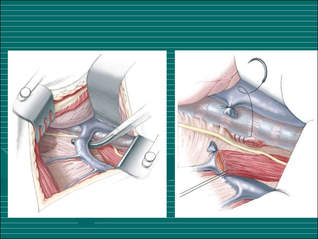
12. Treatment
• Operation includes TEF ligation, transection,and restoration with end-to-end anastamosis.
13.
14.
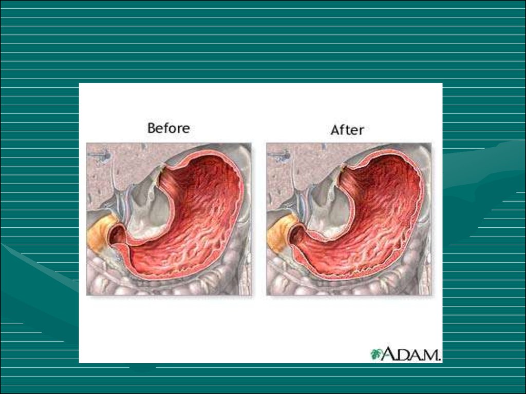
15. Hypertrophic Pyloric Stenosis
16.
• Infantile hypertrophic pyloric stenosis(IHPS) is a common surgical condition
encountered in early infancy, occurring in
2~3 per 1,000 live births. It is
characterized by hypertrophy of the
circular muscle, causing pyloric narrowing
and elongation. Boys are affected four
times more than girls.
17. Cause of hypertrophic circular muscle
• abnormal peptidergic innervation,• abnormality of nitrergic innervation,
• abnormalities of extracellular matrix
proteins,
• abnormalities of smooth-muscle cells
• abnormalities of intestinal hormones.
18. Clinic
• Age is 3-6 weeks (1 month of age)• A 4 week old infant presents with non-bilious
vomiting and hypochloremic, hypokalemic,
metabolic alkalosis.
• Projectile vomiting
• Dehydration
• “Hour-glass deformity sign”
19.
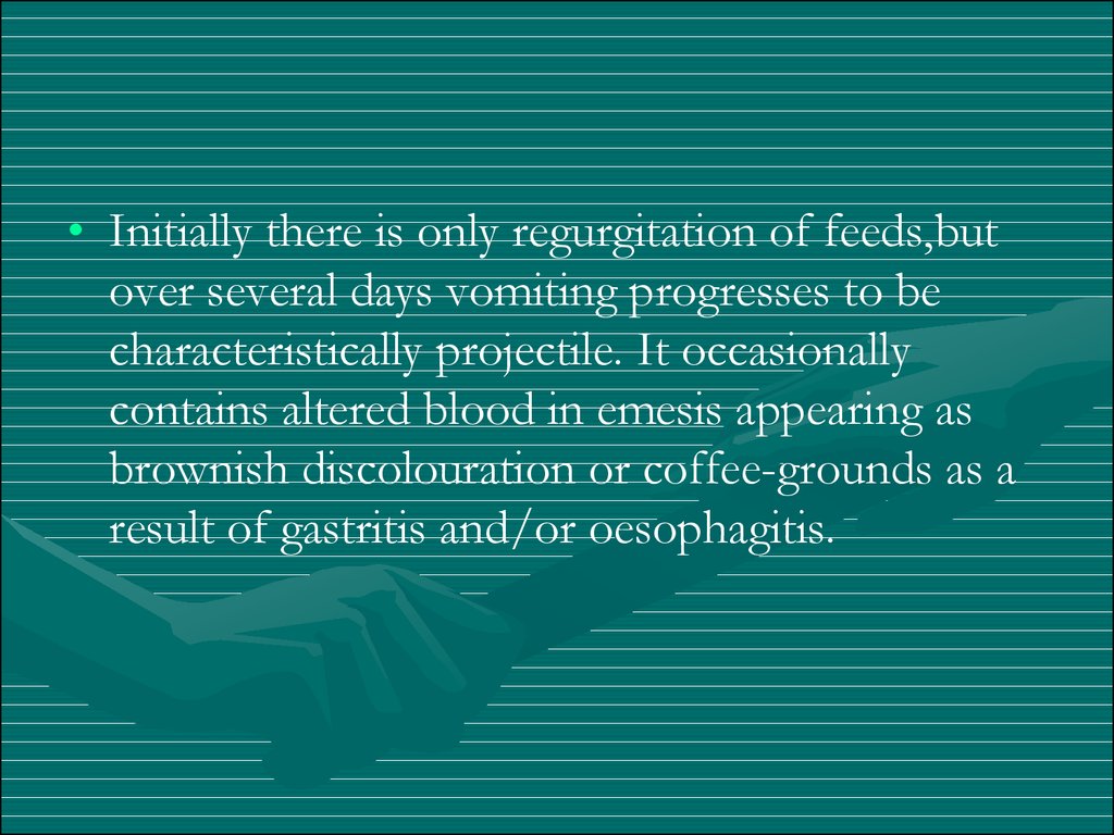
• Initially there is only regurgitation of feeds,butover several days vomiting progresses to be
characteristically projectile. It occasionally
contains altered blood in emesis appearing as
brownish discolouration or coffee-grounds as a
result of gastritis and/or oesophagitis.
20. X-ray symptom
Increas of stomach
Gastric peristalsis
“Beak symptom” or pylorus narrowing
Deceleration evacuation of contrast (2 – 5 h.)
Aerated intestinal canal
21.
22.
23. Differential diagnosis
Congenital pyloric stenosis
Stomach impassability
Duodenal obstruction
Vomiting syndrome
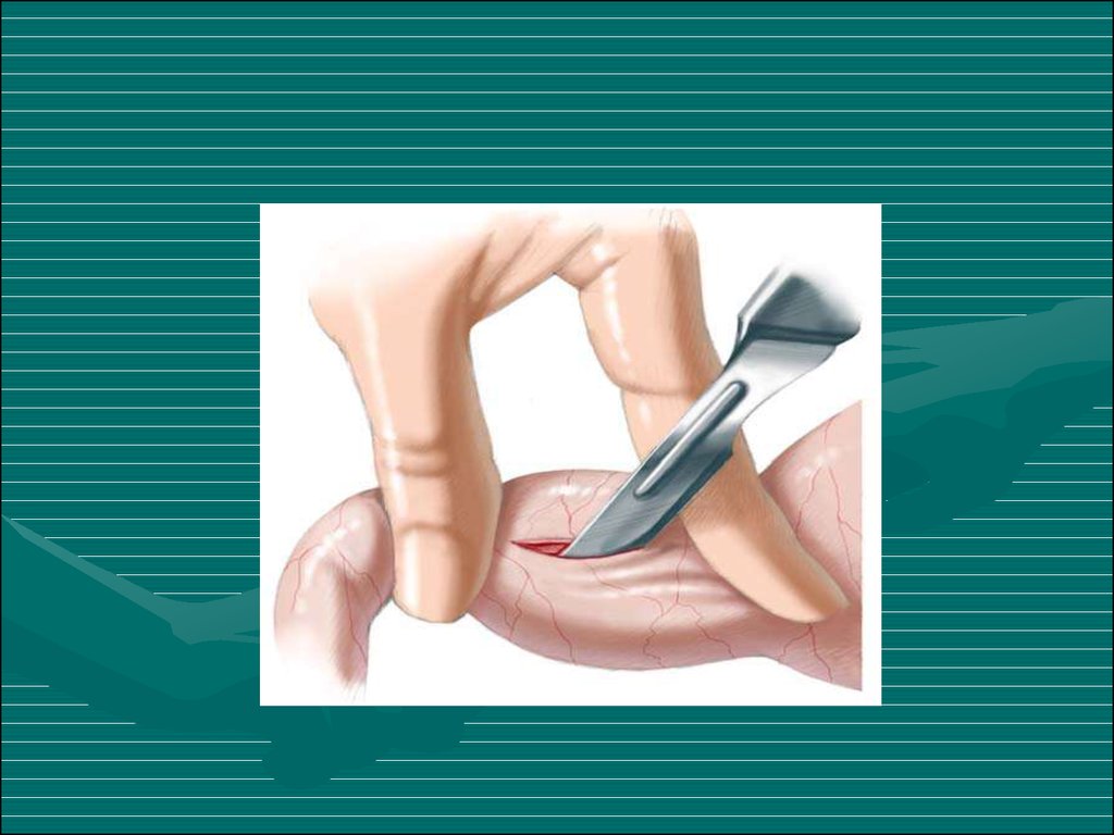
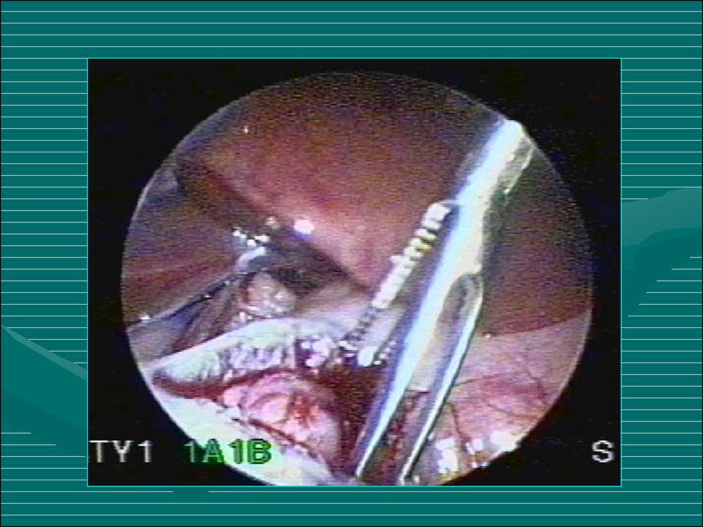
24. Treatment
• The operation for pyloric stenosis is not anemergency and should never be undertaken until
serum electrolytes have returned to normal.
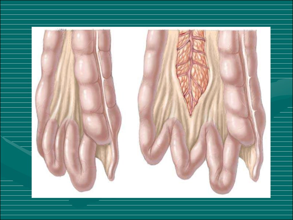
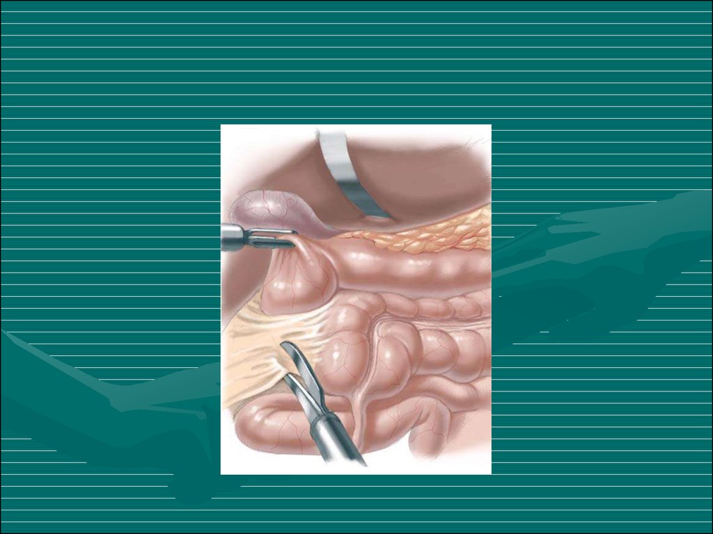
Ramstedt’s pyloromyotomy is the universally
accepted operation for pyloric stenosis.
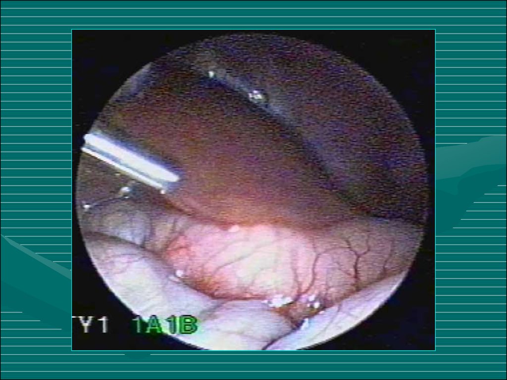
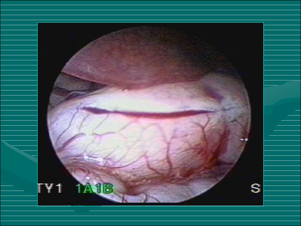
• Recently, laparoscopic pyloromyotomy has been
advocated. The main advantage of the
laparoscopic pyloromyotomy is the superior
cosmetic result.
25.
26.
27.
28.
29.
30.
31. Duodenal obstruction
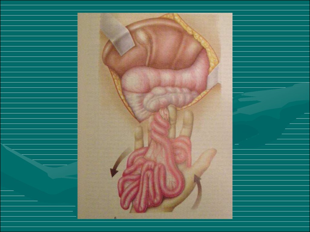
• During the embryonic period theduodenojejunal loop rotates 270° around
the superior mesenteric artery axis in an
anticlockwise direction. The caecocolic loop,
which initially lies inferiorly to the superior
mesenteric artery, also rotates 270° in an
anticlockwise direction. Finally the caecum and
ascending colon become fixed to the posterior
peritoneum. If this process is interrupted at any
point then malrotation or non-rotation results.
32.
33.
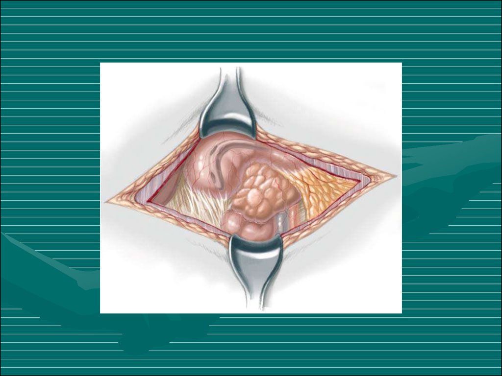
• Duoenal obstruction, with the possibility ofvascular compromise, is due to either an
associated volvulus or extrinsic compression
from peritoneal Ladd's bands.
• Acute bowel obstruction due to Ladd’s
bands or intermittent midgut volvulus can
present with vomiting, typically bilious, as
the commonest presenting feature
accompanied by colicky abdominal pain and
abdominal distention.
34.
35.
36.
• An infant with abdominal tenderness andblood per rectum is suggestive of bowel
ischaemia due to midgut volvulus.
• All symptomatic patients with positive
investigative findings should undergo urgent
laparotomy. Management of the
asymptomatic patient is more controversial.
37.
38. Differential diagnosis
Pylorospasm
Pyloric stenosis
Congenital diaphragmatic hernia
Helminthic invasion
Helminthic cholecystitis
39. Treatment
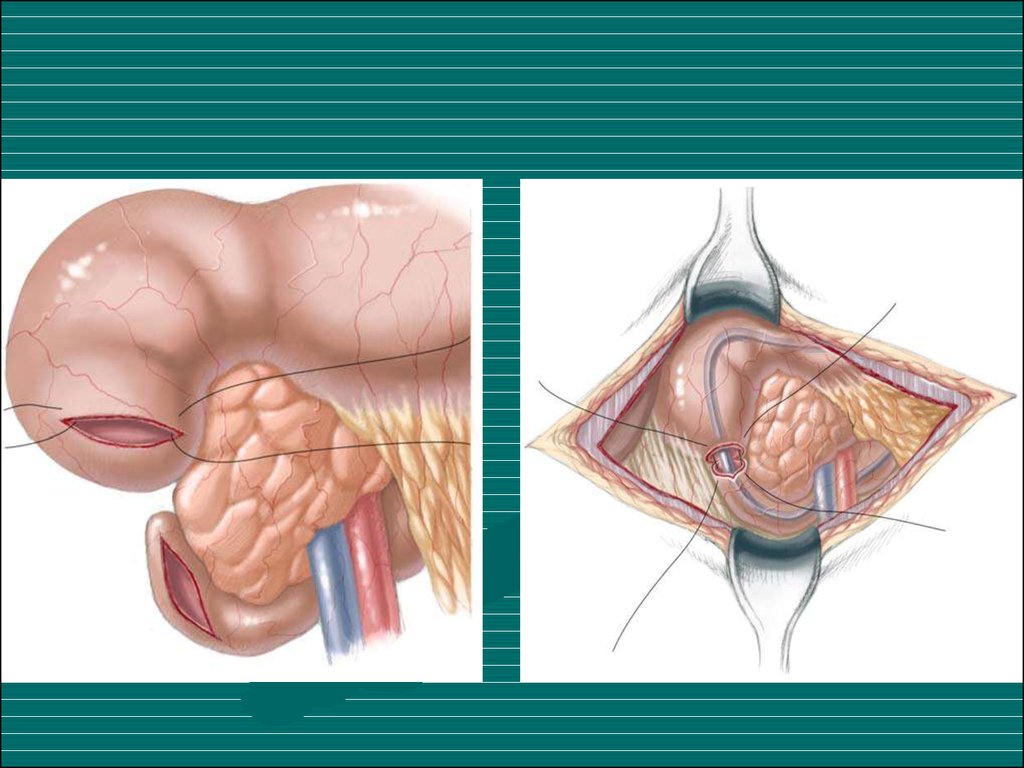
• Duodenoduodenostomy is the procedure ofchoice for patients with duodenal atresia,
stenosis and annular pancreas. The two
surgical techniques, either side-to-side
duodenoduodenostomy or proximal
transverse to distal longitudinal – “diamondshape” anastomosis – may be performed.
Diamond-shaped duodenoduodenostomy
has been reported to allow earlier feeding,
earlier discharge and good long-term results.
40.
41. Hirschprung’s disease
• Hirschsprung’s disease (HD) is characterised by anabsence of ganglion cells in the distal bowel and
extending proximally for varying distances. The absence
of ganglion cells has been attributed to failure of
migration of neural crest cells. The earlier the arrest of
migration, the longer the aganglionic segment.
• The pathophysiology of Hirschsprung’s disease is not
fully understood. There is no clear explanation for the
occurrence of spastic or tonically contracted aganglionic
segment of bowel.
42. Сlassification (Lenushkin, 1989)
Anatomic forms:
Clinic forms
1.
2.
3.
4.
5.
Rectal
Rectosigmoid
Segmental
Subtotal
Total form
1. Compensated
2. Subcompensated
3. Decompensated
43. Clinic
• Of all cases of HD, 80–90% produce clinical symptomsand are diagnosed during the neonatal period.
• The usual presentation of HD in the neonatal period is
with constipation, abdominal distension and vomiting
during the first few days of life.
• The diagnosis of HD is usually based on clinical
history, radiological studies, anorectal manometry and
in particular on histological examination of the rectal
wall biopsy specimens.
44.
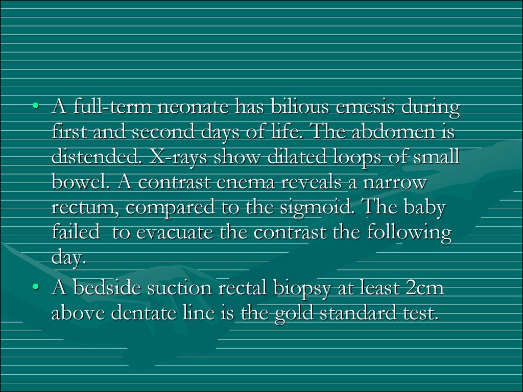
• A full-term neonate has bilious emesis duringfirst and second days of life. The abdomen is
distended. X-rays show dilated loops of small
bowel. A contrast enema reveals a narrow
rectum, compared to the sigmoid. The baby
failed to evacuate the contrast the following
day.
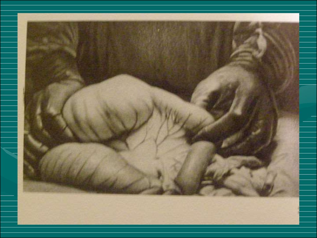
• A bedside suction rectal biopsy at least 2cm
above dentate line is the gold standard test.
45.
46.
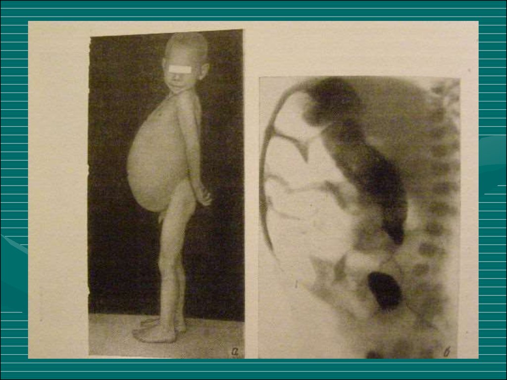
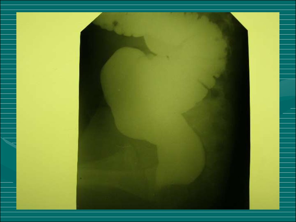
Diagnostic work-up includes:• Contrast enema showing a contracted rectum
with dilated bowel above.
• Failure to evacuate contrast 24h later can be
diagnostic.
• Rectal biopsy is required to confirm absence of
ganglion cells and nerve hypertrophy.
47.
48.
49. Surgical treatment
• Soave endo-rectal pull through with removal ofthe diseased distal bowel with coloanal
anastamosis
• Children who present acutely ill may need staged
procedure with colostomy.
• Need to do intraoperative frozen section to help
determine the anatomic location of transition
zone.
50.
51.
52. Anorectal anomalies
• Anorectal malformations, represent a widespectrum of defects. Surgical techniques useful
to repair the most common types of anorectal
malformations seen by a general pediatric
surgeon are presented following an order of
complexity from the simplest to the most
complex.
53. Сlassification
Full atresiaInferior
High
54.
Atresia with fistulaExternal fistula
Internal fistula
55.
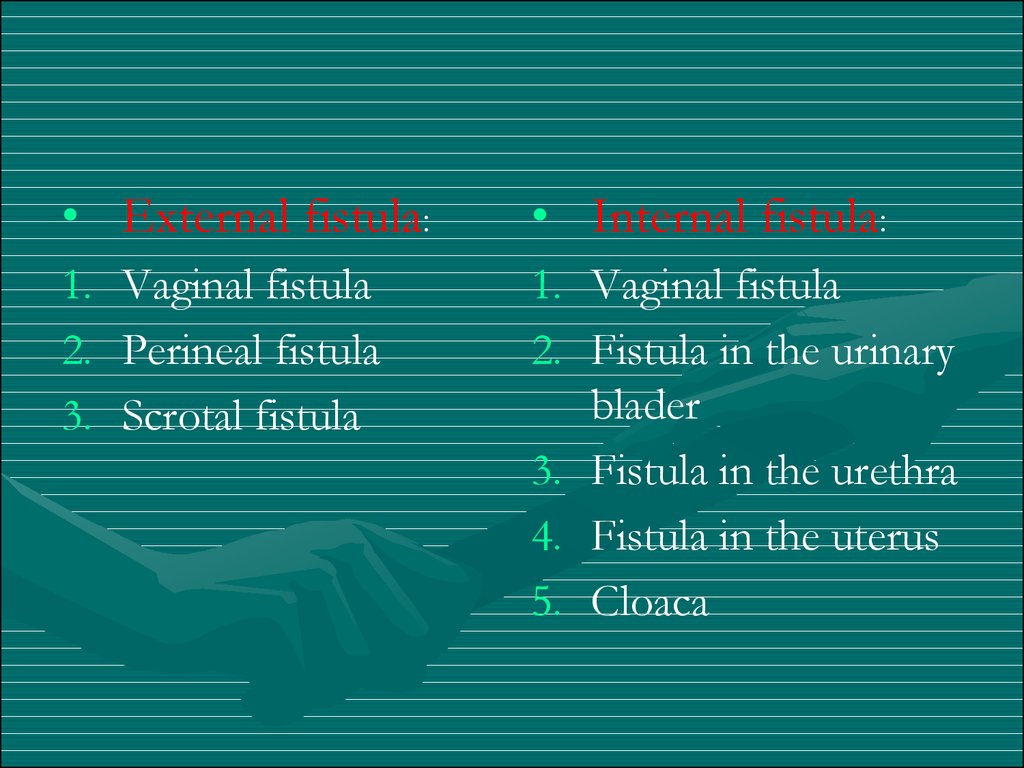
• External fistula:• Internal fistula:
1. Vaginal fistula
2. Perineal fistula
3. Scrotal fistula
1. Vaginal fistula
2. Fistula in the urinary
blader
3. Fistula in the urethra
4. Fistula in the uterus
5. Cloaca
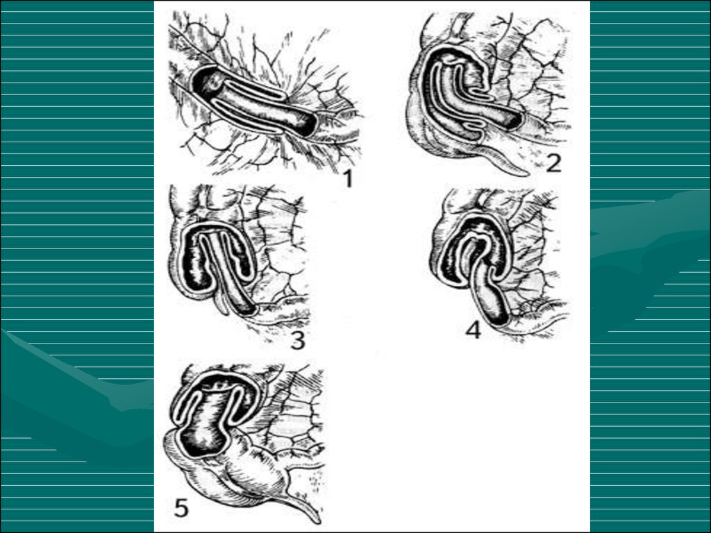
56. Perineal Fistula
• This malformation represents the simplest ofthe spectrum. In this defect, the rectum opens
immediately anterior to the centre of the
sphincter, yet, the anterior rectal wall is
intimately attached to the posterior urethra. The
anal orifice is frequently strictured. These
patients will have bowel control with and
without an operation.
57.
58. Rectourethral Fistula.
• This group of patients include two specificcategories: (a) rectourethral bulbar fistula
(Fig 3), and (b) rectoprostatic fistula (Fig 4).
These two variants represent the majority of
male patients with anorectal malformations.
Rectourethral bulbar fistula patients, in our
experience have an 80% chance of having
bowel control by the age of 3,whereas the
rectoprostatic fistula patients only have a
60% chance.
59.
60. Imperforate Anus Without Fistula
• This particular malformation is unique.When wesay imperforated anus without fistula, we do not
have to refer to the height of the defect because
in all cases the rectum is located approximately
1–2 cm above the perineal skin, at the level of
bulbar urethra. This malformation only happens
in 5% of all cases and half of these have Down’s
syndrome.
61.
62.
63.
64. Rectoperineal Fistula.
• This defect is equivalent to the recto-perinealfistula in males already described. Bowel control
exists in 100% of our patients and less than 10%
of them have associated defects. The patients
are faecally continent with and without an
operation.
• Constipation is a constant sequela and should be
treated energetically.
65.
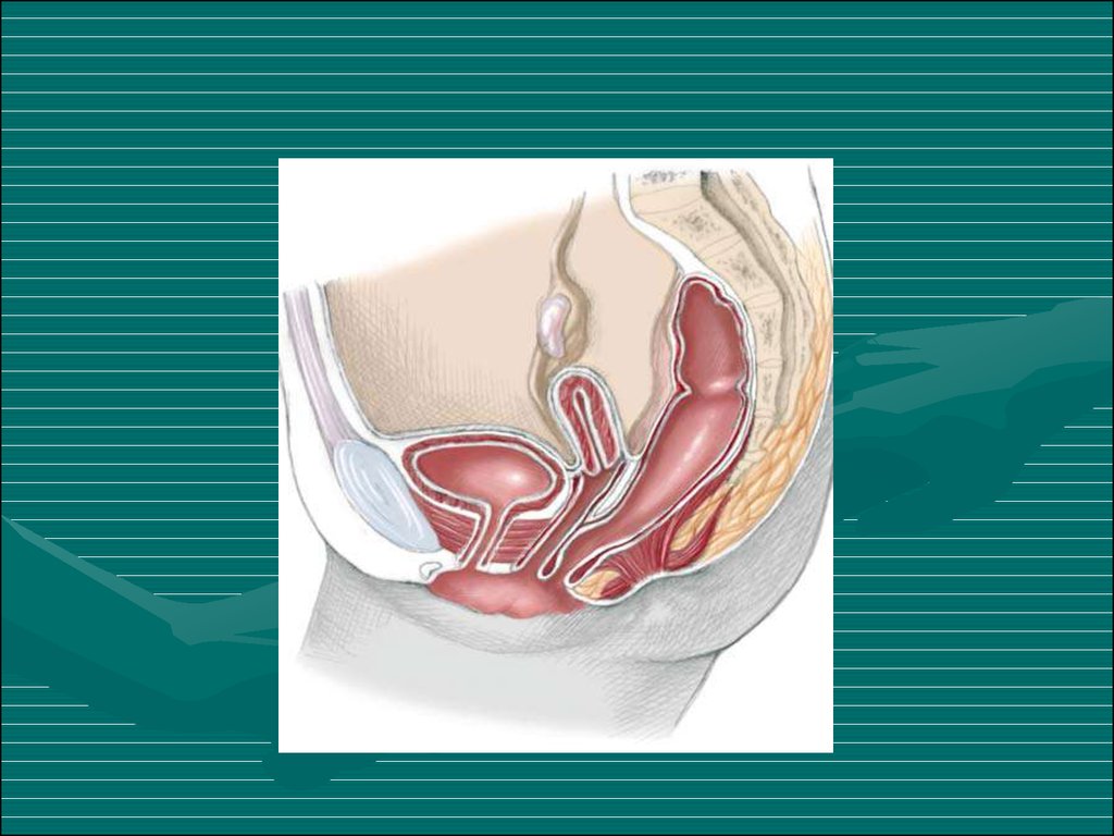
66. Cloaca.
• A cloaca is defined as a malformation in whichthe rectum, vagina and urethra are congenitally
fused, forming a common channel and opening
in a single perineal orifice at the same location
where the normal female urethra is located.
These three structures share common walls that
are very difficult to separate.
67.
68.
INTUSSUSCEPTION69. INTUSSUSCEPTION DEFINITION
• Telescoping of a proximal segment of theintestine (intussusceptum) into a distal segment
(intussuscipiens)
70.
71. INTUSSUSCEPTION ANATOMIC LOCATIONS
• ILEOCOLIC– MOST COMMON IN CHILDREN
• ILEO-ILEOCOLIC
– SECOND MOST COMMON
• ENTEROENTERIC
– ILEO-ILEAL, JEJUNO-JEJUNAL
– MORE COMMON IN ADULTS
– MAY NOT BE SEEN ON BARIUM
ENEMA
• CAECOCOLIC, COLOCOLIC
– MORE COMMON IN ASIAN
CHILDREN
72.
73. PATHOPHYSIOLOGY
Precipitating mechanism unknown
Obstruction of intussusceptum mesentery
Venous and lymphatic obstruction
Ischemic necrosis occurs in both
intussusceptum and intussuscipiens
• Pathologic bacterial translocation
74. ETIOLOGIES
• Majority of pediatric intussusceptions idiopathic (85-90%)– LYMPHOID HYPERPLASIA POSSIBLE
ETIOLOGY
• Mechanical abnormalities may act as “lead points”
– CONGENITAL MALFORMATIONS
(MECKEL’S DIVERTICULUM, DUPLICATIONS)
– NEOPLASMS (LYMPHOMA, LYMPHOSARCOMA)
– POLYPOSIS
– TRAUMA (POST-SURGICAL, HEMATOMA)
– MISCELLANEOUS (APPENDICITIS, PARASITES)
75. EPIDEMIOLOGY
Incidence 2 - 4 / 1000 live births
Usual age group 3 months - 3 years
Greatest incidence 6-12 months
No clear hereditary association
No seasonal distribution
Frequently preceded by viral infection
– ADENOVIRUS
76. INTUSSUSCEPTION CLINICAL CHARACTERISTICS
• Early Symptoms– PAROXYSMAL ABDOMINAL PAIN
– SEPARATED BY PERIODS OF APATHY
– POOR FEEDING AND VOMITING
• Late Symptoms
– WORSENING VOMITING, BECOMING BILIOUS
– ABDOMINAL DISTENTION
– HEME POSITIVE STOOLS
– FOLLOWED BY “RASPBERRY JELLY” STOOL
– DEHYDRATION (PROGRESSIVE)
• Unusual Symptoms
– DIARRHEA
77. PHYSICAL EVALUATION
Moderately to severely ill
Irritable, limited movement
Most are at least 5-10% dehydrated
80% have palpable abdominal masses
Paucity of bowel sounds
Rectal examination (blood, mass)
Abdominal rigidity
“Knocked Out” syndrome
78. INTUSSUSCEPTION STAGES
• I. Bright clinical manifestation• II. Pseudodysenteric stage
• III. Peritonitis
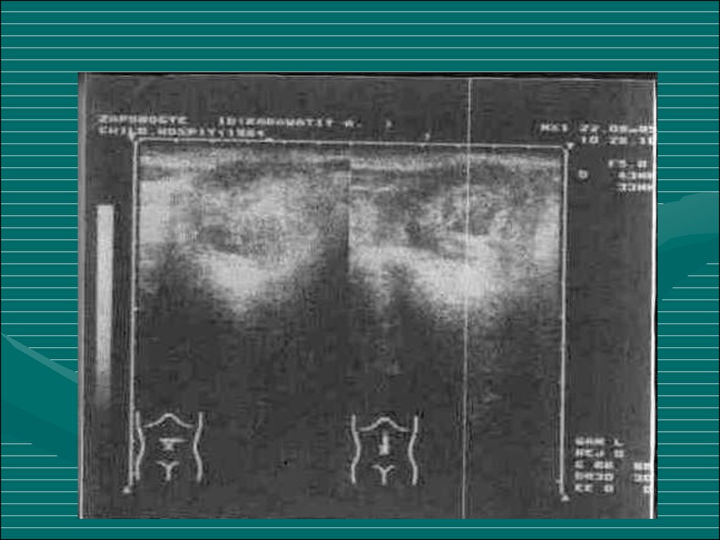
79. Ultrasonic diagnostics
80.
81.
82.
83. RADIOGRAPHIC EVALUATION
• Plain radiographs (acute abdominal series)• Plain films suggestive in majority, but
cannot rule out diagnosis
– PAUCITY OF LUMINAL AIR IN
INTESTINAL
– SMALL BOWEL DISTENTION, AIR
FLUID LEVELS
– LUMINAL AIR CUTOFFS (CECUM,
TRANSVERSE COLON)
84.
85.
86. TREATMENT
• Obstructive surgical emergency• Pediatric surgeon notified immediately
• Supportive Therapy
– AGGRESSIVE FLUID RESUSCITATION
– ELECTROLYTES
– NASOGASTRIC TUBE PLACEMENT AND
DRAINAGE
– ANTIBIOTICS IF ISCHEMIC BOWEL
SUSPECTED
• Arrange radiographic evaluation
87. INTUSSUSCEPTION PNEUMATIC REDUCTION
• Theoretical Advantages– LESS INFLAMMATION IF PERFORATION OCCURS
• Method
– AIR INSUFFLATION LIMITED TO MAXIMUM “RESTING “
PRESSURE OF 120 mmHg
– MAXIMUM PRESSURE MAINTAINED FOR 3 MIN
– USUALLY 3 ATTEMPTS AT REDUCTION
• Success Rate (75-90%)
– MUST OBSERVE AIR IN THE TERMINAL ILEUM
– LESS RECURRENCES (5-10%)
– LOW PERFORATION RATE (1%)
88. INTUSSUSCEPTION NON-OPERATIVE REDUCTION CONTRAINDICATIONS
• Absolute Contraindications– PERITONEAL SIGNS
– SUSPECTED PERFORATION
• Relative Contraindications
– SYMPTOMS > 24-48 HRS
– RECTAL BLEEDING
– POOR PROGNOSTIC
INDICATORS
89. INTUSSUSCEPTION FAILURE OF NON-OPERATIVE REDUCTION
• Factors associated with failure– SYMPTOMS > 48 HRS
– RECTAL BLEEDING
– SMALL BOWEL OBSTRUCTION
RADIOGRAPHICALLY
– ILEOILEOCOLIC OR SMALL BOWEL TYPES
– PRESENCE OF MECHANICAL LEAD POINT
– AGE < 3 MONTHS
• Operative Reduction
90. Acquired intestinal obstruction
Acquired intestinal obstructions are a partialor complete blockage of the small or large
intestine, resulting in failure of the
contents of the intestine to pass through
the bowel normally.
91.
• Intestinal obstructions can be mechanical ornonmechanical.
• Mechanical obstruction is caused by the bowel
twisting on itself (volvulus) or telescoping into
itself (intussusception). Mechanical obstruction
can also result from hernias, fecal impaction,
abnormal tissue growth, the presence of foreign
bodies in the intestines, or inflammatory bowel
disease (Crohn's disease).
92. Clinic
1. Abdominal pain
2. Vomiting
3. Constipation
4. Intoxication syndrome
93. Diagnosis
1.2.
3.
4.
X-ray examination
Ultrasonic diagnostics
Computed tomography
Diagnostic testing will include a complete blood
count (CBC), electrolytes (sodium, potassium,
chloride) and other blood chemistries, blood
urea nitrogen (BUN), and urinalysis.
Coagulation tests may be performed if the child
requires surgery.
94. Treatment
1. Preoperative preparation:a. inserting a nasogastric tube to suction out the
contents of the stomach and intestines
b. Intravenous fluids will be infused to prevent
dehydration and to correct electrolyte
imbalances that may have already occurre
95.
Thank you forattention!