


 medicine
Similar presentations:


Tibial Plateau / Proximal Tibial Fracture: Classification, Diagnosis, and Trentment
1.
2.
3.
4.
5.
6.
7.
8.
9.
10.
11.
12.
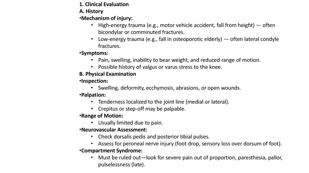
1. Clinical EvaluationA. History
•Mechanism of injury:
• High-energy trauma (e.g., motor vehicle accident, fall from height) — often
bicondylar or comminuted fractures.
• Low-energy trauma (e.g., fall in osteoporotic elderly) — often lateral condyle
fractures.
•Symptoms:
• Pain, swelling, inability to bear weight, and reduced range of motion.
• Possible history of valgus or varus stress to the knee.
B. Physical Examination
•Inspection:
• Swelling, deformity, ecchymosis, abrasions, or open wounds.
•Palpation:
• Tenderness localized to the joint line (medial or lateral).
• Crepitus or step-off may be palpable.
•Range of Motion:
• Usually limited due to pain.
•Neurovascular Assessment:
• Check dorsalis pedis and posterior tibial pulses.
• Assess for peroneal nerve injury (foot drop, sensory loss over dorsum of foot).
•Compartment Syndrome:
• Must be ruled out—look for severe pain out of proportion, paresthesia, pallor,
pulselessness (late).
13.
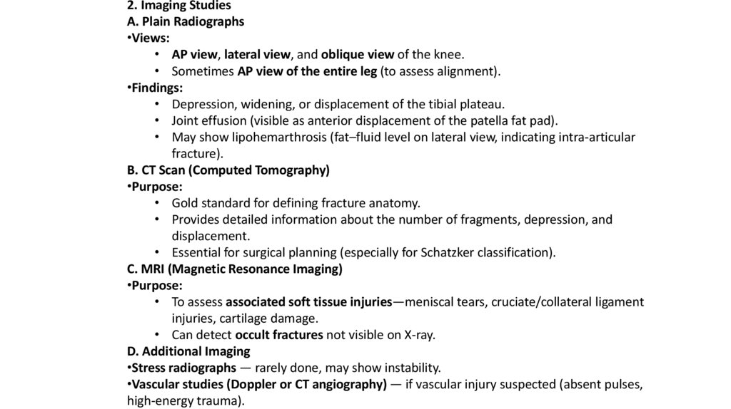
2. Imaging StudiesA. Plain Radiographs
•Views:
• AP view, lateral view, and oblique view of the knee.
• Sometimes AP view of the entire leg (to assess alignment).
•Findings:
• Depression, widening, or displacement of the tibial plateau.
• Joint effusion (visible as anterior displacement of the patella fat pad).
• May show lipohemarthrosis (fat–fluid level on lateral view, indicating intra-articular
fracture).
B. CT Scan (Computed Tomography)
•Purpose:
• Gold standard for defining fracture anatomy.
• Provides detailed information about the number of fragments, depression, and
displacement.
• Essential for surgical planning (especially for Schatzker classification).
C. MRI (Magnetic Resonance Imaging)
•Purpose:
• To assess associated soft tissue injuries—meniscal tears, cruciate/collateral ligament
injuries, cartilage damage.
• Can detect occult fractures not visible on X-ray.
D. Additional Imaging
•Stress radiographs — rarely done, may show instability.
•Vascular studies (Doppler or CT angiography) — if vascular injury suspected (absent pulses,
high-energy trauma).
14.
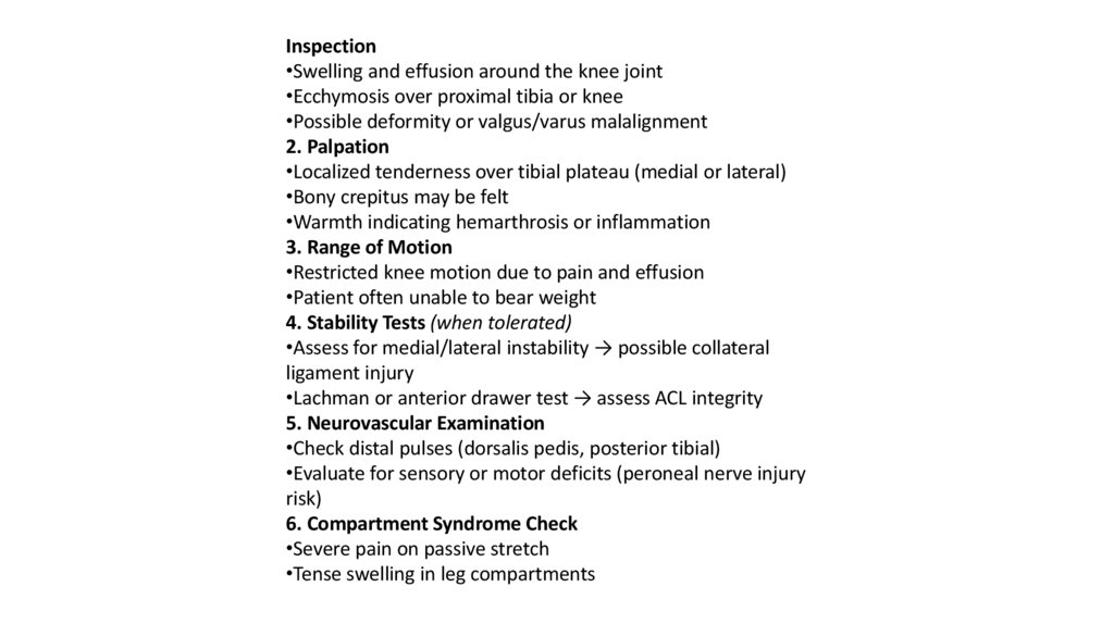
Inspection•Swelling and effusion around the knee joint
•Ecchymosis over proximal tibia or knee
•Possible deformity or valgus/varus malalignment
2. Palpation
•Localized tenderness over tibial plateau (medial or lateral)
•Bony crepitus may be felt
•Warmth indicating hemarthrosis or inflammation
3. Range of Motion
•Restricted knee motion due to pain and effusion
•Patient often unable to bear weight
4. Stability Tests (when tolerated)
•Assess for medial/lateral instability → possible collateral
ligament injury
•Lachman or anterior drawer test → assess ACL integrity
5. Neurovascular Examination
•Check distal pulses (dorsalis pedis, posterior tibial)
•Evaluate for sensory or motor deficits (peroneal nerve injury
risk)
6. Compartment Syndrome Check
•Severe pain on passive stretch
•Tense swelling in leg compartments