


 medicine
Similar presentations:


Intro to Cardiothoracic Surgery
1.
Intro to Cardiothoracic SurgeryCooper University Hospital
School of Perfusion
By:
Michael F. Hancock, CCP
2.
Cardiovascular Perfusionist• Perfusion = the pumping
of fluid through an organ
or tissue
• The Perfusion
Department is involved
in the circulation,
manipulation, and
salvage of the patient’s
blood supply
3.
The Perfusionist• Utilize extracorporeal technology to support
patients undergoing cardiac surgery and other
high risk operations
4.
Cardiac Surgery– The Heart
• Responsible for pumping blood into your systemic
circulation and Perfusing the body
• Most cardiac operations require the heart to be
opened or manipulated in a way that prevents it from
carrying out its normal function
– The body tissue still needs oxygen and nutrients
• How do we support the patient during Open Heart
Surgery???
– Blood Pressure
– Oxygenation
– Temperature
5.
Cardiopulmonary Bypass• Utilizing the Heart and Lung
Machine, we:
– Drain the patients whole venous
blood
– Oxygenate and remove CO2
– Add drugs/fluids to maintain
hemodynamic stability
– Warm or cool the blood
– Return the oxygenated blood to
the patient to nourish their tissue
– Provide a hyperkalemic solution
to arrest the heart to allow the
surgeon to operate on a
bloodless, motionless heart
6.
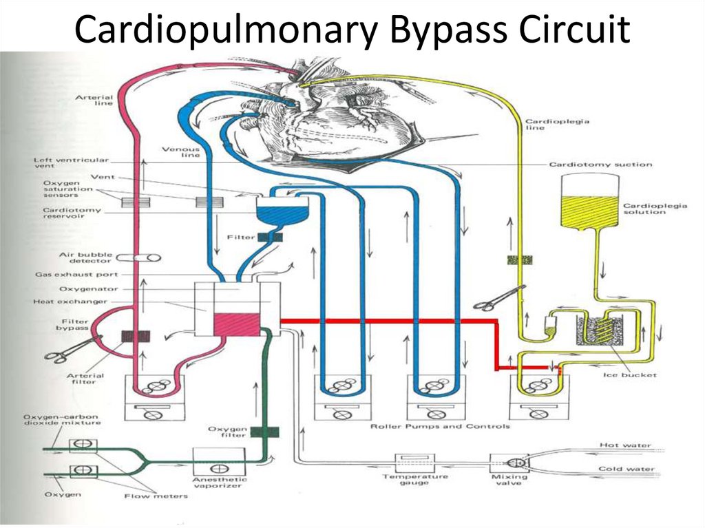
Cardiopulmonary Bypass Circuit7.
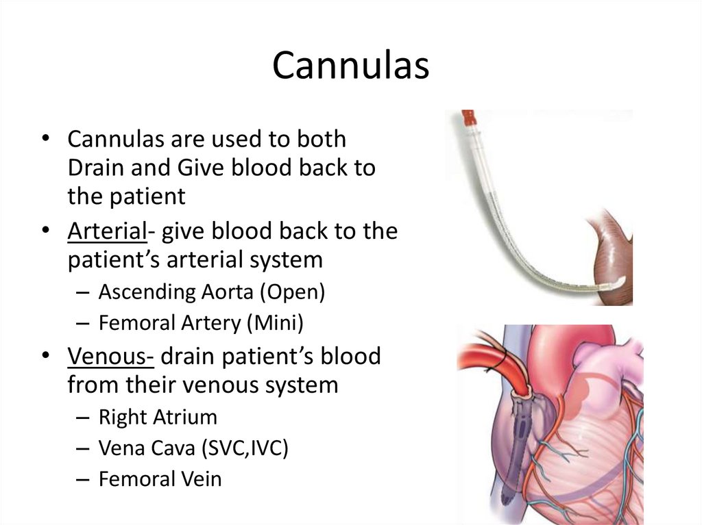
Cannulas• Cannulas are used to both
Drain and Give blood back to
the patient
• Arterial- give blood back to the
patient’s arterial system
– Ascending Aorta (Open)
– Femoral Artery (Mini)
• Venous- drain patient’s blood
from their venous system
– Right Atrium
– Vena Cava (SVC,IVC)
– Femoral Vein
8.
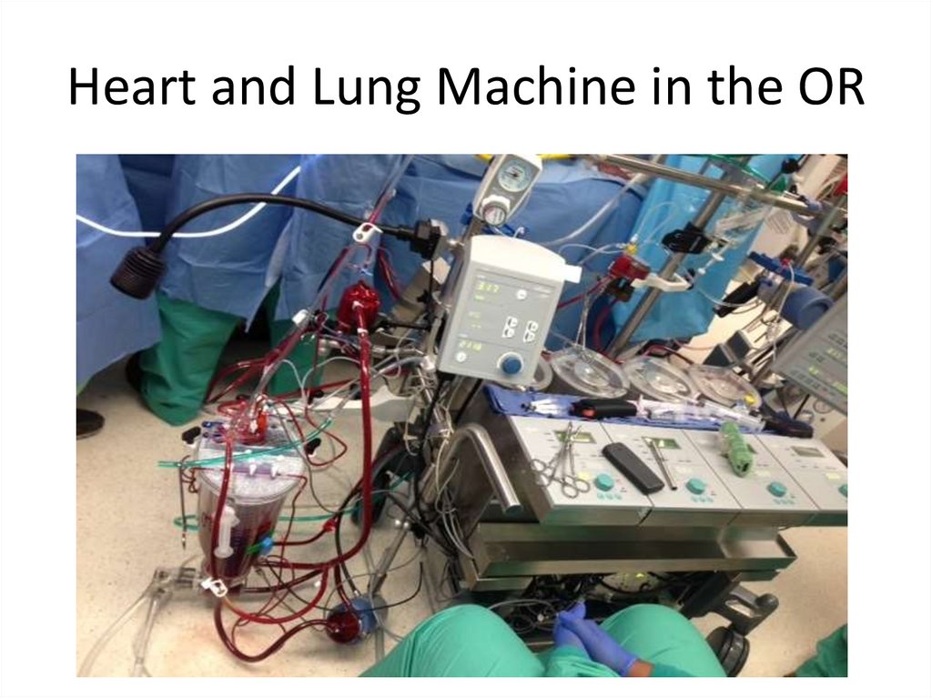
Heart and Lung Machine in the OR9.
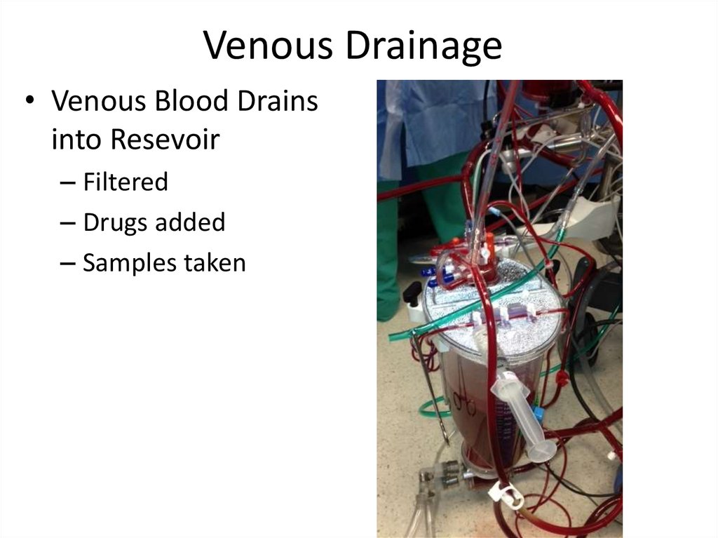
Venous Drainage• Venous Blood Drains
into Resevoir
– Filtered
– Drugs added
– Samples taken
10.
Centrifugal Pump Head• Provides the driving force for the blood to be propelled into
the oxygenator and then back to the patient, simulating
their cardiac output
– Blood comes out of the venous resevoir
– Into the centrifugal head
– Pumped by the pump into the heat exchanger and oxygenator
11.
Arterial Line• Blood comes out of
the oxygenator
• Into the arterial filter
to be filtered
• Back to the patient
12.
Arresting the Heart• Cardioplegia- a hyperkalemic (↑ K+)
solution delivered to the coronary arteries
to “stop” the “heart”
– Provides a blood less, motionless field
for the surgeon to operate on
– Solution given at 2⁰ C to lower the
metabolic demand of the heart
– We give maintenance doses of
cardioplegia every 15-20 minutes to
keep the heart arrested and to provide
nourishment, preventing permanent
tissue damage
13.
Responsibilities of the Perfusionist• Adequately Perfuse the patient– Give blood back to them at a rate comparable to their native
cardiac output (4-6 LPM)
• Maintain hemodynamic stability– Keep their BP high enough perfuse end organs
– Ensure adequate oxygenation and CO2 removal of the blood
– Maintain a normal pH
– Keep their Hemoglobin/Hematocrit adequate
• Effective Communication in the OR– Follow the commands of the cardiothoracic surgeon
– Work closely with the anesthesia, nursing and other members
of the Heart Team
• Keep the patient SAFE!!
– Prevent air emboli, circuit malfunctions, or any other potentially
harmful event
14.
Types of Cardiac Cases• Coronary Artery Bypass Grafting (CABGs)
• Valve Repair/Replacements
• Aneurysm/Dissection Repair
• Other Misc. Cases
– To be continued…
15.
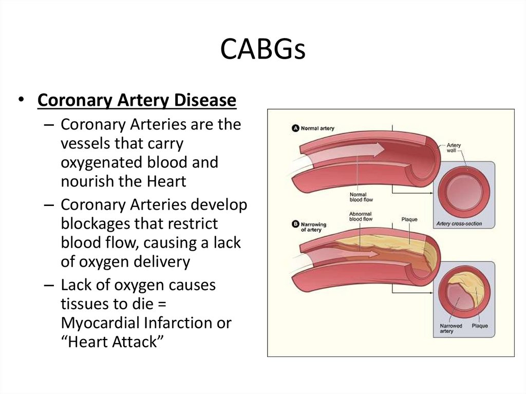
CABGs• Coronary Artery Disease
– Coronary Arteries are the
vessels that carry
oxygenated blood and
nourish the Heart
– Coronary Arteries develop
blockages that restrict
blood flow, causing a lack
of oxygen delivery
– Lack of oxygen causes
tissues to die =
Myocardial Infarction or
“Heart Attack”
16.
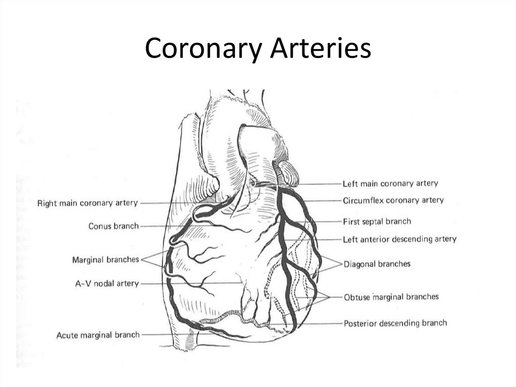
Coronary Arteries17.
Bypass Grafting• Goal is to divert
blood flow around
the blockage and
perfuse distal to the
blockage
• Need a conduit to
divert the blood
flow
– Internal Mammery
Artery (L or R)
– Saphenous Vein
(harvested from the
leg)
– Radial Artery (rare)
18.
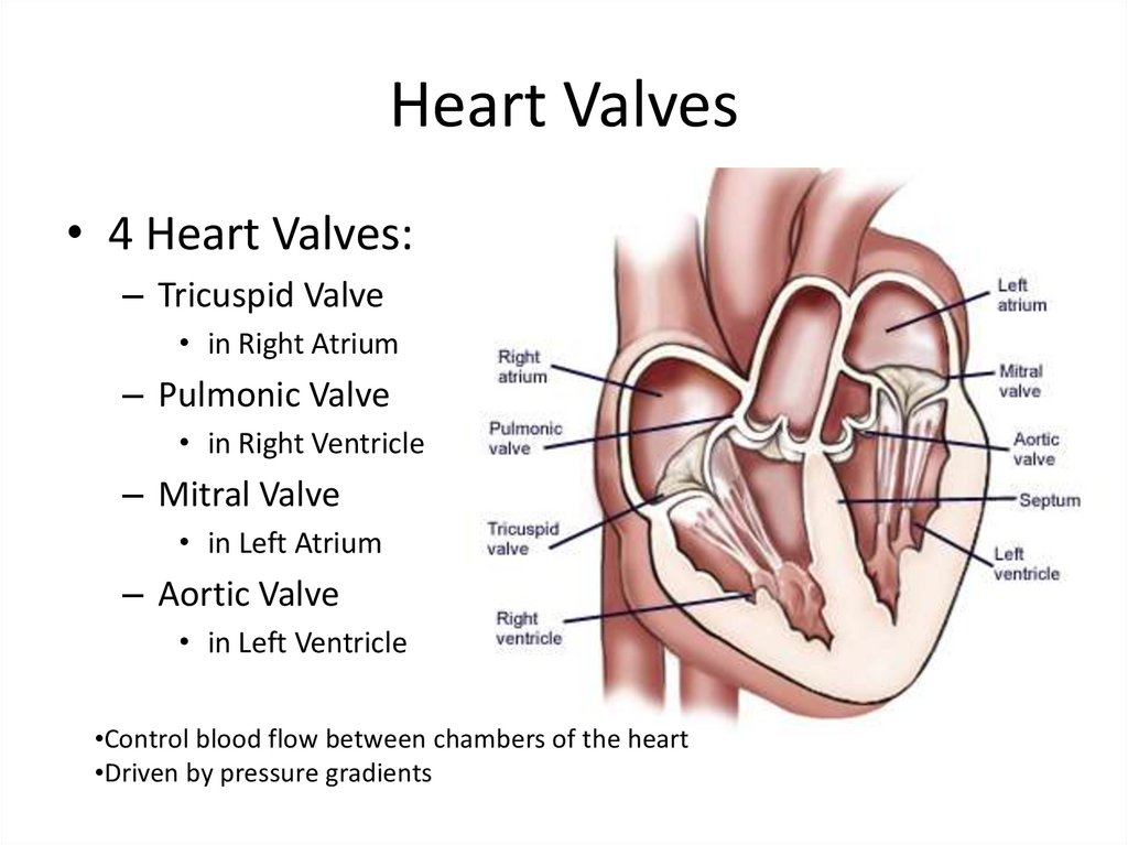
Heart Valves• 4 Heart Valves:
– Tricuspid Valve
• in Right Atrium
– Pulmonic Valve
• in Right Ventricle
– Mitral Valve
• in Left Atrium
– Aortic Valve
• in Left Ventricle
•Control blood flow between chambers of the heart
•Driven by pressure gradients
19.
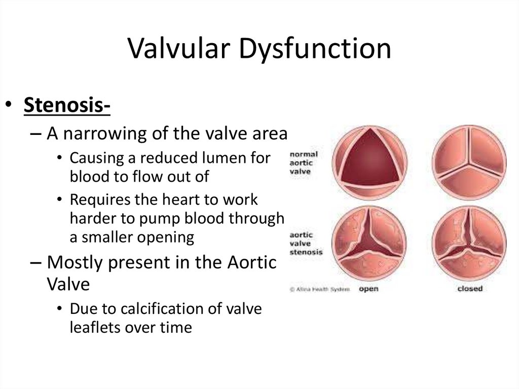
Valvular Dysfunction• Stenosis– A narrowing of the valve area
• Causing a reduced lumen for
blood to flow out of
• Requires the heart to work
harder to pump blood through
a smaller opening
– Mostly present in the Aortic
Valve
• Due to calcification of valve
leaflets over time
20.
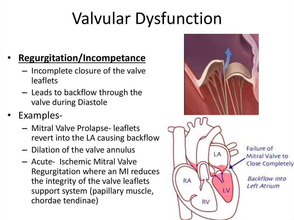
Valvular Dysfunction• Regurgitation/Incompetance
– Incomplete closure of the valve
leaflets
– Leads to backflow through the
valve during Diastole
• Examples– Mitral Valve Prolapse- leaflets
revert into the LA causing backflow
– Dilation of the valve annulus
– Acute- Ischemic Mitral Valve
Regurgitation where an MI reduces
the integrity of the valve leaflets
support system (papillary muscle,
chordae tendinae)
21.
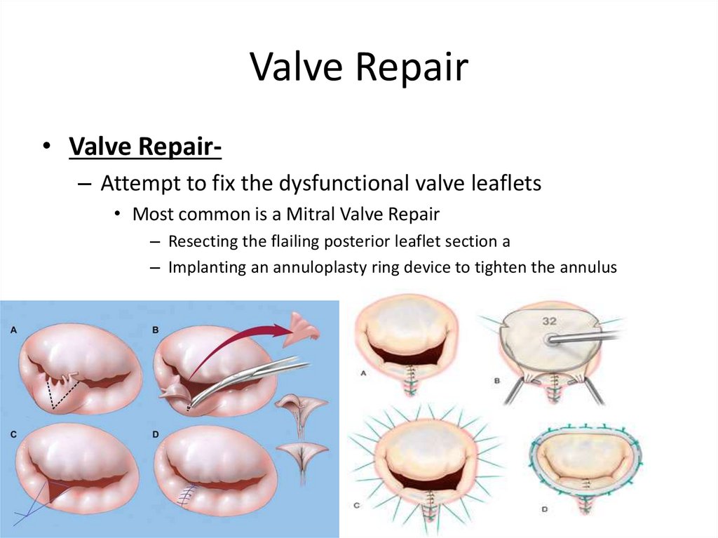
Valve Repair• Valve Repair– Attempt to fix the dysfunctional valve leaflets
• Most common is a Mitral Valve Repair
– Resecting the flailing posterior leaflet section a
– Implanting an annuloplasty ring device to tighten the annulus
22.
Valve Replacement• Old dysfunctional valve removed and a new
valve implanted
• Two Options for Replacement:
– Tissue Valve- bioprosthetic, usually bovine or
porcine
• Last ~15 years
• Do not require anticoagulation
– Mechanical Valve
• Last “forever”
• DO require anticoagulation (Coumadin)
23.
Valve Replacement• Gain Access– Median Sternotomy
• In this example…
• Cannulate and Initiate
CPB
• Arrest the Heart with
Cardioplegia
• Open Aorta
(Aortotomy)
24.
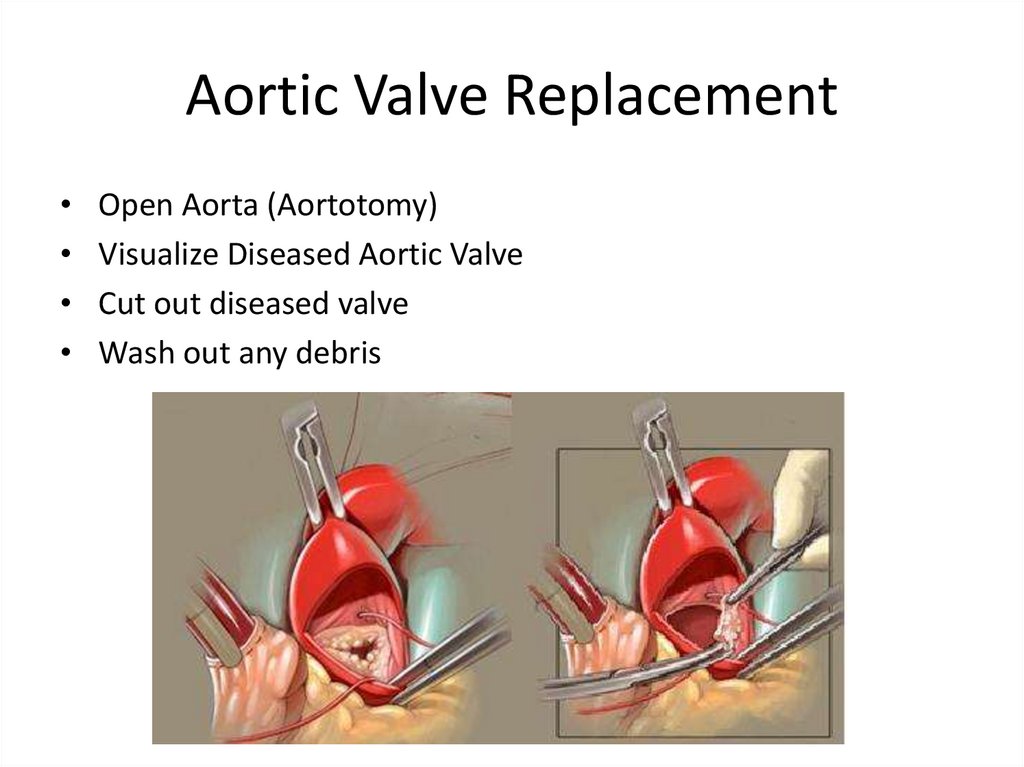
Aortic Valve ReplacementOpen Aorta (Aortotomy)
Visualize Diseased Aortic Valve
Cut out diseased valve
Wash out any debris
25.
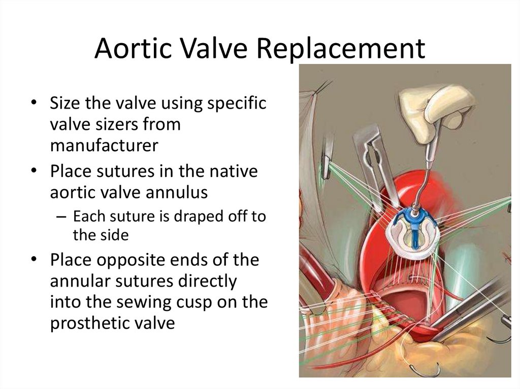
Aortic Valve Replacement• Size the valve using specific
valve sizers from
manufacturer
• Place sutures in the native
aortic valve annulus
– Each suture is draped off to
the side
• Place opposite ends of the
annular sutures directly
into the sewing cusp on the
prosthetic valve
26.
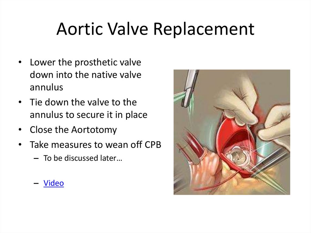
Aortic Valve Replacement• Lower the prosthetic valve
down into the native valve
annulus
• Tie down the valve to the
annulus to secure it in place
• Close the Aortotomy
• Take measures to wean off CPB
– To be discussed later…
– Video
27.
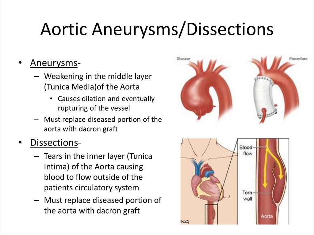
Aortic Aneurysms/Dissections• Aneurysms– Weakening in the middle layer
(Tunica Media)of the Aorta
• Causes dilation and eventually
rupturing of the vessel
– Must replace diseased portion of the
aorta with dacron graft
• Dissections– Tears in the inner layer (Tunica
Intima) of the Aorta causing
blood to flow outside of the
patients circulatory system
– Must replace diseased portion of
the aorta with dacron graft
28.
Techniques of Exposing the Heart• “Open” Procedures– Provides maximum exposure to the surgeon
– Median Sternotomy
• Sawing through the sternum
• Minimally Invasive Procedures
– Anterior Thoracotomy
• Done for Mini AVR
– Mini Sternotomy
• Done for Mini AVR
– Right Thoracotomy
• Done for Mini MVR
– Left Thoracotomy
• Done for MIDCAB
– Minimally invasive CABG, one or two jumps using LIMA
29.
Median Sternotomy• Provides the best
exposure
• We cannulate
“Centrally”
– Ascending Aorta
– Right Atrium
30.
Right Anterior Thoracotomy• Provides access for AVR
only
– No access to lateral part
of the heart
• Cannulate Femorally
ideally
– Can use central aortic
cannula (straight)
– Can use central venous
cannula (gets in the way)
31.
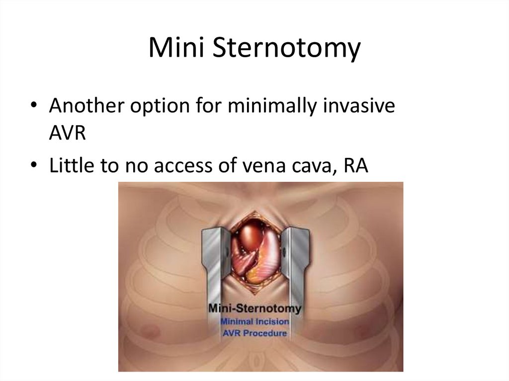
Mini Sternotomy• Another option for minimally invasive
AVR
• Little to no access of vena cava, RA
32.
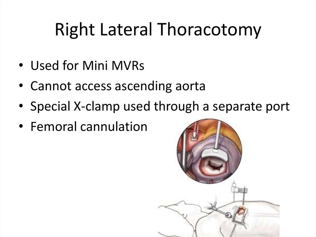
Right Lateral Thoracotomy• Used for Mini MVRs
• Cannot access ascending aorta
• Special X-clamp used through a separate port
• Femoral cannulation
33.
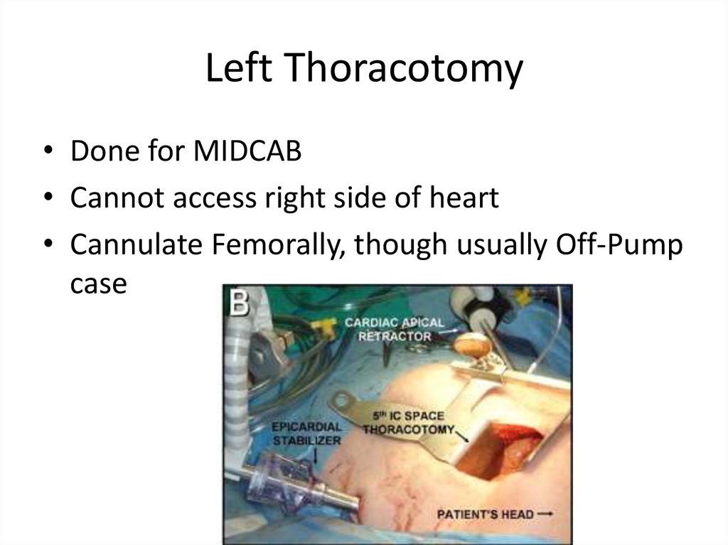
Left Thoracotomy• Done for MIDCAB
• Cannot access right side of heart
• Cannulate Femorally, though usually Off-Pump
case
34.
Trans-cathetar Aortic ValveReplacements (TAVR)
• Aortic valves inserted percutaneously
• Two access points:
– Trans-Femoral
• Through the femoral artery
– Trans-Apical
• Directly into the apex of the LV
• Bioprosthetic valves are deployed over top of
the patients native stenotic Aortic Valve
35.
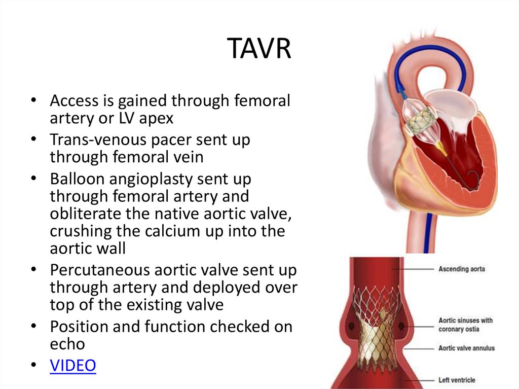
TAVR• Access is gained through femoral
artery or LV apex
• Trans-venous pacer sent up
through femoral vein
• Balloon angioplasty sent up
through femoral artery and
obliterate the native aortic valve,
crushing the calcium up into the
aortic wall
• Percutaneous aortic valve sent up
through artery and deployed over
top of the existing valve
• Position and function checked on
echo
• VIDEO
36.
Other Perfusion Services• Autologous Blood
Salvage- “Cell Saver”
– Used in cases where
blood loss is significant
• Collect patients whole
blood
• Process it in a large
centrifuge to isolate RBCs
• Wash the RBCs with saline
• Give back concentrated
RBCs
– Provides an alternative to
giving donor blood
37.
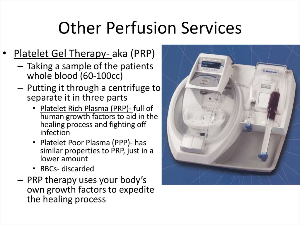
Other Perfusion Services• Platelet Gel Therapy- aka (PRP)
– Taking a sample of the patients
whole blood (60-100cc)
– Putting it through a centrifuge to
separate it in three parts
• Platelet Rich Plasma (PRP)- full of
human growth factors to aid in the
healing process and fighting off
infection
• Platelet Poor Plasma (PPP)- has
similar properties to PRP, just in a
lower amount
• RBCs- discarded
– PRP therapy uses your body’s
own growth factors to expedite
the healing process
38.
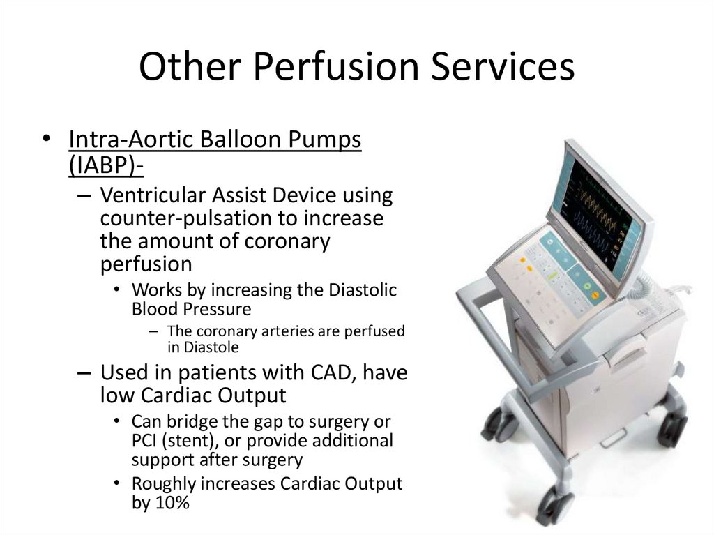
Other Perfusion Services• Intra-Aortic Balloon Pumps
(IABP)– Ventricular Assist Device using
counter-pulsation to increase
the amount of coronary
perfusion
• Works by increasing the Diastolic
Blood Pressure
– The coronary arteries are perfused
in Diastole
– Used in patients with CAD, have
low Cardiac Output
• Can bridge the gap to surgery or
PCI (stent), or provide additional
support after surgery
• Roughly increases Cardiac Output
by 10%
39.
Other Perfusion Services• Extracorporeal Membrane Oxygenation
(ECMO)– Utilizes a smaller “closed” (no resevoir) bypass
circuit to provide long term support
– Can provide purely respiratory support (V-V)
• Drain from a Vein, give back to a vein after oxygenating
• Don’t bypass anything, just oxygenate the blood
– Can provide cardiac and respiratory support (V-A)
• Drain from a Vein, give back to an artery
• Bypass the heart and lungs
40.
ECMO Circuit• Circuit– Drainage cannula
– Tubing
– Centrifugal Pump
– Oxygenator
– Heater-Cooler
– Return cannula
• Take blood
• Oxygenate it
• Give it back