


 medicine
Similar presentations:


Позитронно-эмиссионная томография
1.
Позитронно-эмиссионнаятомография
Подготовили
студентки группы ЛД4-С21
Сизова Дарина, Тригуб Мария, Москаленко Дарья.
2.
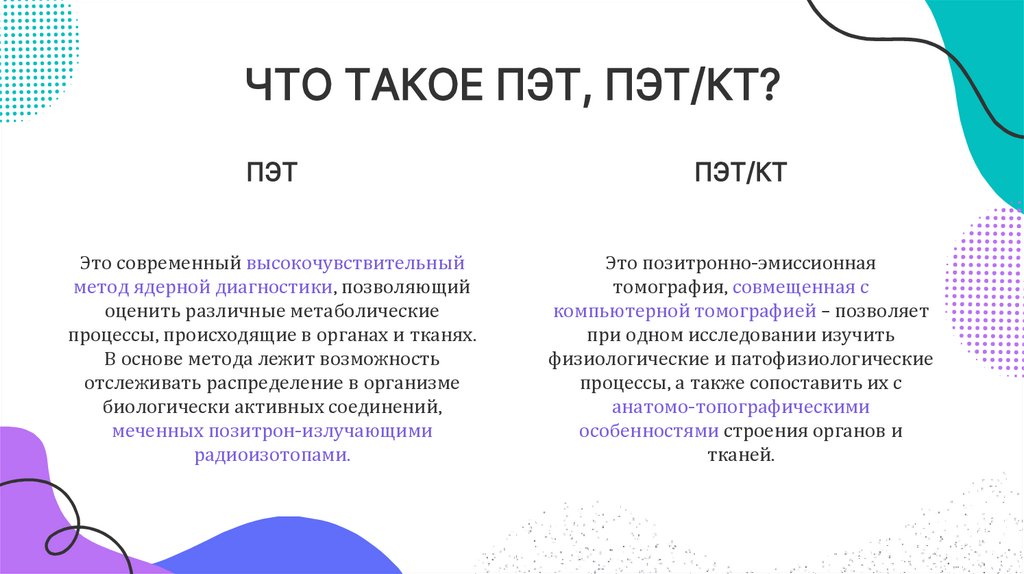
ЧТО ТАКОЕ ПЭТ, ПЭТ/КТ?ПЭТ
ПЭТ/КТ
Это современный высокочувствительный
метод ядерной диагностики, позволяющий
оценить различные метаболические
процессы, происходящие в органах и тканях.
В основе метода лежит возможность
отслеживать распределение в организме
биологически активных соединений,
меченных позитрон-излучающими
радиоизотопами.
Это позитронно-эмиссионная
томография, совмещенная с
компьютерной томографией – позволяет
при одном исследовании изучить
физиологические и патофизиологические
процессы, а также сопоставить их с
анатомо-топографическими
особенностями строения органов и
тканей.
3.
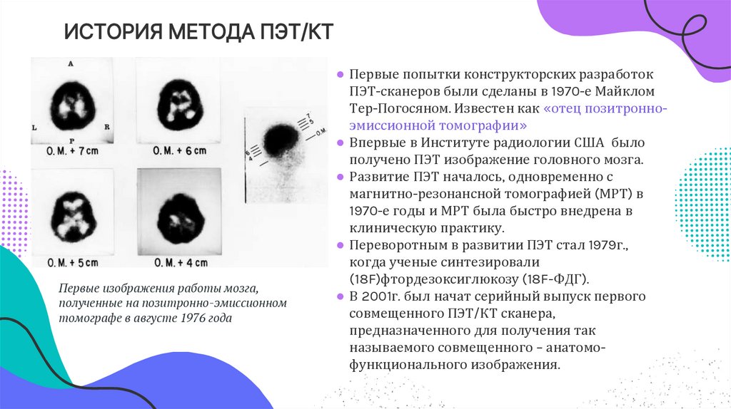
ИСТОРИЯ МЕТОДА ПЭТ/КТПервые изображения работы мозга,
полученные на позитронно-эмиссионном
томографе в августе 1976 года
● Первые попытки конструкторских разработок
ПЭТ-сканеров были сделаны в 1970-е Майклом
Тер-Погосяном. Известен как «отец позитронноэмиссионной томографии»
● Впервые в Институте радиологии США было
получено ПЭТ изображение головного мозга.
● Развитие ПЭТ началось, одновременно с
магнитно-резонансной томографией (МРТ) в
1970-е годы и МРТ была быстро внедрена в
клиническую практику.
● Переворотным в развитии ПЭТ стал 1979г.,
когда ученые синтезировали
(18F)фтордезоксиглюкозу (18F-ФДГ).
● В 2001г. был начат серийный выпуск первого
совмещенного ПЭТ/КТ сканера,
предназначенного для получения так
называемого совмещенного – анатомофункционального изображения.
4.
передовымПЭТ томография относится к
методам,
позволяет обнаружить болезнь даже тогда, когда человек не
испытывает никаких симптомов, что позволяет вовремя
провести лечение, повысить шансы на полное выздоровление.
5.
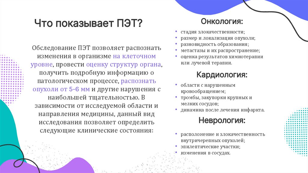
Что показывает ПЭТ?Обследование ПЭТ позволяет распознать
изменения в организме на клеточном
уровне, провести оценку структур органа,
получить подробную информацию о
патологическом процессе, распознать
опухоли от 5-6 мм и другие нарушения с
наибольшей тщательностью. В
зависимости от исследуемой области и
направления медицины, данный вид
исследования позволяет определить
следующие клинические состояния:
Онкология:
• стадия злокачественности;
• размер и локализация опухоли;
• разновидность образования;
• метастазы и их распространение;
• оценка результатов химиотерапии
или лучевой терапии.
Кардиология:
• области с нарушенным
кровообращением;
• тромбы, закупорки крупных и
мелких сосудов;
• динамика после лечения инфаркта.
Неврология:
• расположение и злокачественность
внутричерепных опухолей;
• эпилептические участки;
• изменения в сосудах.
6.
Contents of this templateYou can delete this slide when you’re done editing the presentation
Fonts
To view this template correctly in PowerPoint, download and install the fonts we used
Used and alternative resources
An assortment of graphic resources that are suitable for use in this presentation
Thanks slide
You must keep it so that proper credits for our design are given
Colors
All the colors used in this presentation
Icons and infographic resources
These can be used in the template, and their size and color can be edited
Editable presentation theme
You can edit the master slides easily. For more info, click here
For more info:
You can visit our sister projects:
SLIDESGO | BLOG | FAQs
FREEPIK | FLATICON | STORYSET | WEPIK | VIDEVO
7.
Table of contents01
02
03
Abstract
Introduction
Case presentation
04
05
06
Discussion
Conclusion
Roadmap
8.
01Abstract
9.
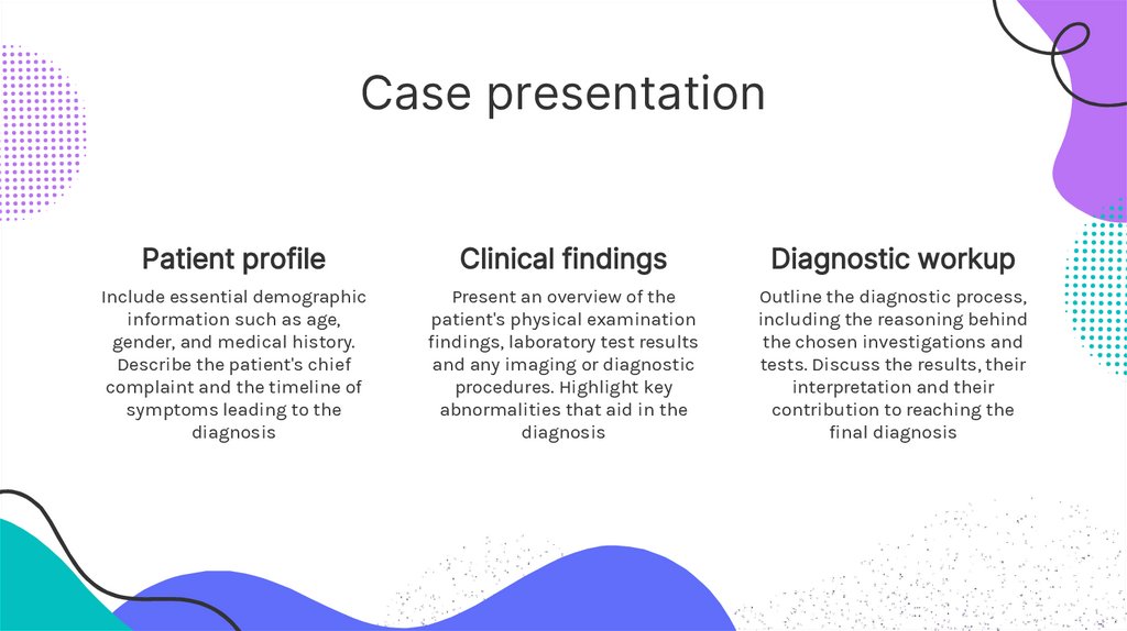
Case presentationPatient profile
Clinical findings
Diagnostic workup
Include essential demographic
information such as age,
gender, and medical history.
Describe the patient's chief
complaint and the timeline of
symptoms leading to the
diagnosis
Present an overview of the
patient's physical examination
findings, laboratory test results
and any imaging or diagnostic
procedures. Highlight key
abnormalities that aid in the
diagnosis
Outline the diagnostic process,
including the reasoning behind
the chosen investigations and
tests. Discuss the results, their
interpretation and their
contribution to reaching the
final diagnosis
10.
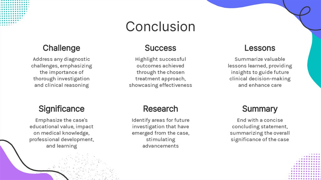
ConclusionChallenge
Success
Lessons
Address any diagnostic
challenges, emphasizing
the importance of
thorough investigation
and clinical reasoning
Highlight successful
outcomes achieved
through the chosen
treatment approach,
showcasing effectiveness
Summarize valuable
lessons learned, providing
insights to guide future
clinical decision-making
and enhance care
Significance
Research
Summary
Emphasize the case's
educational value, impact
on medical knowledge,
professional development,
and learning
Identify areas for future
investigation that have
emerged from the case,
stimulating
advancements
End with a concise
concluding statement,
summarizing the overall
significance of the case
11.
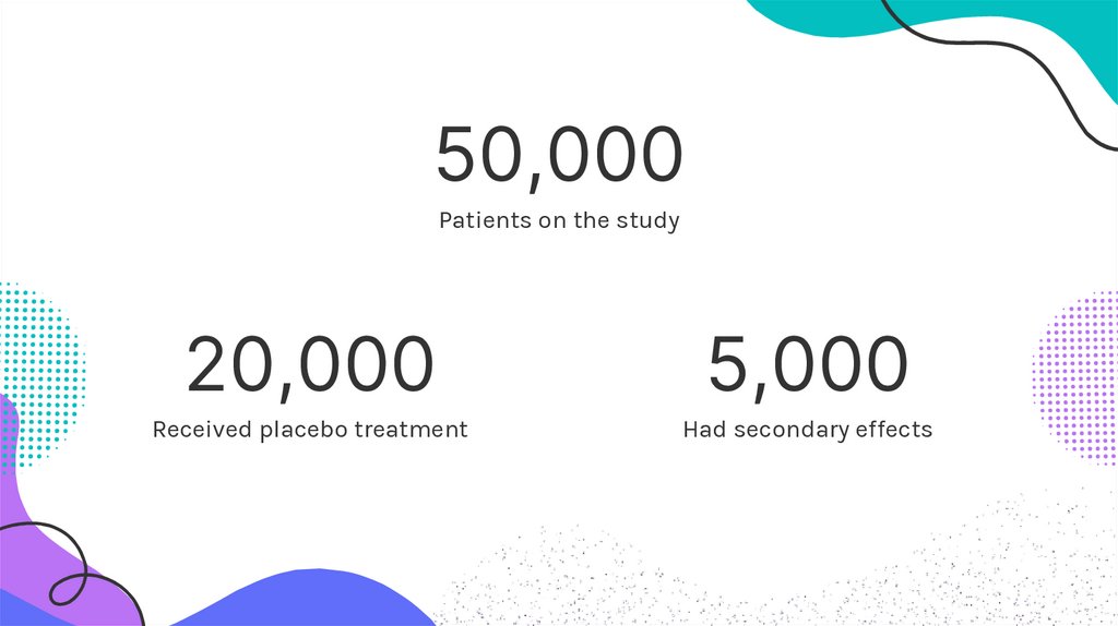
50,000Patients on the study
20,000
5,000
Received placebo treatment
Had secondary effects
12.
A picture is worth athousand words
Using an image in your case report enhances visual
representation and provides a concise and
impactful way to present key findings or illustrate
important aspects of the case
13.
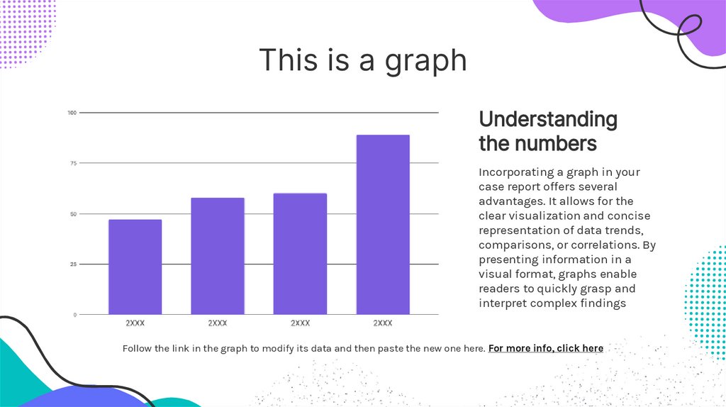
This is a graphUnderstanding
the numbers
Incorporating a graph in your
case report offers several
advantages. It allows for the
clear visualization and concise
representation of data trends,
comparisons, or correlations. By
presenting information in a
visual format, graphs enable
readers to quickly grasp and
interpret complex findings
Follow the link in the graph to modify its data and then paste the new one here. For more info, click here
14.
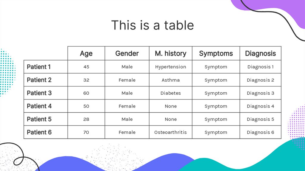
This is a tableAge
Gender
M. history
Symptoms
Diagnosis
Patient 1
45
Male
Hypertension
Symptom
Diagnosis 1
Patient 2
32
Female
Asthma
Symptom
Diagnosis 2
Patient 3
60
Male
Diabetes
Symptom
Diagnosis 3
Patient 4
50
Female
None
Symptom
Diagnosis 4
Patient 5
28
Male
None
Symptom
Diagnosis 5
Patient 6
70
Female
Osteoarthritis
Symptom
Diagnosis 6
15.
Milestones reachedInitial presentation
with chief complaint
Diagnostic evaluation
and investigations
Confirmation of
the diagnosis
Initiation of
appropriate treatment
Monitoring response
for treatment
Resolution of
presenting symptoms
Follow-up assessment
for outcomes
Case discussion and
lessons learned
16.
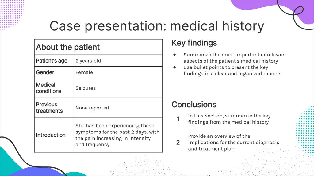
Case presentation: medical historyAbout the patient
Patient’s age
2 years old
Gender
Female
Medical
conditions
Seizures
Previous
treatments
None reported
Introduction
She has been experiencing these
symptoms for the past 2 days, with
the pain increasing in intensity
and frequency
Key findings
Summarize the most important or relevant
aspects of the patient's medical history
Use bullet points to present the key
findings in a clear and organized manner
Conclusions
1
In this section, summarize the key
findings from the medical history
2
Provide an overview of the
implications for the current diagnosis
and treatment plan
17.
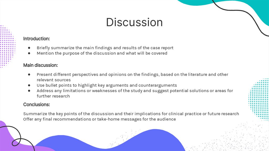
DiscussionIntroduction:
Briefly summarize the main findings and results of the case report
Mention the purpose of the discussion and what will be covered
Main discussion:
Present different perspectives and opinions on the findings, based on the literature and other
relevant sources
Use bullet points to highlight key arguments and counterarguments
Address any limitations or weaknesses of the study and suggest potential solutions or areas for
further research
Conclusions:
Summarize the key points of the discussion and their implications for clinical practice or future research
Offer any final recommendations or take-home messages for the audience
18.
Outcomes and recommendationsStart
1
2
3
Outcome
Outcome
Outcome
A. Write your
recommendation
B. Write your
recommendation
C. Write your
recommendation
D. Write your
recommendation
A. Write your
recommendation
B. Write your
recommendation
C. Write your
recommendation
D. Write your
recommendation
A. Write your
recommendation
B. Write your
recommendation
C. Write your
recommendation
D. Write your
recommendation
19.
Photoshowcase
You can replace the images on the screen
with your own work. Just right-click on
any of them and select “Replace image”
20.
ThanksDo you have any questions?
youremail@freepik.com
+91 620 421 838
yourwebsite.com
CREDITS: This presentation template was created by Slidesgo, and
includes icons by Flaticon and infographics & images by Freepik
Please keep this slide for attribution
21.
Icon pack22.
Alternative resourcesDid you like the resources on this template? Get them for free at our other websites:
Vectors
Abstract organic shapes banners
23.
ResourcesDid you like the resources on this template? Get them for free at our other websites:
Vectors
Flat medical instagram stories
Photos
Close up parent holding cute baby
Doctor measuring new born baby head
Doctor holding cute baby front view
Back view doctors holding a new born baby
Adult taking care of baby weight
Black mother taking car of her child
Side view smiley doctor checking patient
Icons
Icon Pack: Healthcare and Medical | Lineal
24.
Instructions for useIf you have a free account, in order to use this template, you must credit Slidesgo by keeping the Thanks slide. Please
refer to the next slide to read the instructions for premium users.
As a Free user, you are allowed to:
Modify this template.
Use it for both personal and commercial projects.
You are not allowed to:
Sublicense, sell or rent any of Slidesgo Content (or a modified version of Slidesgo Content).
Distribute Slidesgo Content unless it has been expressly authorized by Slidesgo.
Include Slidesgo Content in an online or offline database or file.
Offer Slidesgo templates (or modified versions of Slidesgo templates) for download.
Acquire the copyright of Slidesgo Content.
For more information about editing slides, please read our FAQs or visit our blog:
https://slidesgo.com/faqs and https://slidesgo.com/slidesgo-school
25.
Instructions for use (premium users)As a Premium user, you can use this template without attributing Slidesgo or keeping the "Thanks" slide.
You are allowed to:
Modify this template.
Use it for both personal and commercial purposes.
Hide or delete the “Thanks” slide and the mention to Slidesgo in the credits.
Share this template in an editable format with people who are not part of your team.
You are not allowed to:
Sublicense, sell or rent this Slidesgo Template (or a modified version of this Slidesgo Template).
Distribute this Slidesgo Template (or a modified version of this Slidesgo Template) or include it in a database or in
any other product or service that offers downloadable images, icons or presentations that may be subject to
distribution or resale.
Use any of the elements that are part of this Slidesgo Template in an isolated and separated way from this
Template.
Register any of the elements that are part of this template as a trademark or logo, or register it as a work in an
intellectual property registry or similar.
For more information about editing slides, please read our FAQs or visit our blog:
https://slidesgo.com/faqs and https://slidesgo.com/slidesgo-school
26.
Fonts & colors usedThis presentation has been made using the following fonts:
Inter
(https://fonts.google.com/specimen/Inter)
Karla
(https://fonts.google.com/specimen/Karla)
#333333
#ffffff
#04bfbf
#606ef9
#7a5dde
#afafcc
#ba73f4
27.
StorysetCreate your Story with our illustrated concepts. Choose the style you like the most, edit its
colors, pick the background and layers you want to show and bring them to life with the
animator panel! It will boost your presentation. Check out how it works.
Pana
Amico
Bro
Rafiki
Cuate
28.
Use our editable graphic resources...You can easily resize these resources without losing quality. To change the color, just ungroup the resource and click
on the object you want to change. Then, click on the paint bucket and select the color you want. Group the resource again
when you’re done. You can also look for more infographics on Slidesgo.
29.
30.
31.
JANUARYFEBRUARY
MARCH
APRIL
MAY
JUNE
PHASE 1
Task 1
Task 2
PHASE 2
Task 1
Task 2
JANUARY
PHASE 1
Task 1
Task 2
FEBRUARY
MARCH
APRIL
32.
33.
34.
...and our sets of editable iconsYou can resize these icons without losing quality.
You can change the stroke and fill color; just select the icon and click on the paint bucket/pen.
In Google Slides, you can also use Flaticon’s extension, allowing you to customize and add even more icons.
35.
Educational IconsMedical Icons
36.
Business IconsTeamwork Icons
37.
Help & Support IconsAvatar Icons
38.
Creative Process IconsPerforming Arts Icons