


 biology
Similar presentations:


Morphology of the viruses
1.
MORPHOLOGY OF THE VIRUSESDepartment of Microbiology, Virology and Immunology
2.
Introduction to the VirusesIn 1898, Friedrich Loeffler and Paul Frosch found
evidence that the cause of foot-and-mouth disease in
livestock was an infectious particle smaller than any
bacteria. This was the first clue to the nature of
viruses, genetic entities that lie somewhere in the grey
area between living and non-living states.
They have small size and “filterability” (ability to pass
through filters that can hold back bacteria) that led to
their recognition as a separate class of infectious
agents.
3.
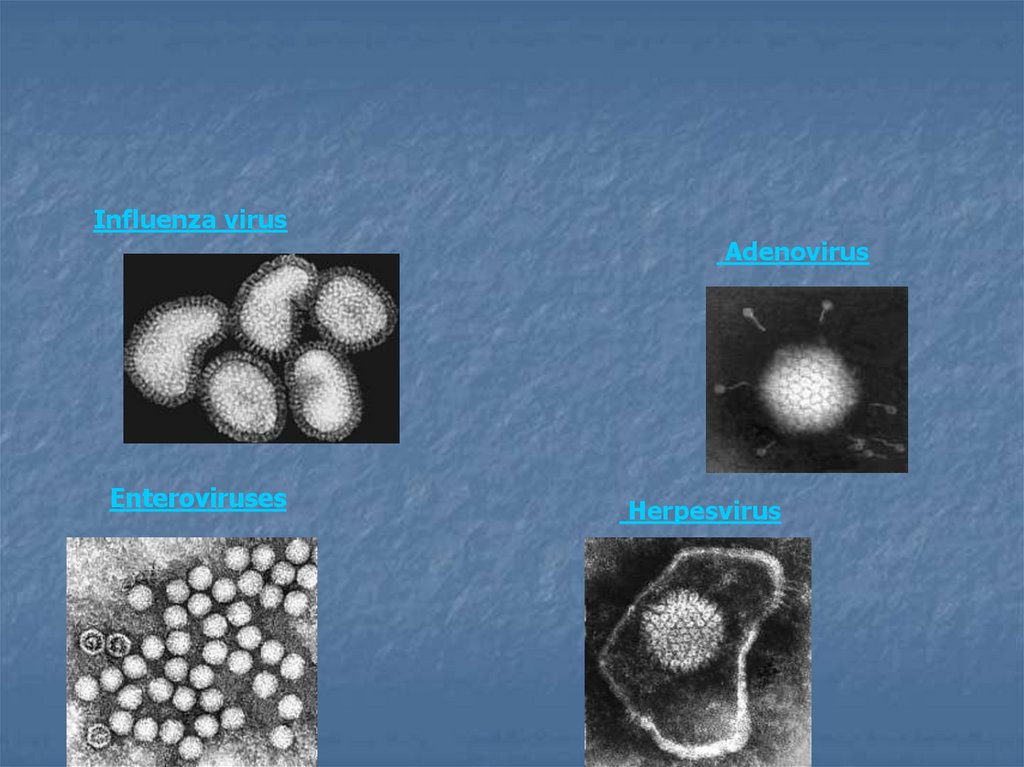
Influenza virusAdenovirus
Enteroviruses
Herpesvirus
4.
Introduction to the VirusesViruses- smallest known infective agents infecting unicellular
organisms (mycoplasmas, bacteria, algae) and all higher plants and
animals.
They are not microorganisms because they are not cell.
Do not always contain DNA.
Viruses cannot produce energy or synthesis molecules.
Do not undergo binary fission.
Bacteria
Mycoplasmas
Rickettsiae
Chlamydiae
Viruses
Growgh on
non-living
media
+
+
-
Binary DNA and Ribosomes Sensitivity
fission RNA
to Antibiotics
+
+
+
+
-
+
+
+
+
-
+
+
+
+
-
+
+
+
+
-
Sensitivity
to Interferon
+
+
5.
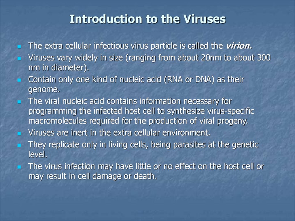
Introduction to the VirusesThe extra cellular infectious virus particle is called the virion.
Viruses vary widely in size (ranging from about 20nm to about 300
nm in diameter).
Contain only one kind of nucleic acid (RNA or DNA) as their
genome.
The viral nucleic acid contains information necessary for
programming the infected host cell to synthesize virus-specific
macromolecules required for the production of viral progeny.
Viruses are inert in the extra cellular environment.
They replicate only in living cells, being parasites at the genetic
level.
The virus infection may have little or no effect on the host cell or
may result in cell damage or death.
6.
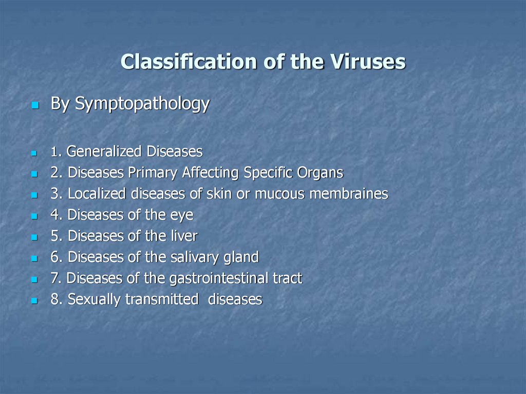
Classification of the VirusesBy Symptopathology
1. Generalized Diseases
2. Diseases Primary Affecting Specific Organs
3. Localized diseases of skin or mucous membraines
4. Diseases of the eye
5. Diseases of the liver
6. Diseases of the salivary gland
7. Diseases of the gastrointestinal tract
8. Sexually transmitted diseases
7.
Classification of the VirusesBy the type of Nucleic Acid
DNA-containing Viruses
Parvoviruses
Hepadnoviruses
Papovaviruses
Adenoviruses
Herpesviruses
Poxviruses
RNA-containing Viruses
Picornaviruses
Reoviruses
Arboviruses
Togaviruses
Arenaviruses
Coronaviruses
Retroviruses
Orthomixoviruses
Paramixoviruses
Rhabdoviruses
Bunyaviruses etc.
8.
Morphology of the VirusesVirus particle contains:
1.
Nucleic Acid: DNA or RNA, single stranded or double-stranded.
Most of RNA viruses are SS; most DNA viruses are DS.
Carries genetic information for replication
Mol. weight: DNA viruses: 1.5-160 (million daltons)
RNA viruses: 2-15
Some viruses have transcriptase for the multiplication.
2 Protein coat or capsid
Composed of capsomers held together. Made up of one or more
polypeptide chains.
Functions: a) pritect Nu acid, b) Involved in attachment of viral particle
on susceptible cell, c) Responsible for viral simmetry, d)
Determines antigenic character of virus
Capsid + Nu acid= Nucleocapsid
9.
Morphology of the VirusesEnvelope
Loosely fitting and surrounds nucleocapsid. Only on some viruses.
Contains lipids, protein and carbohydrate e.g. lipo-protein,glucoprotein.
Glycoprotein's – important of viral antigenic determinants.
Envelope partially derived from outer membrane of susceptible host
cell.
In some viruses appears spiky under E.M.; forms surface antigens
involved in HI and Neut tests.
Most RNA viruses are enveloped and most DNA viruses are nonenveloped.
10.
Morphology of the VirusesSimmetry
1.
Cubic or icosahedral: 12
vertices and 20 faces, each
equilateral triangle. Made up of
protein shells with Nu acid
inside.
2.
Helical: Elongated and spiral.
Capsomers arranged round
spiral of Nu acid. Most helical
viruses possess outer envelope.
3.
Complex: Does not conform to
(1) or (2) e.g. poxviruses.
particles neither cubical or
helical symmetry
11.
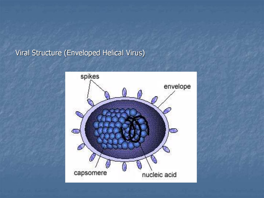
Viral Structure (Enveloped Helical Virus)12.
Cultivation of the VirusesAnimal Inoculation- The earliest method for the virus
cultivation. Monkeys, mice, infant (suckling) mice.
Chick Embryos- First used by Goodpasture 1931 and the
method was developed by Burnet. Viruses growth in an
embryonated chick egg, on the chorionallantoic membrane
for detection of the visible lesions (pocks) (variola or
vaccinia), allantoic cavity (influenza and some
paramixoviruses), amnionic sac (influenza).
13.
Cultivation of the VirusesTissue Cultures- The availability of cells growth in vitro has
facilitated the identification and cultivation of newly isolated and
previously known viruses. There 3 basic types of cell culture
1. Primary cell cultures: These are normal cells freshly taken
from the body and cultured. In general they are unable to grow for
more than a few passages in culture. Examples: monkey kidney,
human embryonic kidney, human amnion and etc.
2. Diploid cell lines are secondary cultures. These are cells of a
single type that retain the original diploid chromosome number (up
to 50 passages). Diploid strains developed from human fibroblasts.
3. Continuous cell lines: These are cells derived from cancer
cells, that are capable of continuous serial cultivation indefinitely.
Examples: derived from human cancers- HeLa, Hep-2, KB and etc.
14.
Detection of virus growth in cells culturesCytopathic effect (CPE) or necrosis of cells- morphological
changes in cultured cells in which viruses drow. These changes can
readily observed by microscopic examination of the cultures. It
helps in the presumptive identification of virus isolates (polio,
herpes, measels, adenoviruses and etc.).
Metabolic inhibition- when viruses grow in cell culture, cell
metabolism inhibited and there is no acid production. This can made
out by the colour of the indicator (phenol red) incorporated in the
medium (enteroviruses).
Hemadsorption- when the hemagglutinating viruses grow in cell
cultures, their presence can be indicated by the addition of guinea
pig erythrocytes to the cultures. If the viruses are multiplying the
RBC will adsorb onto surface of cells
15.
Detection of virus growth in cells culturesInterference by a non-cytopathogenic virus (rubella) with
replication and cytophatic effect of a second, indicator virus
(echovirus)
Morphologic transformation by an oncogenic virus
(sarcoma virus), tumour forming induce cell “transformation” and
loss of contact inhibition so that growth appears in a piled-up
fashion producing “microtumours”.
16.
Viral ReplicationViral replication can result in:
Abortive infection
Restrictive infection
Productive infection
The common stages of viral replication include:
1) Attachment
2) Penetration
3) Synthesis
4) Assembly
5) Maturation
6) Release
17.
Viral ReplicationATTACHMENT TO THE HOST CELL
Attachment (adsorption)
Often depends on specific attachment sites (viral receptors)
Usually glycoproteins located on the host cell
ENTRY INTO HOST CELLS: PENETRATION AND UNCOATING
After the virus attaches to the host cell, it can enter the cell by
several mechanisms:
1) Transfer of the entire viral particle across the cell membrane by
endocytosis
2) Transfer of only the viral genome through the cell membrane
3) Fusion of the viral envelope with the host cell membrane
Uncoating
18.
Viral ReplicationEXPRESSION AND REPLICATION-ds/ssDNA, AND
+ss/-ss/ds RNA VIRUSES
After the viral nucleic acid is released inside the host cell:
The transcription and translation processes of the host cell are
redirected for the production of viral proteins and nucleic acids The
different types of nucleic acid genomes are expressed and replicated
in several ways DNA genomes undergo replication-using processes
similar to cellular replication RNA genomes may be +ssRNA; Can be
read directly as an mRNA or reverse transcribed by reverse
transcriptase into DNA RNA genomes may also be -ssRNA; The RNA
must first be used as a template to form +mRNAs
19.
Viral ReplicationASSEMBLY, MATURATION, AND RELEASE
Two stages
Early synthesis
Results in the formation of large quantities of viral nucleic acids
Late synthesis
Results in the formation of large quantities of viral proteins
After sufficient quantities of viral proteins and nucleic acids are
formed, the viral particles are assembled
Completion of the capsid or maturation of the viral particle
After maturation, the completed particles are referred to as virions
20.
Viral ReplicationRelease from the host cell by:
Host cell lysis
Referred to as the lytic cycle, which results, in the death
of the host cell
Budding through cytoplasmic or vesicle membranes
21.
22.
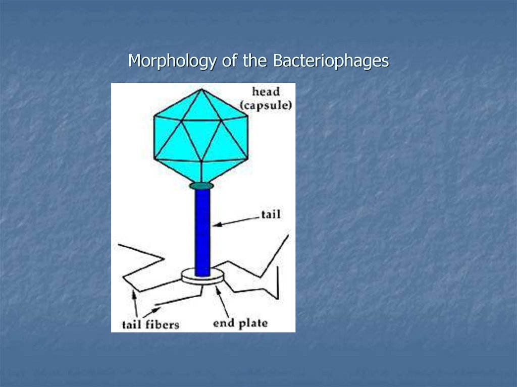
Bacteriofages23.
Bacteriophages or phages are viruses that infect bacteria.d”Herelle 1917 observed that filtrates of feces cultures from
dysentery patients induced transmissible lysis of a broth culture of a
dysentery bacillus.
Phages occur widely in nature in close association with bacteria.
Phages play an important role in the transmission of genetic
information between bacteria by the process of transduction
The specificity of the host range of phages is the basis of phage
typing methods, by which bacteria can be identified and typed
24.
Morphology of the Bacteriophages25.
REPLICATION OF BACTERIOPHAGEBacteriophages can be:
Lytic-replicating by a lytic cycle Temperate-undergoing a state of
lysogeny
Prophage
ASSAYING FOR LYTIC BACTERIOPHAGE
Plaque assay:
Lawn of bacteria in exponential growth on solid agar with a soft
agar overlay containing bacteriophage
Infection of the bacterial cells results in a clear zone (plaques)
26.
GROWTH CURVE OF LYTIC PHAGEThe typical growth curve of a lytic phage includes:
1) Eclipse phase (represents the time required for the
sinthesis of the phage paricles)
2) Latent phase (the interval between the infection of a
bacterial cell and first release of infectious phage particles)
3) Simultaneous release phase