


 medicine
Similar presentations:


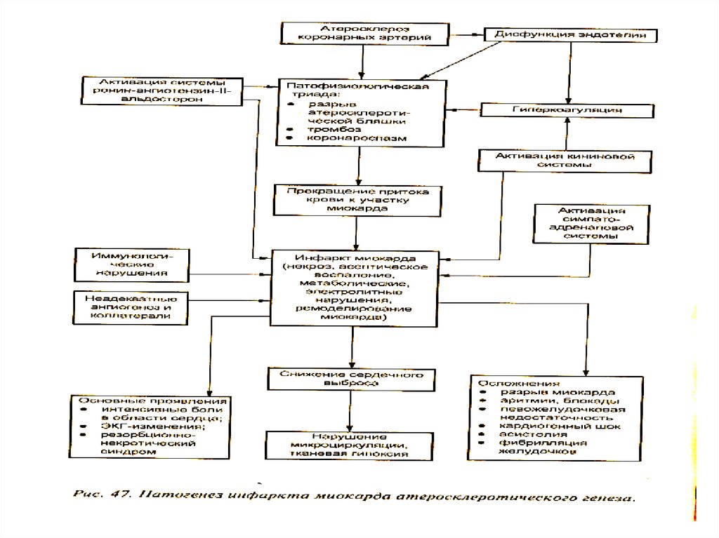
Патофизиология коронарной недостаточности
1. ПАТОФИЗИОЛОГИЯ КОРОНАРНОЙ НЕДОСТАТОЧНОСТИ
ЛЕКТОР - ПРОФ. В.В.МИХАЙЛОВ2.
• ОСОБЕННОСТИ МЕТАБОЛИЗМА,КРОВОСНАБЖЕНИЯ И
ВЕГЕТАТИВНОЙ ИННЕРВАЦИИ
МИОКАРДА
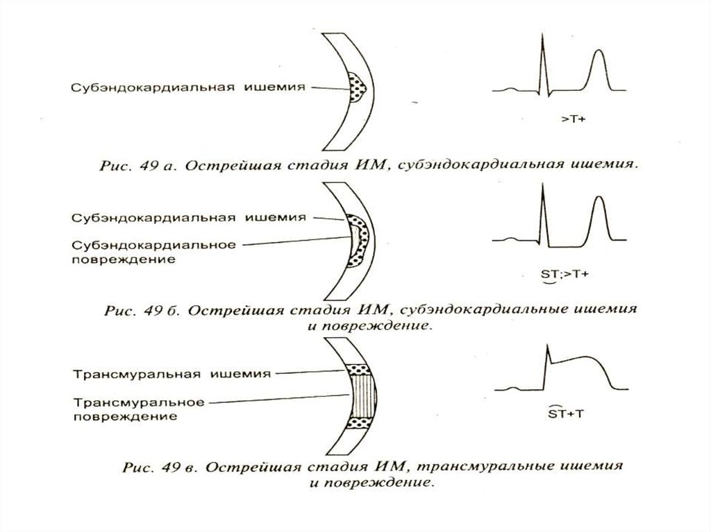
• ПАТОГЕНЕЗ ТРАНЗИТОРНОЙ
ИШЕМИИ МИОКАРДА
• ПАТОГЕНЕЗ НЕОБРАТИМОЙ ИШЕМИИ
МИОКАРДА
3.
4.
5.
6.
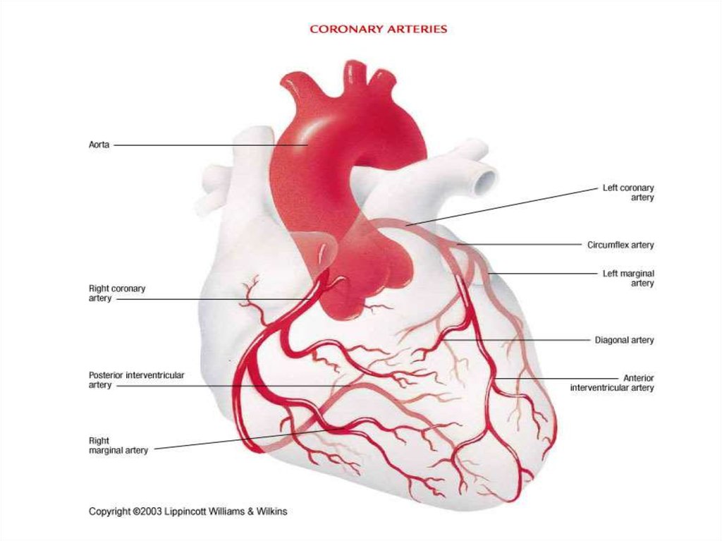
Here is a normal coronary artery seen in cross section at low power.The three major coronary branches (left anterior descending, left
circumflex, and right coronary arteries) supply blood to the heart. The
intima is so thin that it is indistinct. The media with smooth muscle
forms the bulk of the artery. The adventitia is outside the media and
merges with surrounding epicardial fat and connective tissue. The
lumen is large, without any narrowing by atheromatous plaque. The
muscular arterial wall is of normal proportion.
7.
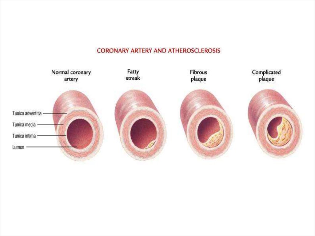
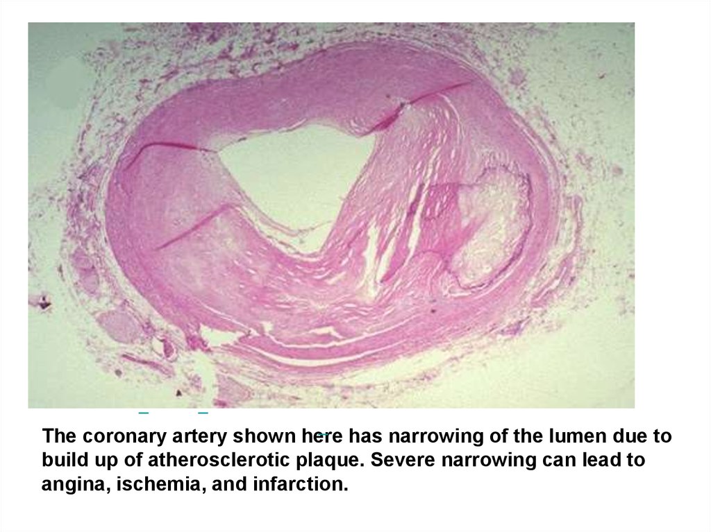
The coronary artery shown here has narrowing of the lumen due tobuild up of atherosclerotic plaque. Severe narrowing can lead to
angina, ischemia, and infarction.
8.
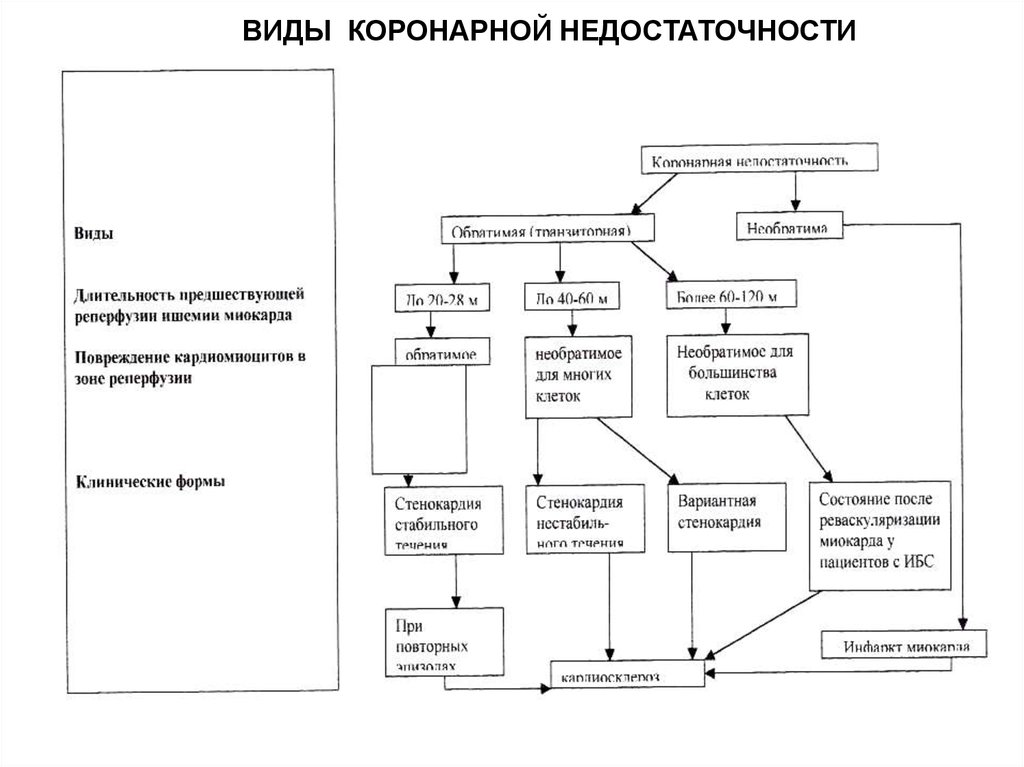
ВИДЫ КОРОНАРНОЙ НЕДОСТАТОЧНОСТИ9.
10. ТРАНЗИТОРНАЯ ИШЕМИЯ МИОКАРДА МОЖЕТ ВЫЗЫВАТЬСЯ * ФИКСИРОВАННОЙ КОРОНАРНОЙ ОБСТРУКЦИЕЙ * ДИНАМИЧЕСКОЙ КОРОНАРНОЙ ОБСТРУКЦИЕЙ * ВАЗОСПАСТИЧ
ТРАНЗИТОРНАЯ ИШЕМИЯМИОКАРДА МОЖЕТ
ВЫЗЫВАТЬСЯ
* ФИКСИРОВАННОЙ КОРОНАРНОЙ
ОБСТРУКЦИЕЙ
* ДИНАМИЧЕСКОЙ КОРОНАРНОЙ
ОБСТРУКЦИЕЙ
* ВАЗОСПАСТИЧЕСКОЙ
ОБСТРУКЦИЕЙ
11.
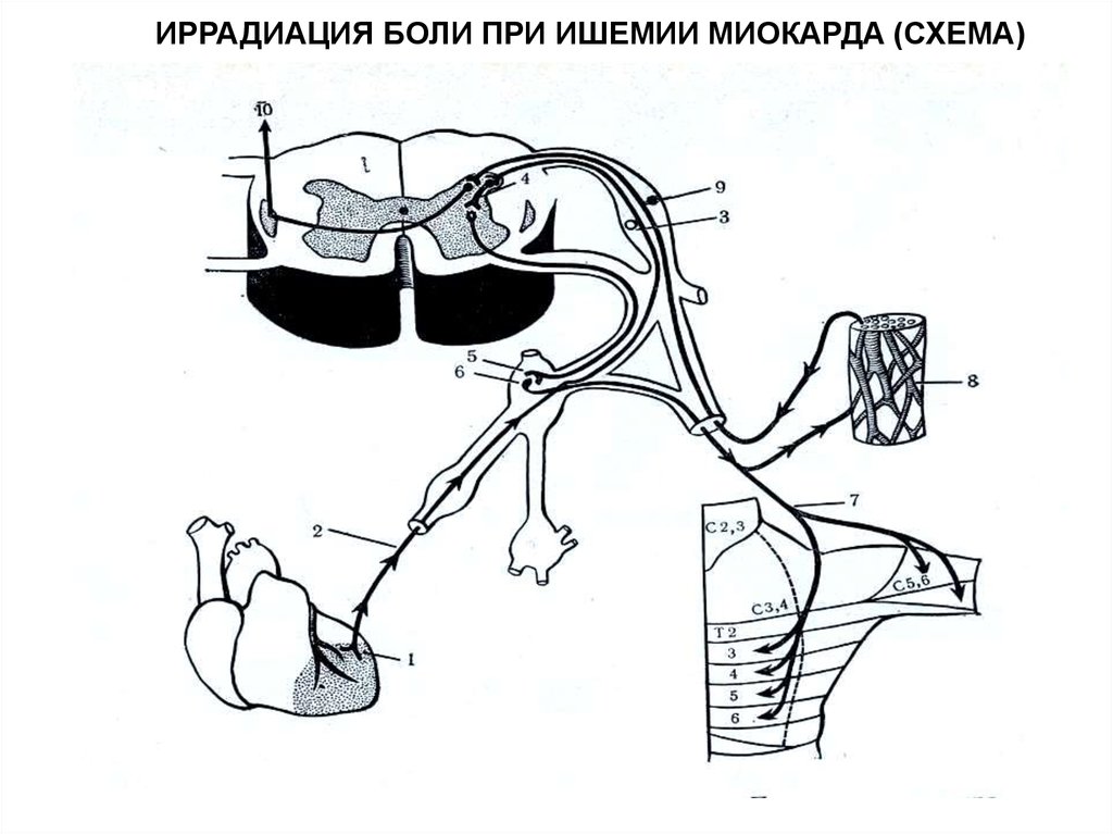
ИРРАДИАЦИЯ БОЛИ ПРИ ИШЕМИИ МИОКАРДА (СХЕМА)12.
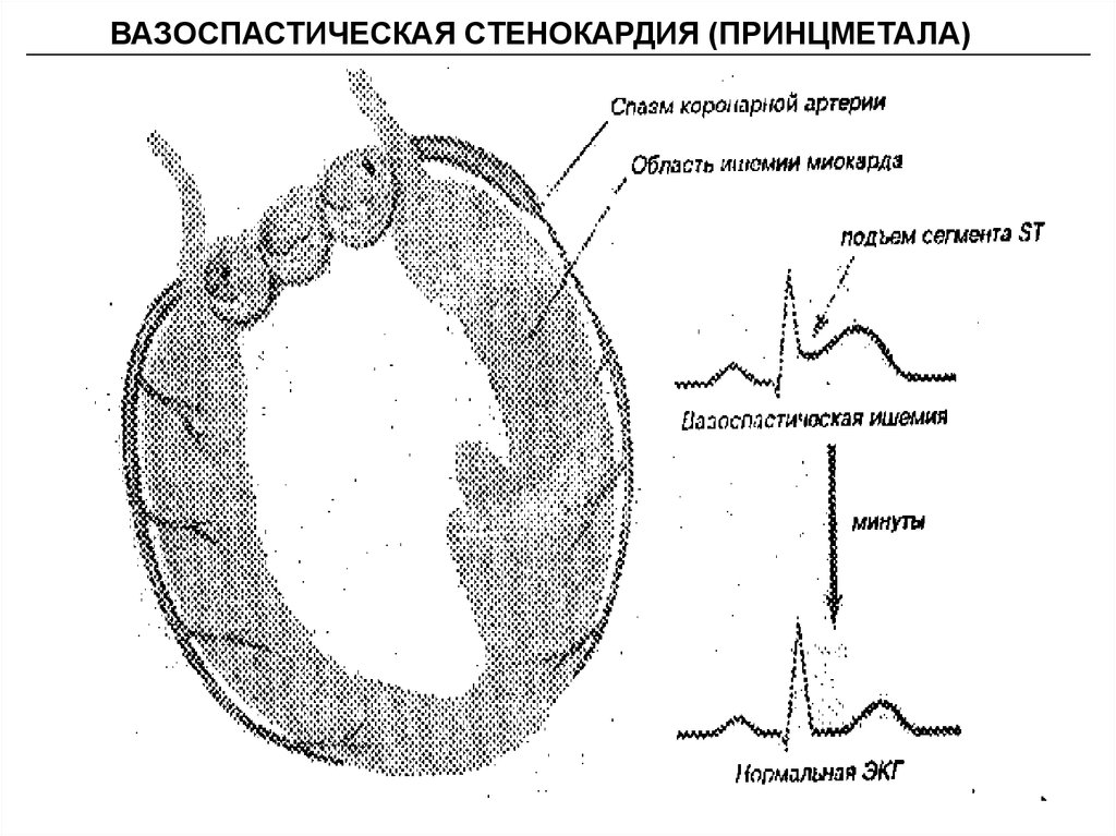
ВАЗОСПАСТИЧЕСКАЯ СТЕНОКАРДИЯ (ПРИНЦМЕТАЛА)13.
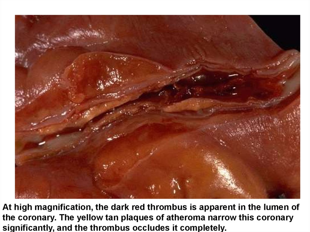
At high magnification, the dark red thrombus is apparent in the lumen ofthe coronary. The yellow tan plaques of atheroma narrow this coronary
significantly, and the thrombus occludes it completely.
14.
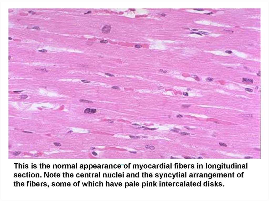
This is the normal appearance of myocardial fibers in longitudinalsection. Note the central nuclei and the syncytial arrangement of
the fibers, some of which have pale pink intercalated disks.
15.
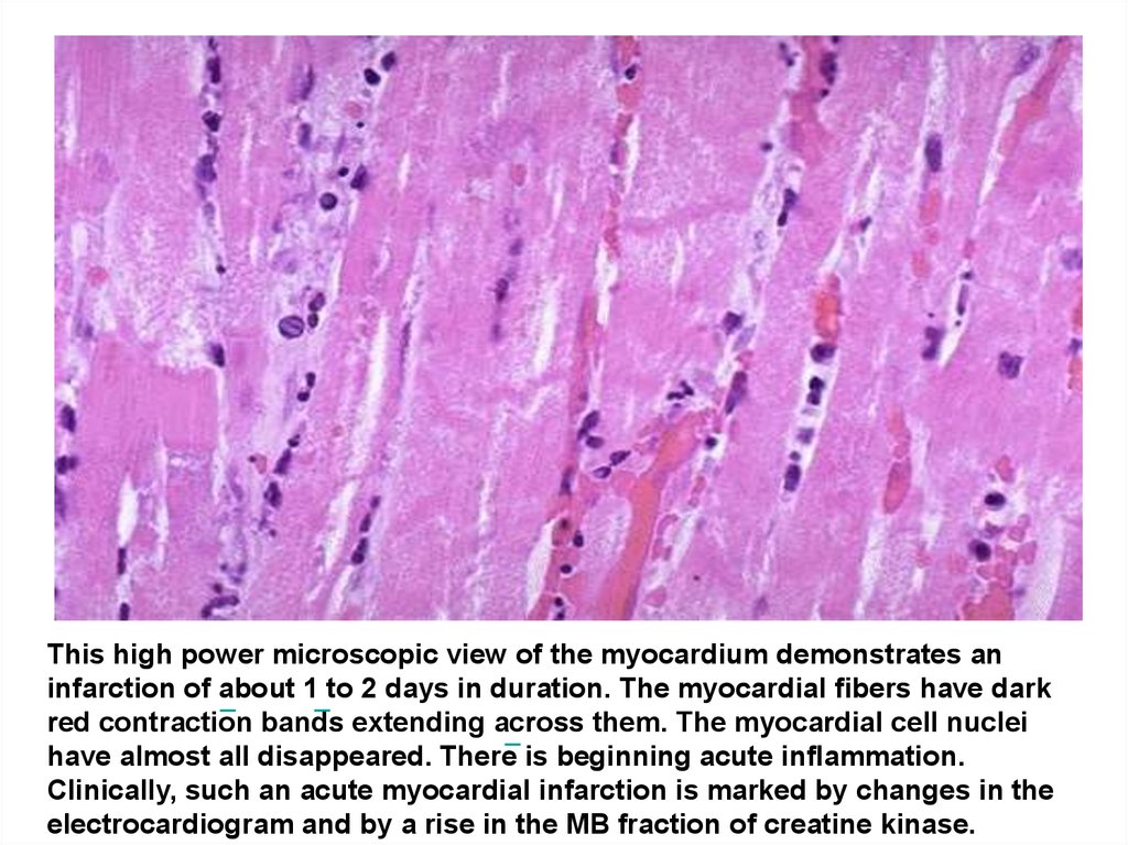
This high power microscopic view of the myocardium demonstrates aninfarction of about 1 to 2 days in duration. The myocardial fibers have dark
red contraction bands extending across them. The myocardial cell nuclei
have almost all disappeared. There is beginning acute inflammation.
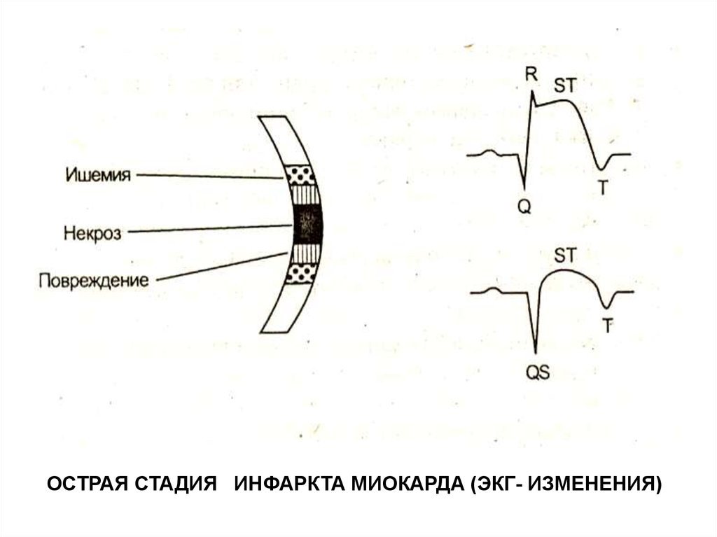
Clinically, such an acute myocardial infarction is marked by changes in the
electrocardiogram and by a rise in the MB fraction of creatine kinase.
16.
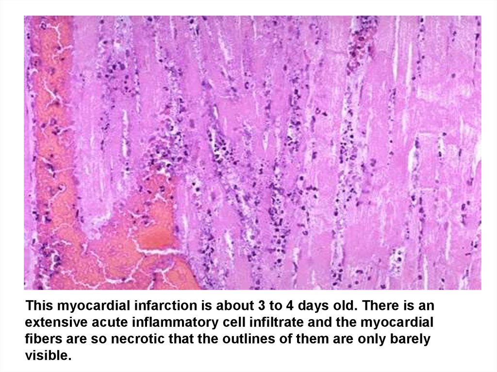
This myocardial infarction is about 3 to 4 days old. There is anextensive acute inflammatory cell infiltrate and the myocardial
fibers are so necrotic that the outlines of them are only barely
visible.
17.
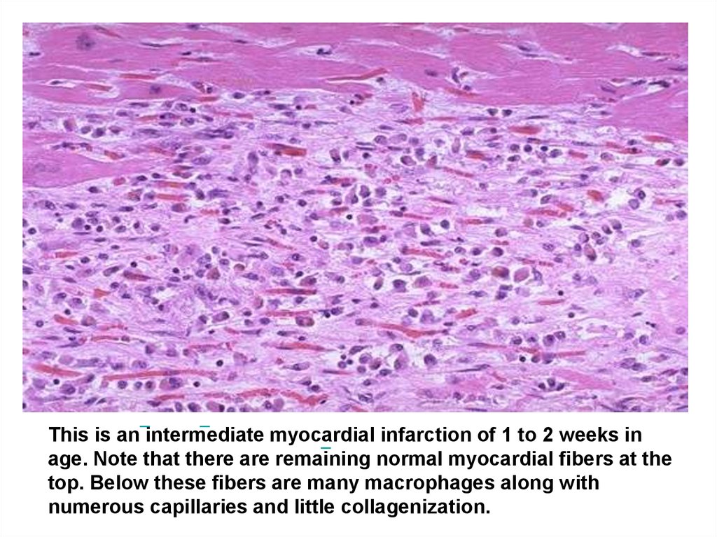
This is an intermediate myocardial infarction of 1 to 2 weeks inage. Note that there are remaining normal myocardial fibers at the
top. Below these fibers are many macrophages along with
numerous capillaries and little collagenization.
18.
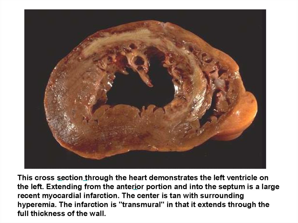
This cross section through the heart demonstrates the left ventricle onthe left. Extending from the anterior portion and into the septum is a large
recent myocardial infarction. The center is tan with surrounding
hyperemia. The infarction is "transmural" in that it extends through the
full thickness of the wall.
19.
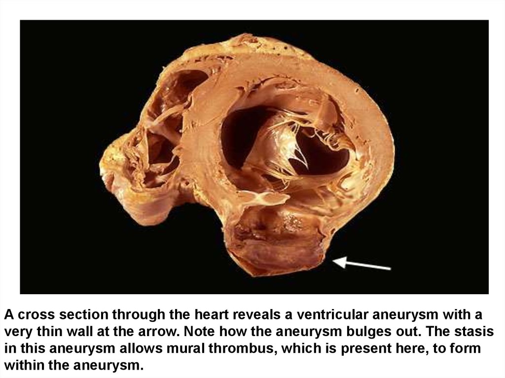
A cross section through the heart reveals a ventricular aneurysm with avery thin wall at the arrow. Note how the aneurysm bulges out. The stasis
in this aneurysm allows mural thrombus, which is present here, to form
within the aneurysm.