


 medicine
Similar presentations:


")


Medical academy name after s.i.georgievsky department of histology and embryology
1.
MEDICAL ACADEMY NAME AFTER S.I.GEORGIEVSKYDEPARTMENT OF HISTOLOGY AND EMBRYOLOGY
Lecturer : Associated professor:
LUGIN IGOR ANATOLIEVICH
2.
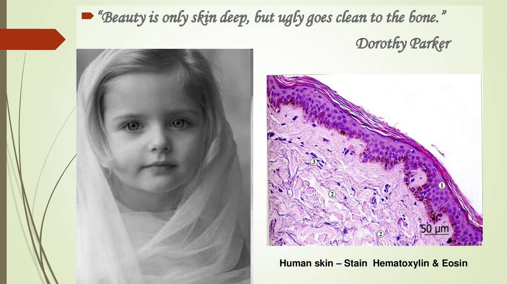
“Beauty is only skin deep, but ugly goes clean to the bone.”Dorothy Parker
Human skin – Stain Hematoxylin & Eosin
3.
A person is as old as his connective tissueILYA MECHNIKOV
4.
4Is a complex of mesenchyme
derivatives, consisting of cells differones
and big quantity of extracellular matrix
(fibers and ground substance),
participating in supporting gomeostasis
of body internal environment and
differing from other tissues by lesser need
in aerobic oxidation processes
5.
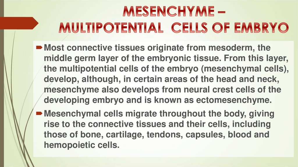
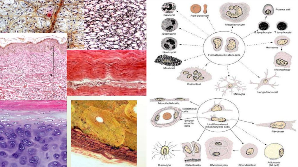
Most connective tissues originate from mesoderm, themiddle germ layer of the embryonic tissue. From this layer,
the multipotential cells of the embryo (mesenchymal cells),
develop, although, in certain areas of the head and neck,
mesenchyme also develops from neural crest cells of the
developing embryo and is known as ectomesenchyme.
Mesenchymal cells migrate throughout the body, giving
rise to the connective tissues and their cells, including
those of bone, cartilage, tendons, capsules, blood and
hemopoietic cells.
6.
6Stain Hematoxylin & Eosin
7.
7Connective tissue, group of tissues in the body
that maintain the form of the body and its
organs and provide cohesion and internal
support. The connective tissues include several
types of fibrous tissue that vary only in their
density and cellularity, as well as the more
specialized and recognizable variants—
bone, ligaments, tendons, cartilage,
and adipose (fat), reticular, elastic and
mucous connective tissue.
8.
89.
MATURE CONNECTIVE TISSUEis classified as connective tissue proper, and
specialized connective tissue
Connective tissue is composed of cells and
extracellular matrix (ECM) consisting of ground
substance and fibers ( collagen fibers, reticular
fibers and elastic fibers
Extracellular matrix (ECM) is dominated in all
types of connective tissues
10.
СОЕДИНИТЕЛЬНЫЕ ТКАНИСобственно
соединительные ткани
Со специальными
свойствами
Волокнистые
Хрящевые
ткани
Дентин
Костные ткани
Цемент
Неоформленная
(неориентированная) — дерма кожи
ПВСТ
РВСТ
Скелетные ткани
10
11.
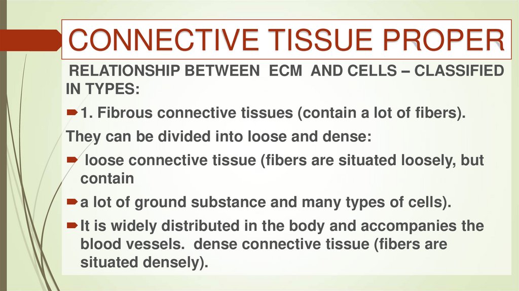
CONNECTIVE TISSUE PROPERRELATIONSHIP BETWEEN ECM AND CELLS – CLASSIFIED
IN TYPES:
1. Fibrous connective tissues (contain a lot of fibers).
They can be divided into loose and dense:
loose connective tissue (fibers are situated loosely, but
contain
a lot of ground substance and many types of cells).
It is widely distributed in the body and accompanies the
blood vessels. dense connective tissue (fibers are
situated densely).
12.
DENSE CONNECTIVE TISSUEcan be divided into regular and irregular:
regular (fibers situated regularly, in parallel)
as in tendons, ligaments, fascia;
irregular (fibers are situated irregularly, in all
three directions), as
in dermis (reticular layer) and capsule of
organs.
13.
SPI
II
III
4
3
III
1
2
7
5
6
ГИСТИОН
13
14.
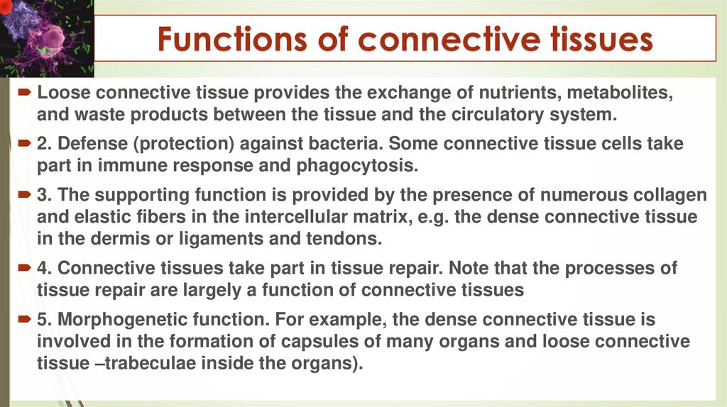
Functions of connective tissuesLoose connective tissue provides the exchange of nutrients, metabolites,
and waste products between the tissue and the circulatory system.
2. Defense (protection) against bacteria. Some connective tissue cells take
part in immune response and phagocytosis.
3. The supporting function is provided by the presence of numerous collagen
and elastic fibers in the intercellular matrix, e.g. the dense connective tissue
in the dermis or ligaments and tendons.
4. Connective tissues take part in tissue repair. Note that the processes of
tissue repair are largely a function of connective tissues
5. Morphogenetic function. For example, the dense connective tissue is
involved in the formation of capsules of many organs and loose connective
tissue –trabeculae inside the organs).
15.
КЛАССИФИКАЦИЯ КЛЕТОК РВСТКЛЕТКИ РЕЗИДЕНТЫ
(ФИКСИРОВАННЫЕ)
КЛЕТКИ МИГРАНТЫ
(БЛУЖДАЮЩИЕ)
АДК
АДИПОЦИТ
ФИБРОБЛАСТ
ПЛАЗМОЦИТ
ЛАБРОЦИТ
МАКРОФАГИ
ФИБРОЦИТ
15
ЛЕЙКОЦИТЫ
16.
17.
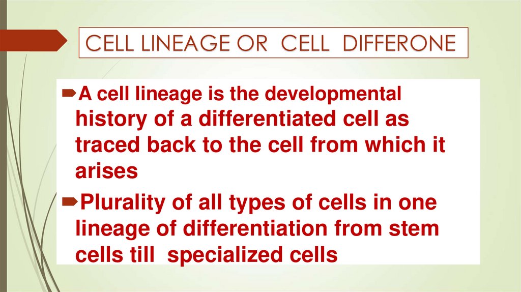
CELL LINEAGE OR CELL DIFFERONEA cell lineage is the developmental
history of a differentiated cell as
traced back to the cell from which it
arises
Plurality of all types of cells in one
lineage of differentiation from stem
cells till specialized cells
18.
19.
DETERMINATION / DIFFERENTIATIONDevelopment proceeds in a series of stages in which precursor cells are first restricted in their
developmental potential (determination) and subsequently express their genetic information as
specific tissues (differentiation)
DETERMINATION – IS RESTRICTION OF CELL ABILITY FOR
TRANSFORMATION IN DIFFERENT LINEAGE AND
EXPRESSION OF GENETIC INFORMATION AS SPECIALIZED
LINEGES OF CELLS IN TISSUE
DIFFERENTIATION – The process by which cells become
progressively more specialized; a normal process through
which cells mature. This process of specialization for the cell
comes at the expense of its breadth of potential
20.
21.
21FIBROBLASTS AND FIBROCYTES
22.
23.
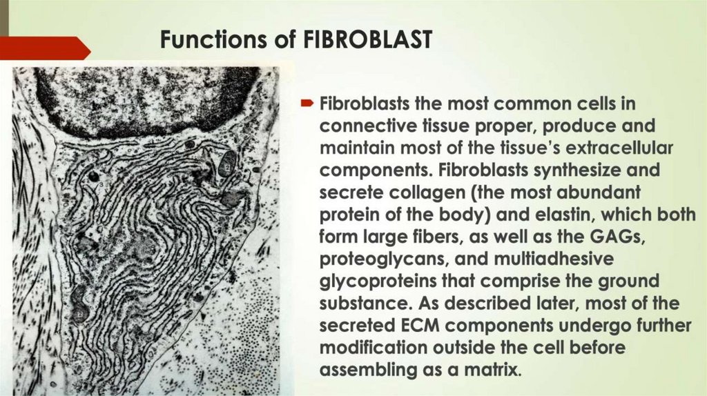
STRUCTURE AND FUNCTIONSThe fibroblast is a spindle-shaped or stellate cell. Its
nucleus is round with a lot of euchromatin (active
chromatin).
well-developed ER in which the collagen (for collagen
fibers), elastin (for the elastic fibers), as well as
proteoglycans, and glycoproteins (for ground substance)
are synthesized.
Fibroblast produces extracellular matrix constantly. It is a
reason why the borderlines of fibroblasts under the light
microscope are indistinct
24.
УЛЬТРАСТРУКТУРНАЯ ОРГАНИЗАЦИЯПРИ УЛЬТРАСТРУКТУРНОМ
АНАЛИЗЕ (ЭЛЕКТРОННОМИКРОСКОПИЧЕСКИЙ МЕТОД
ВЫЯВЛЯЮТСЯ:
мелкие вакуоли,
гранулы, клеточный центр,
митохондрии, аппарат
Гольджи
гранулы содержат
гликозаминогликаны,
щелочнофосфатные
протеины, в цитоплазме
имеется РНК
24
25.
25МИОФИБРОБЛАСТЫ
Миофибробласты
клетки, сходные
морфологически с
фибробластами,
сочетающие в себе
способность к синтезу
не только
коллагеновых, но и
сократительных белков,
особенно
многочисленны в
соед.ткан.миометрия,
ран.пов. кожи.
26.
МПГ26
27.
2728.
2829.
30.
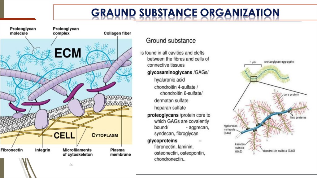
30ФИБРОНЕКТИН
FIBRONECTIN
Основной гликопротеин
взаимосвязи клеток и МКВ
31.
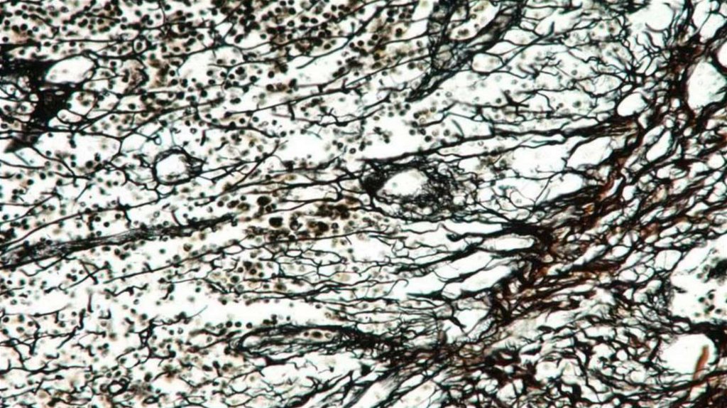
COLLAGEN FIBERSCollagen fibers are the most abundant
type of connective tissue fiber
They are flexible and have a remarkably
high tensile strength. In the light
microscope, collagen fibers typically
appear as wavy structures of variable
width and indeterminate length
They stain readily with eosin and other
acidic dyes. They can also be colored
with the dye aniline blue used in
Mallory’s connective tissue stain or with
the dye light green used in Masson’s
stain
31
32.
ELECTRON MICROSCOPICEXAMINATION OF COLLAGEN
When examined with the TEM, collagen fibers
appear as bundles of fine, threadlike subunits.
These subunits are collagen fibrils
Within an individual fiber, the collagen fibrils are
relatively uniform in diameter. In different
locations and at different stages of development,
however, the fibrils differ in size. In developing or
immature tissues, the fibrils may be as small as
15 or 20 nm in diameter. . In dense, regular
connective tissue of tendons or other tissues that are
subject to considerable stress, they may measure up to
300 nm in diameter.
1- dark line, 2- light line, 3- protofibrilles
33.
3334.
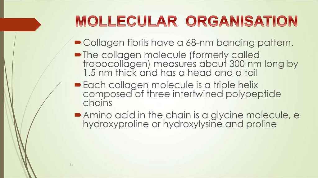
Collagen fibrils have a 68-nm banding pattern.The collagen molecule (formerly called
tropocollagen) measures about 300 nm long by
1.5 nm thick and has a head and a tail
Each collagen molecule is a triple helix
composed of three intertwined polypeptide
chains
Amino acid in the chain is a glycine molecule, e
hydroxyproline or hydroxylysine and proline
34
35.
GOLLAGEN SYNTHESIS ANDDEGRADETION
The formation of collagen fibers involves the uptake of amino acids (proline,
lysine, etc.) by fibroblasts, the production of polypeptide chains in the RER.
Modifications of the polypeptide chains occur within the cisternae of RER
and the Golgi complex. The resultant molecules are called procollagen,
packing into secretory vesicles and leaving the cell by exocytosis. Extracellular
events include the formation of tropocollagen molecules, 280 nm
long, with a head and a tail, which is aggregated into collagen protofibrils.
Forming protofibrils these molecules become aligned head to tail in
overlapping rows with a gap between the molecules within each row (64-nm
banding pattern). A collagen protofibrils are connected by glycoproteins
into the collagen fibers. Then the fibers are connected by glycoproteins to
36.
GOLLAGEN SYNTHESIS ANDDEGRADETION
The formation of collagen fibers involves the
uptake of amino acids (proline, lysine, etc.) by
fibroblasts, the production of polypeptide chains in
the RER.
Modifications of polypeptide chains occur within
the cisternae RER
And Golgi complex
The resultant mollecules are called procollagen
packing into secretory vesicles and leaving the
cell by exocytosis
37.
38.
EXTRACELLULAR AGRIGATIONSExtracellular events include the formation of
tropocollagen molecules, 280 nm long, with a head
and a tail, which is aggregated into collagen
protofibrils.
Forming protofibrils these molecules become aligned
head to tail in overlapping rows with a gap between
the molecules within each row (64-nm banding
pattern).
A collagen protofibrils are connected by
glycoproteins into the collagen fibers
39.
40.
4041.
Assembly of Collagen1- tropocollagen molecules, 280 nm
2- collagen protofibrils
3 - collagen fibers
4 – bundles of collagen fibers
42.
43.
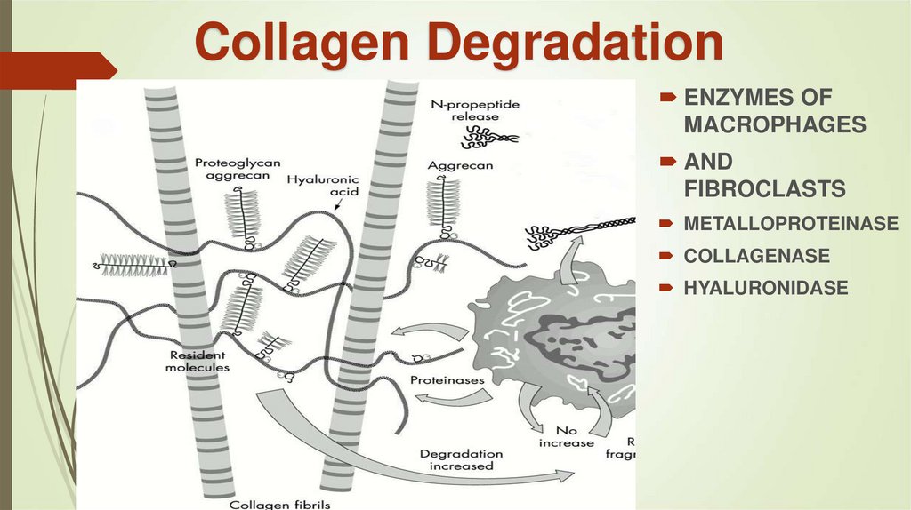
Collagen DegradationENZYMES OF
MACROPHAGES
AND
FIBROCLASTS
METALLOPROTEINASE
COLLAGENASE
HYALURONIDASE
43
44.
45.
46.
ELASTIC FIBERSElastic fibers are elastic: they can stretch after
distension. Elastic fibers are thinner than collagen
fibers, they can not compose bundles, but can
branching.
They are composed of elastin core and microfibrils
sheath.
Elastin is synthesized by the same pathway as
collagen
46
47.
48.
4849.
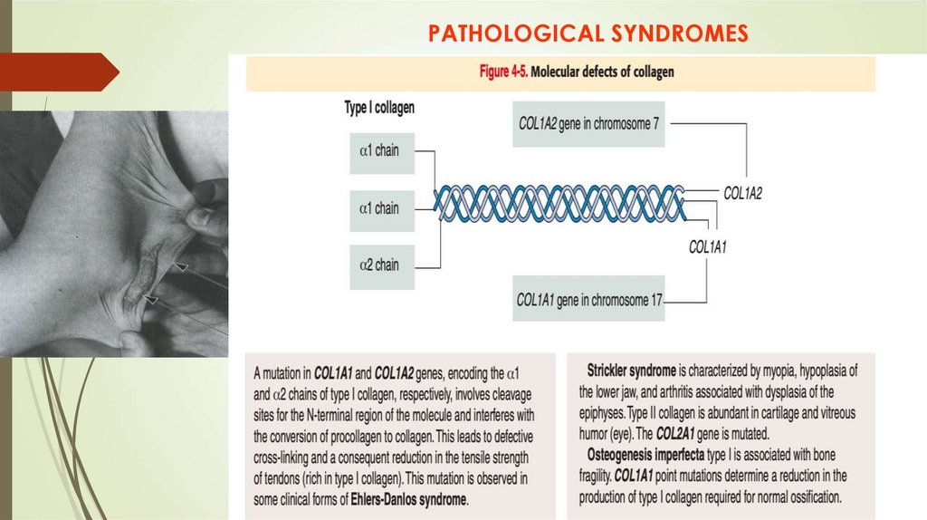
PATHOLOGICAL SYNDROMES50.
51.
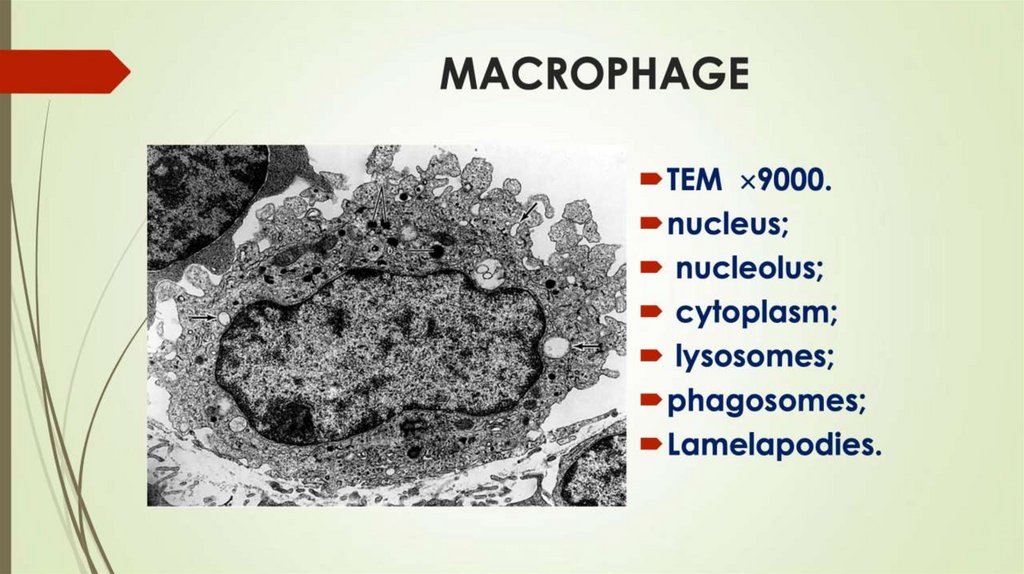
MACROPHAGEMacrophages (“big eaters”) can phagocyte (catch and digest) bacteria
and foreign particles
Macrophages are phagocytotic cells derived from monocytes.
Therefore they contain many lysosomes and phagolysosomes, as well as all other
organelles. They can actively move in the intercellular matrix and contain winding
borderline, because of numerous
folds or finger-like processes.
Macrophages take part in the processing and presentation of foreign
antigens on their surface to lymphocytes that stimulate their antigen dependent
proliferation and differentiation – Antigen Presenting Cells
They are derived from the blood monocytes, which leave the bloodstream and
migrate to the connective tissues to turn into macrophages
52.
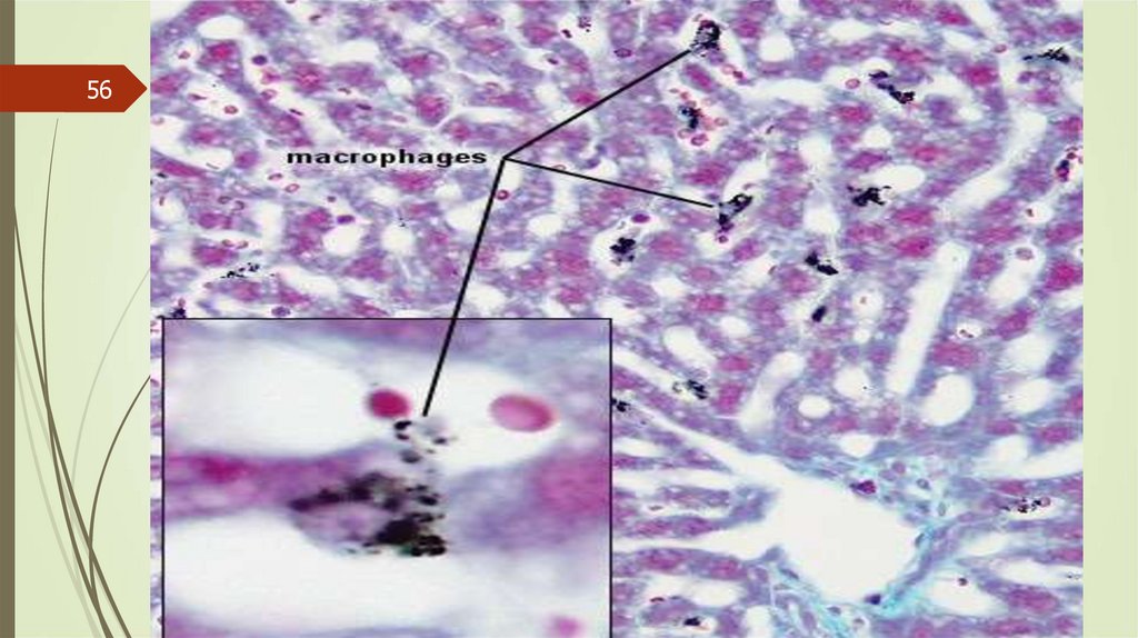
Acid phosphatase – marker for histochemistry identification - MacrophageLysosomes are abundant in the cytoplasm and
can be revealed by staining for acid phosphatase
activity (both in the light microscope and with the
TEM); a positive reaction is a further aid in
identification of the macrophage.
With the TEM, the surface of the. macrophage
exhibits numerous folds and fingerlike
projections
53.
54.
55.
THE SYSTEM OF MONONUCLEAR PHAGOCYTESWANDERING CELLS
RESIDENT CELLS
Hoffbauer cells
HISTIOCYTE
Dendritic cells
Serous
Space Cells
Kupfer Cells
Dust Cells
Langergans cell
Osteoclast
Microglial cells
55
Interstitial Macrophage
Exudative
Macrofage
56.
5657.
PLASMA CELLThey derived from B-lymphocytes. A plasma cell
contains a well developed rER (the reason for high
basophilia of cytoplasm).
Light microscopy shows radially arranged clumps of
heterochromatin adjacent to the nuclear membrane
which gives the cartwheel appearance to the nucleus.
Besides, the well-developed Golgi region gives the light
macula near the nucleus.
Plasma cells produce immunoglobulins – antibodies and
responsible for humoral immunity.
58.
59.
60.
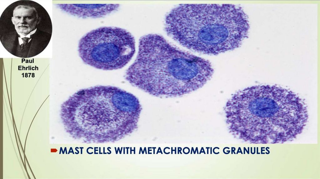
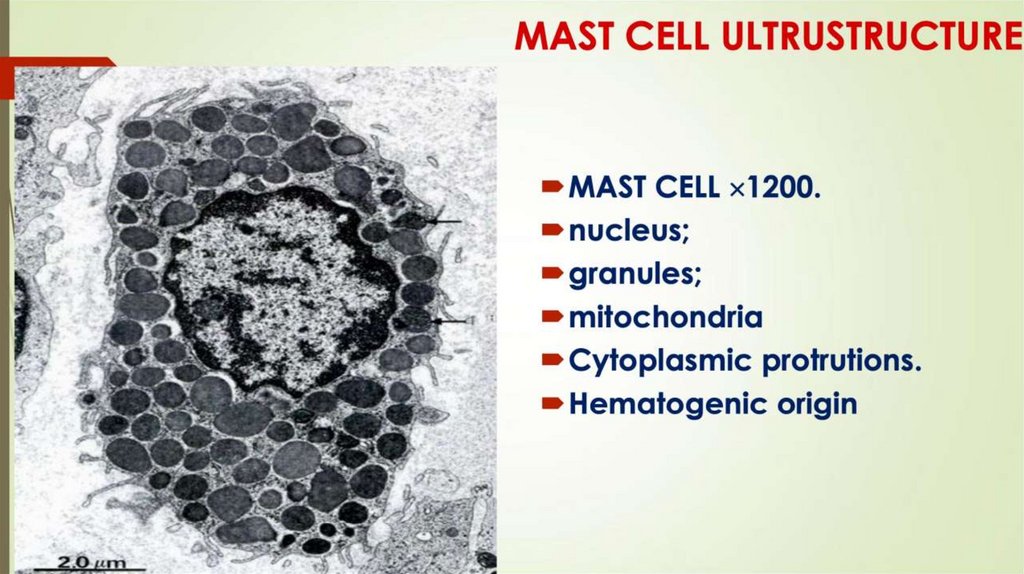
Mast cells (tissue basophils)As a rule, mast cells are located around small blood vessels. They
contain and release the big granules with heparin, histamine, and
other mediators that initiate the inflammatory and allergic reactions
The allergic reaction is known to be mediated by IgE.
They are produced by plasma cells in response to certain antigens
called allergens. Mast cells and blood basophils have a high affinity
for IgE. The combination of antigen with IgE triggers the release of
mediators due to the degranulation
(discharge of granules), producing an allergic disease
61.
62.
63.
64.
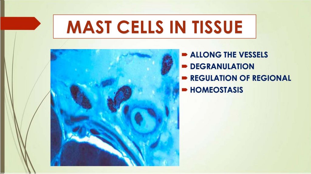
ГЕПАРИНMAST CELLS
IN TISSUE
обладает противосвёртывающим
действием,
способствует рассасыванию
тромбов,
снижает проницаемость
межклеточного вещества,
проявляет противовоспалительное
действие
64
65.
LEUKOCYTIC INFILTRATION IN CONNETIVE TISSUEDURING INFLAMATION
65
66.
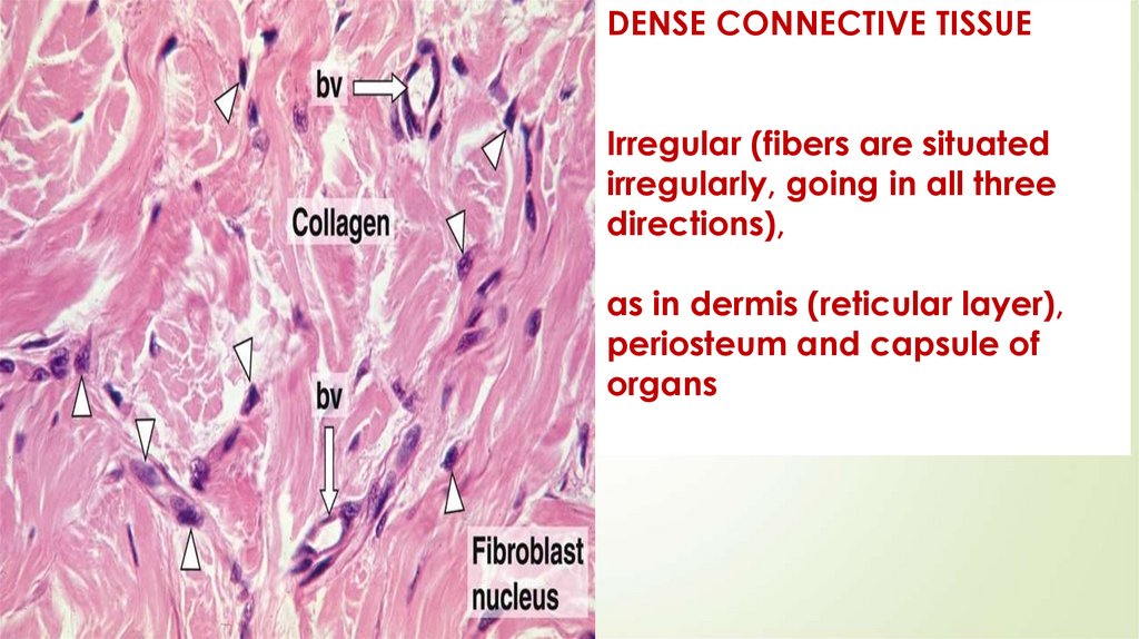
DENSE CONNECTIVE TISSUEIrregular (fibers are situated
irregularly, going in all three
directions),
as in dermis (reticular layer),
periosteum and capsule of
organs
66
67.
TENDONTendon consists of bundles of type 1 (collagen
fibers) surrounded
by fibrocytes. Several bundles of type 1 are
combined into bundles of type 2, surrounding
loose connective tissue layer endotendinum.
Several bundles of type 2 are combined into
bundles of type 3,
surrounding thicker loose connective tissue layer
peritendinum
The big tendon surrounded by a loose connective
tissue layer is epitendinum
68.
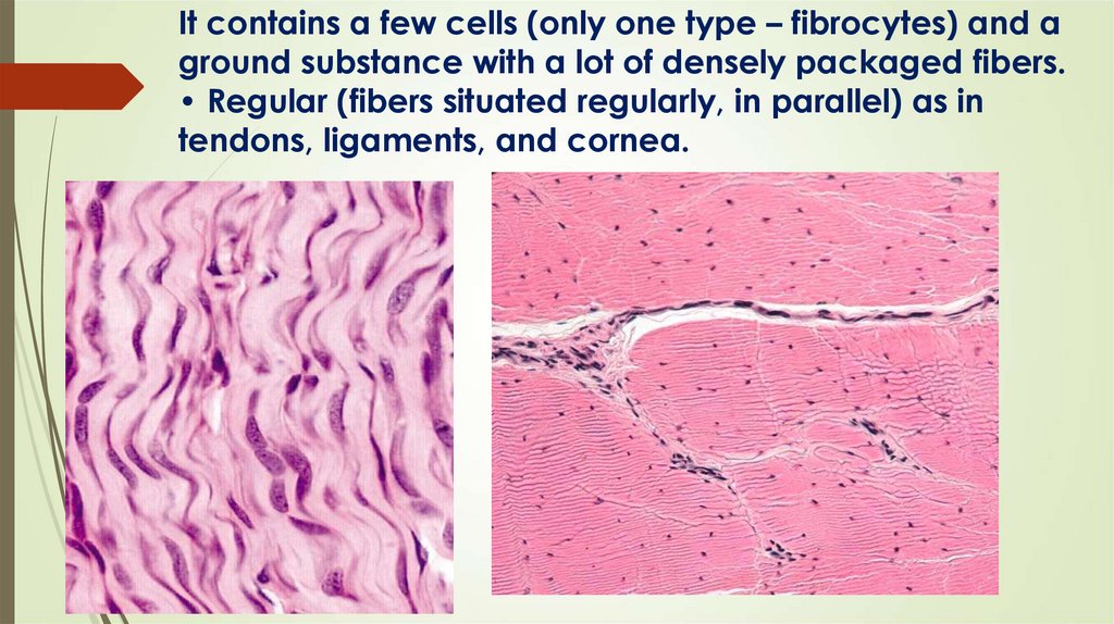
It contains a few cells (only one type – fibrocytes) and aground substance with a lot of densely packaged fibers.
• Regular (fibers situated regularly, in parallel) as in
tendons, ligaments, and cornea.
68
69.
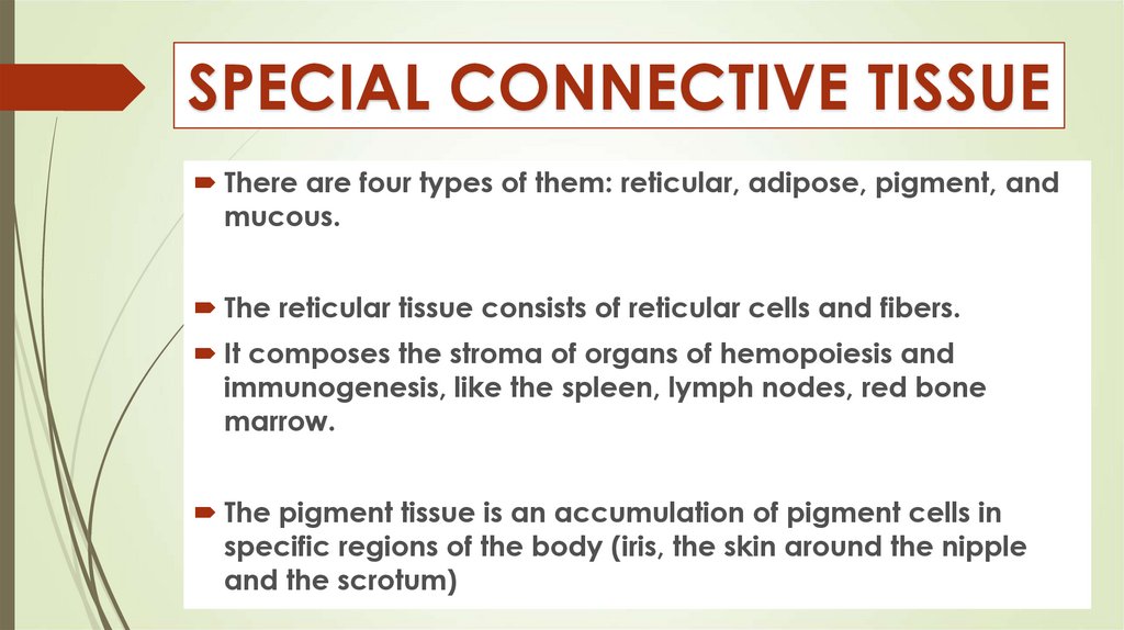
SPECIAL CONNECTIVE TISSUEThere are four types of them: reticular, adipose, pigment, and
mucous.
The reticular tissue consists of reticular cells and fibers.
It composes the stroma of organs of hemopoiesis and
immunogenesis, like the spleen, lymph nodes, red bone
marrow.
The pigment tissue is an accumulation of pigment cells in
specific regions of the body (iris, the skin around the nipple
and the scrotum)
70.
MELANOCYTES70
71.
ADIPOSE TISSUEThe adipose tissue is divided into two types: white and
brown.
White adipose tissue is composed of large fat cells or
adipocytes.
Every cell contains a large single lipid droplet of fat
inclusion which pushes the nucleus and organelles to
the periphery. If special staining (Sudan) is used, the lipid
droplet stains orange or black. If the paraffin sections
are used, the adipose cells look empty, because the fat
dissolved in alcohol and xylene during the making of
histological preparation.
72.
73.
STRUCTURE AND FUNCTIONSFat cells are organized into groups called lobules. The
lobules of fat
cells are separated by loose connective tissue septa,
which conducts blood vessels and nerves into adipose
tissue
It is situated under the skin (hypodermis), especially in
females, in the mammary glands, around the kidney,
in the mesentery and omentum of the abdominal
cavity
74.
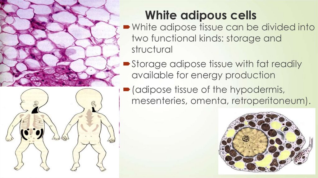
74White adipous cells
White adipose tissue can be divided into
two functional kinds: storage and
structural
Storage adipose tissue with fat readily
available for energy production
(adipose tissue of the hypodermis,
mesenteries, omenta, retroperitoneum).
75.
Structural adipose tissueStructural adipose tissue with the role of an elastic
pad, mechanical support, and protection (adipose
tissue in the orbits of the eye, articulations, palms,
soles, cheeks, etc.).
Note that this adipose tissue remains practically
unchanged during fasting.
76.
Functions of White Adipose tissue1. Reserve of calorie-rich
material (fat storage);
2. Thermo isolation (heat
insulator);
3. Store of water;
4. Replacement of involuted
organs – thymus, red bone
marrow, mammary gland;
5. Elastic pad (in palms and
soles);
6. Mechanical support (around
the kidney).
77.
Brown adipose tissueThe brown adipose tissue appears brown because it is rich
in mitochondria cytochrome pigments and every cell is
surrounded by blood capillaries
The brown adipose tissue has a lobular organization. It is
formed of
small cells filled with many small lipid droplets and
numerous mitochondria; the cell nucleus is located in the
center of this cell
The peculiarity of the metabolism of these cells:
mitochondria are the
produce of heat energy instead of the production of ATP
78.
Brown adipose tissue in bodyIn humans, this tissue is present in large amounts in just
newborns. It is
located in the mediastinum, in the posterior triangle of the
neck, around
the thyroid gland, the carotid arteries, and the kidney.
It acts like a heat generator; it is heating the blood and the
body.
It is very important for the newborns and for the hibernators,
e.g., in bear.
During winter it sleeps in the den. It does not freeze, because
its blood is warmed by brown adipose tissue.
79.
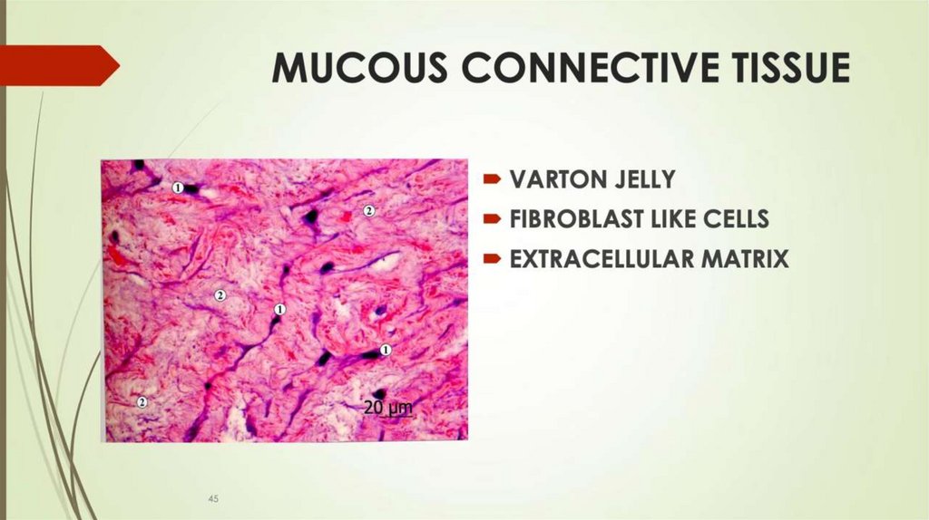
СЛИЗИСТАЯ СОЕДИНИТЕЛЬНАЯТКАНЬ
ВАРТОНОВ СТУДЕНЬ –
видоизмененная РВСТ с
преобладанием МКВ
Основные клетки –
фибробластоподобные
Входит в состав пупочного
канатика
У новорожденных встречается
регионально на ладонях и
подошвах
79