


 medicine
Similar presentations:


")
")


 degenerations, and hyperkeratosis. Intracellular lipidosis")
Reversible damage to cells and tissues. Intracellular accumulation (lipids, proteins, glycogen)
1.
Reversible damage tocells and tissues.
Intracellular
accumulation (lipids,
proteins, glycogen)
2.
Non-lethal cell damage iscalled dystrophy. It reflects
metabolic disorders accompanied
by accumulation or violation of
normal content various substances
in cells, in the extracellular matrix,
in the walls of blood vessels and
stroma of organs.
3.
Reasons dystrophy can be1) hypoxia,
2) genetic damage,
3) toxic substances or
medicines,
4) food imbalance,
5) violation of the blood
composition and
urine in diseases
internal organs.
4.
Distinguish between three types ofintracellular accumulations.
First of all: accumulation natural
endogenous metabolites that are
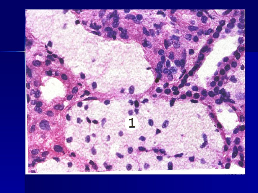
formed in a normal or accelerated
rhythm, but the rate of their
removal is insufficient (fats,
proteins, carbohydrates, pigments).
5.
Secondly: accumulation endogenoussubstances that they cannot be
metabolized. The most common cause of
these clusters is a genetic defect in the
enzyme. As a result, metabolic products
are not used, but are deposited inside
the cell – accumulation diseases
(thesaurismoses) develop. Most often,
these diseases are manifested by the
accumulation of amino acids (cystine,
phenylpyruvic acid, tyrosine).
6.
Third: accumulation exogenousones substances that the cell
can neither break down with
the help of enzymes, nor
transport to another place (coal
or silicon particles).
7.
Fatty degenerations.Adipose dystrophy is the
most common type of
intracellular accumulation. All
classes of lipids can accumulate
in cells: triglycerides, cholesterol
esters, and phospholipids.
8.
Accumulation triglycerides inparenchymal cells is called
steatosis or adipose
dystrophy. Most often,
triglycerides accumulate in the
liver, heart, kidneys, and
muscles.
9.
Fatty liver disease.Accumulation of
triglycerides in the liver can
occur as a result of:
1) Hypoxia
2) Toxic effects
3) Fasting.
10.
Most often, liver steatosis isobserved in alcoholism,
obesity, diabetes mellitus,
hypoxia, toxic effects, and a
lack of protein in food.
11.
Macroscopic picture:The liver is enlarged,
flabby, yellow in the cut,
with a touch of fat.
Figuratively, such a liver is
called "goose" (similar to
the liver of a fattened
goose).
12.
Microscopically - whenstained with hematoxylin
and eosin, fat inclusions
look like transparent
vacuoles of small (smalldrop obesity) or large
(large-drop obesity) sizes.
13.
Large-drop obesity often develops in theperipheral parts of the liver lobules,
small-drop-in the center of the lobules.
When painting with Sudan III vacuoles
are colored in a bright yellow-red color.
As a rule, fatty liver disease (steatosis) is
reversible, but if the damaging factor is
strong, then necrosis of "obese"
hepatocytes can develop.
14.
15.
Myocardial fatty degeneration.Reasons:
1) hypoxia (blood diseases,
cardiovascular insufficiency);
2) intoxication (for alcoholism,
infections, phosphorus or arsenic
poisoning).
16.
The mechanism ofmyocardiocyte obesity is
associated with a decrease in
lipid oxidation, due to the
destruction of mitochondria
under the influence of hypoxia
or toxins.
A special feature is the focal
nature of the lesion.
17.
Macroscopic picture.The size of the heart is enlarged,
the chambers are stretched, the
heart muscle is flabby, clay-yellow
in color. Under the endocardium of
the left ventricle is visible white
striation (focality), which gave
reason to compare the myocardium
with the skin of a tiger ("tiger's
heart").
18.
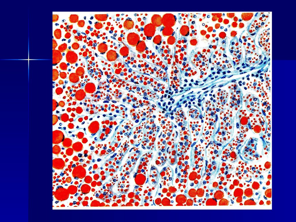
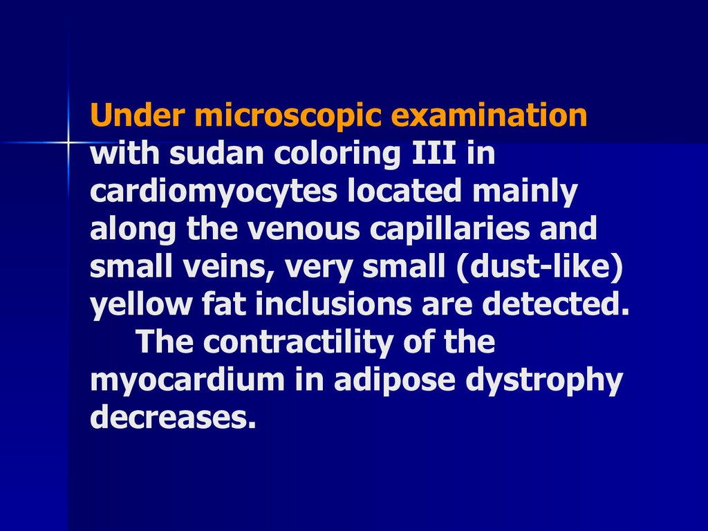
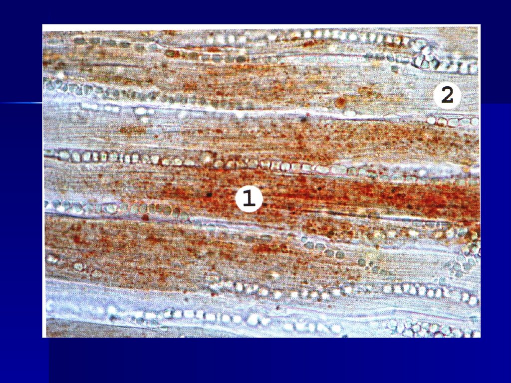
Under microscopic examinationwith sudan coloring III in
cardiomyocytes located mainly
along the venous capillaries and
small veins, very small (dust-like)
yellow fat inclusions are detected.
The contractility of the
myocardium in adipose dystrophy
decreases.
19.
20.
Fatty kidney disease.Most often adipose
dystrophy of the kidneys
occurs in nephrotic
syndrome and chronic renal
failure, less often in
infections and intoxications.
21.
The mechanism ofaccumulation of fat
inclusions is associated
with the occurrence of
hyperlipidemia (for
nephrotic syndrome), and
lipiduria.
22.
Macroscopically - kidneyswith nephrotic syndrome are
enlarged, flabby, with
yellow speckles on the
surface.
In chronic renal failure,
the kidneys are reduced,
with a grainy surface, grayyellow.
23.
Accumulation of cholesterol and itsesters.
These accumulations are typical for
atherosclerosis They are detected in
smooth muscle cells and macrophages
that are part of atherosclerotic plaques
of the intima of the aorta and large
arteries. When stained with hematoxylin
and eosin, these cells are light, as if filled
with foam-hence the name "foam cells".
24.
25.
26.
Congenital disorders of lipidmetabolism (hereditary
fermentopathies). Among the large
number of varieties of these
diseases, the most common are:
1) Xanthomatoses accumulations of foamy cells
containing cholesterol not only in
blood vessels, but also in the skin
and tendons.
27.
2) Cerebrolipidosis (Gaucher'sdisease) – Gaucher's disease
affects the liver, spleen, bone
marrow, and central nervous
system. In all these organs,
clusters of large, light, irregularly
shaped cells loaded with
cerebrolipids (Gaucher cells) are
found.
28.
Depending on the nature ofdamage to intracellular organelles,
various types of protein dystrophies
occur:
First view: hyaline drip
dystrophy It is manifested by the
accumulation of eosinophilic
droplets, vacuoles or masses in the
cytoplasm of cells.
29.
In kidney diseases, theseaccumulations are
associated with proteinuria.
Epithelial cells increase in
volume, their apical edges
are uneven, and the lumen
of the tubules is narrowed.
30.
31.
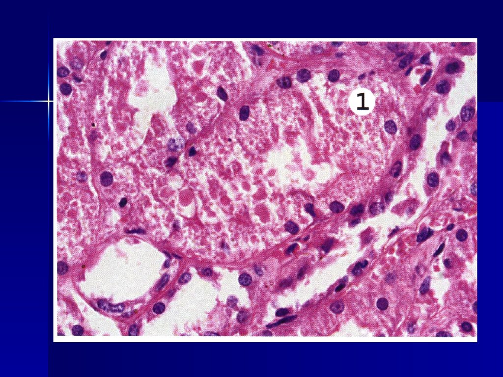
In hepatocytes, eosinophilicinclusions are found in acute
alcoholic hepatitis. These
inclusions are called – alcoholic
hyaline, and hepatocytes
containing it – Mallory's taurus.
32.
Eosinophilic inclusions alsoaccumulate in plasma cells, and
then these cells are called Roussel's
taurus (for rhinoscleroma). As a
result of hyaline droplet dystrophy,
prolonged exposure to damaging
factors may develop coagulation
necrosis.
33.
The second type of proteindystrophy – hydropic dystrophy - it
can also develop in the epithelium
of the renal tubules (with nephrotic
syndrome), in hepatocytes (with
viral B-hepatitis), in nerve and
ganglion cells (with hypoxia and
viral lesions), in the epidermis
(with eczema, with herpes, with
smallpox).
34.
Cells with hydropic dystrophy areswollen, vacuoles are detected in
the cytoplasm of cells (vacuole
dystrophy) or complete filling of
the cytoplasm with water (balloon
dystrophy). The cell nuclei are
displaced by vacuoles to the
basement membrane and are pale
in color.
35.
36.
Hydropic dystrophy occurs 1) due todamage to the membrane-enzyme
systems (in the kidneys) or
2) as a result of perversion of the
protein-synthetic function of cells (viral
hepatitis, herpes).
Hydropic dystrophy can be reversible
at the stage of vacuolization. If balloon
dystrophy develops, the cell dies (focal
or total colliquation necrosis).
37.
With violations of theexchange of nucleoproteins
(purine metabolism) in
patients, an increased
content of uric acid salts in
the blood is found
(hyperuricemia) and urine
(hyperuricuria).
38.
This leads to the depositionof urate microcrystals in the
joints (in synovia, cartilage,
tendons, joint bags), in the
kidneys (in the interstitium,
epithelium of the tubules, in
the lumen of the collecting
tubules and pelvis), in the
ureters and bladder.
39.
The cause may be agenetically determined
violation of the activity of
enzymes involved in uric
acid metabolism.
40.
Contributing factors:excessive consumption of
meat, legumes, and other
foods rich in purine bases.
Gout affects almost
exclusively men aged 35-50
years.
41.
The most vivid goutsyndrome - acute
arthritis, which is chronic,
undulating, with longterm remissions and
exacerbations.
42.
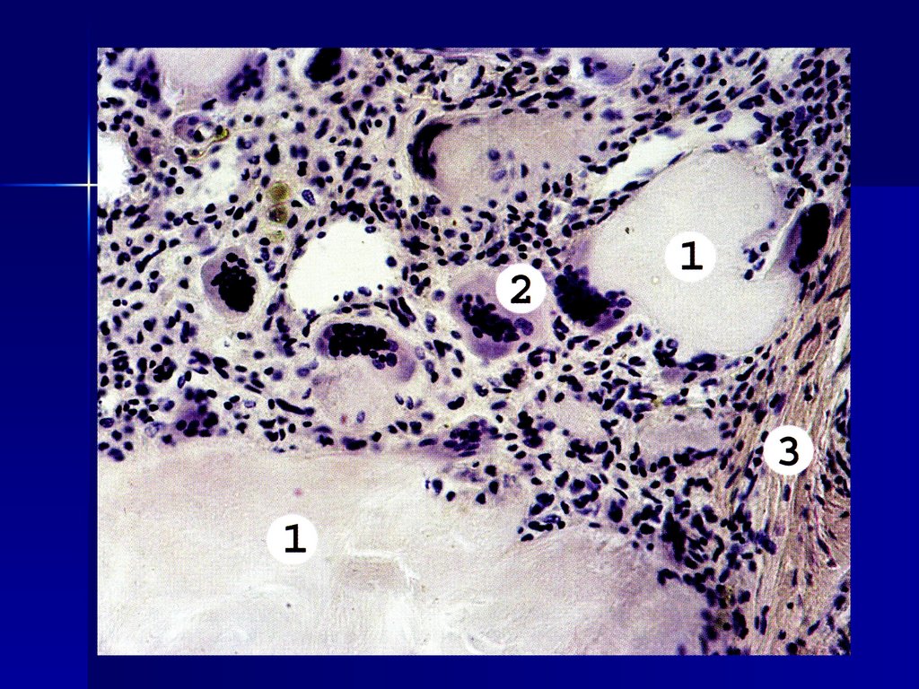
Pat. anatomiya. In the areaof salt deposition, tissue
necrosis occurs and
inflammatory lymphoplasmocytic and
macrophage-histiocytic
infiltration develops with a
large number of giant
foreign body cells.
43.
44.
As salt deposits andovergrowths increase
around the connective tissue
infiltrate, they form gouty
bumps-tophusy, the joints
are deformed.
45.
The deposition of salts in thekidneys leads to the
development of abacterial
pyelonephritis and atrophy
with an outcome in
nephrosclerosis (gouty
shriveled kidney). The
formation of urate stones in
the kidneys is possible.
46.
Carbohydrate dystrophy.Disorders of glucose or glycogen
metabolism lead to intracellular
glycogen accumulations. In
diabetes mellitus, glycogen is found
in the epithelial cells of the kidneys,
as well as in liver cells, B-cells of
the insular apparatus of the
pancreas, etc.
47.
Glycogen also accumulates inthe cells of the liver, kidneys,
gastrointestinal tract, muscles,
and red blood cells during
glycogenosis (storage diseases,
thesaurismoses).