
")


 medicine
Similar presentations:


Introduction in topographic anatomy and operative surgery
1. Introduction in topographic anatomy and operative surgery
Associate-professor Slabyy O.B.2.
Topographical anatomy is a scienceabout the dimensional structure of
healthy human body organs, tissues
and parts of the body
3.
The operative surgery is a scienceabout surgical operations, methods of
surgical operations, the essence of
which comes to mechanical action
upon the organs and tissues with
diagnostic, medical or reconstructive
purpose.
4. M.I. Pyrogov ( 1810-1881)
5. Classification of operations
EmergencyUrgent
Planned
Bloodless
Bloody
Radical
Palliative
Single stage
Stage operations
6.
Operative approach means to makethe wound for the exposure of the
organ to be operated on
7.
Operative method – the main part ofthe operation, performing the action
contained in the name of the
operation
8. General surgical instruments
Scalpels9. Positions of scalpels, forceps
а —scalpels; 1 — position of bow; 2 — position of table knife; 3 —writing pen;4 — amputating knife; б — forceps
10. The scissors
11. The surgical saw
12. Forceps
13. Retractors
14. Instruments for the arrest bleeding
15. Needles
16. Suture material
Absorbable- Plain catgut
- Chromic catgut
- Polyglycolic synthetics
-
-
-
Nonabsorbable
Natural (silk, cotton)
Synthetic braids (Ticron,
Tevdek, Ethibond)
Synthetic monofilament
( nylon, Prolen)
Monofilament stainless
Steel wire
17. Type of sutures
InterruptedContinuous
18. Regions of the Head and Neck
19.
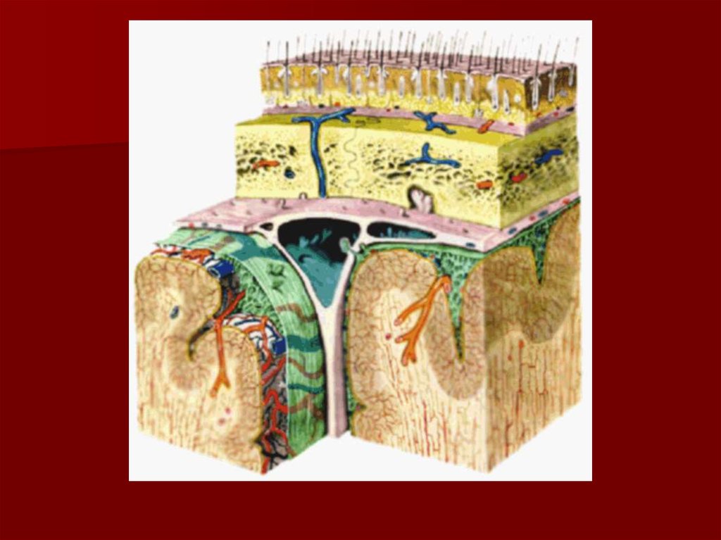
20. Layer Structure of Fronto-parieto-occipital Region
Layer Structure of Fronto-parietooccipital Region1.
2.
3.
4.
5.
6.
7.
Skin;
subcutaneous tissues;
gala aponeurotica;
loose areolar tissue;
periosteum (pericranium);
loose areolar tissue;
bone (internal, external lamina and
diploe).
21.
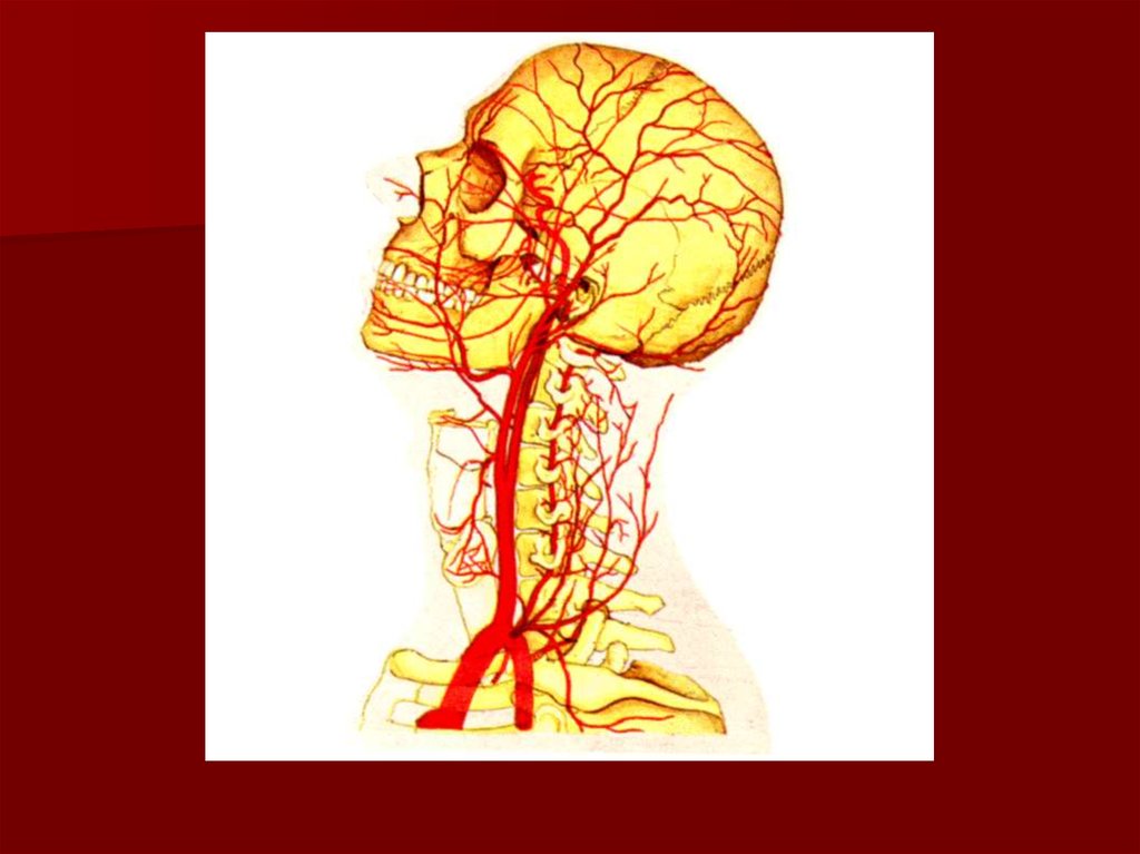
22. Arterial and nerve supply of the Scalp
The supratrochlear and the supraorbital arteriesin company with supratrochlear and the
supraorbital nerves.
The superficial temporal
artery,zygomaticotemporal and auriculotemporal
nerve.
The posterior auricular artery and lesser occipital
nerve (cervical plexus C2)
The occiptal artery and greater occipital nerve
(posterior ramus of the second cervical nerve).
23.
24. The venous drainage of the Scalp
The supratrochlear and supraorbital veins (to from thefacial vein).
The superficial temporal vein (to from the
retromandibular vein).
The postrior auricular vein (to from the external jugular
vein).
The occipital vein (into the suboccipital venous plexus, in
turn into the vertebral veins, occasionally forward into
the internal jugular vein.
The veins of the Scalp freely anastomose with another
and are connected to the diploic veins and the
intracranial venous sinuses by the valveless emissary
veins.
25.
26. Temporal region and parotid regions
27. Layer Structure of Temporal Region
Skin;2.
subcutaneous tissues;
3.
temporal aponeurosis:
- external lamina;
- loose areolar tissue;
- internal lamina;
4. subaponeurotical fat;
5. temporal muscle;
6. submuscular loose areolar tissue;
7. pericranium;
8. temporal bone.
1.