


 medicine
Similar presentations:


Asepsis Department of General Surgery
1. Asepsis Department of General Surgery
Department of General SurgeryMade by students from 309 group
Supervisor: Mikhail Aleksandrovich Kozhevnikov
Irkutsk 2018
2.
The measures to prevent an infection from enteringa wound are referred to as asepsis, while those to
cause the exclusion or destruction of harmful microbes
are generally called antisepsis.
3. History:
HISTORY:The modern concept of asepsis evolved in the 19th century.
Ignaz Semmelweis (washing hands)
Joseph Lister (use of carbolic acid as an antiseptic)
Lawson Tait (went from antisepsis to asepsis)
Ernst von Bergmann (autoclave)
4. source of infection
SOURCE OF INFECTIONSource
Modes of
transmission
EXOGENOUS
ENDOGENOUS
patients with purulent inflammation
chronic infections outside the
area of the operation
carriers of the microbes
chronic infections inside the
area of the operation
animals
oral, intestinal and
respiratory saprophytes
airborne
direct contact
direct contact
haematogenous spread.
implantation
lymphogenous spread.
5. surgical hospital’s structure
SURGICAL HOSPITAL’S STRUCTUREA surgical hospital contains the main functional
blocks which are as follows:
a surgical block
surgery departments
plaster
treatment rooms
dressing-rooms.
An operating unit houses special rooms for
operating on patients. It has to be isolated from
surgery departments on a separate floor or
detachment of the building and be connected with
the them by a corridor.
6. functional zones in the surgical block
FUNCTIONAL ZONES IN THE SURGICAL BLOCKThe sterile
zone (the
operating
theatre,
scrub-up
room, and
the room
for
sterilisatio
n.)
The clean
zone (the
rooms for
personal
hygiene and
changing
clothes of the
staff)
The technical
zone (the
rooms where
apparatus for
airconditioning
or oxygen
supplying and
vacuum
devices are
stored.)
The dirty
zone (the
sister's room,
the room of
the head of
surgery and
the one for
dirty clothes
etc.)
7.
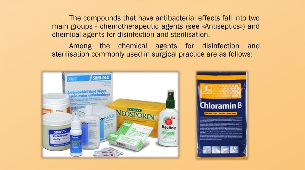
The compounds that have antibacterial effects fall into twomain groups - chemotherapeutic agents (see «Antiseptics») and
chemical agents for disinfection and sterilisation.
Among the chemical agents for disinfection and
sterilisation commonly used in surgical practice are as follows:
8.
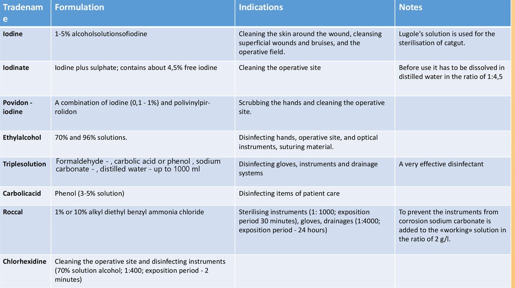
Tradename
Formulation
Indications
Notes
Iodine
1-5% alcoholsolutionsofiodine
Cleaning the skin around the wound, cleansing
superficial wounds and bruises, and the
operative field.
Lugole's solution is used for the
sterilisation of catgut.
Iodinate
Iodine plus sulphate; contains about 4,5% free iodine
Cleaning the operative site
Before use it has to be dissolved in
distilled water in the ratio of 1:4,5
Povidon iodine
A combination of iodine (0,1 - 1%) and polivinylpirrolidon
Scrubbing the hands and cleaning the operative
site.
Ethylalcohol
70% and 96% solutions.
Disinfecting hands, operative site, and optical
instruments, suturing material.
Triplesolution
Formaldehyde - , carbolic acid or phenol , sodium
carbonate - , distilled water - up to 1000 ml
Carbolicacid
Phenol (3-5% solution)
Disinfecting items of patient care
Roccal
1% or 10% alkyl diethyl benzyl ammonia chloride
Sterilising instruments (1: 1000; exposition
period 30 minutes), gloves, drainages (1:4000;
exposition period - 24 hours)
Chlorhexidine
Cleaning the operative site and disinfecting instruments
(70% solution alcohol; 1:400; exposition period - 2
minutes)
Disinfecting gloves, instruments and drainage
systems
A very effective disinfectant
To prevent the instruments from
corrosion sodium carbonate is
added to the «working» solution in
the ratio of 2 g/l.
9. Prevention of microorganisms' contact with the wound
PREVENTION OF MICROORGANISMS' CONTACTWITH THE WOUND
Sterilising instruments, operating sheets, towels and
dressing materials involves the following stages:
preparation of the materials,
preparing for sterilisation itself,
sterilisation,
safe-keeping of the materials sterilised.
All these stages are to be performed in accordance
with specific standards «Sterilisation and disinfection of
materials for medical use».
10. Sterilisation of instruments
STERILISATION OF INSTRUMENTSStage 1 - preparation of the materials - is aimed at thorough
mechanical cleansing of instruments; removal of pyogenic compounds and
destruction of hepatitis viruses. The instruments that were used but not
infected will be washed under running water separately with a brush for 5
minutes. In contrast, blood-stained equipment must be washed immediately
(without subsequent drying!), then soaked in one of special washing
solutions, warmed to a temperature of 50 °C for 15-20 minutes, syringes
being dismantled before washing.
The formulations of the washing solutions are as follow
• Solution A (Perhydrol - 20 g washing detergent - 5 water - 975 ml. )
• Solution B (2,5% hydrogen peroxide - 200 ml washing detergent - 5
water - 795 ml. )
The instruments contaminated with pus or intestinal contents are first
soaked in enamel containers with 5% lysol for 30 minutes, then washed in
the same solution with brush, rinsed with running water and soaked in one
of the washing solutions.
.
11.
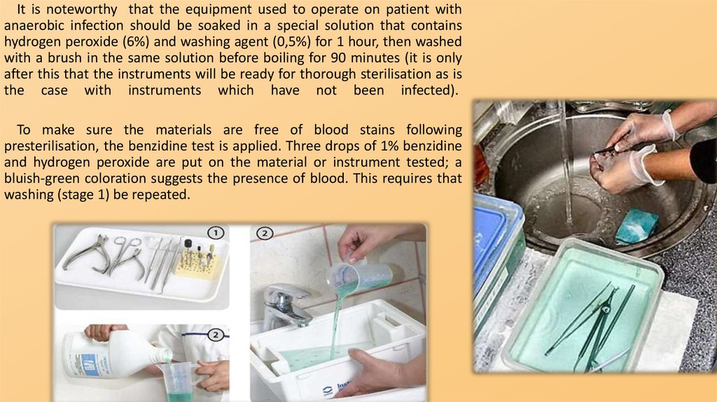
It is noteworthy that the equipment used to operate on patient withanaerobic infection should be soaked in a special solution that contains
hydrogen peroxide (6%) and washing agent (0,5%) for 1 hour, then washed
with a brush in the same solution before boiling for 90 minutes (it is only
after this that the instruments will be ready for thorough sterilisation as is
the case with instruments which have not been infected).
To make sure the materials are free of blood stains following
presterilisation, the benzidine test is applied. Three drops of 1% benzidine
and hydrogen peroxide are put on the material or instrument tested; a
bluish-green coloration suggests the presence of blood. This requires that
washing (stage 1) be repeated.
12.
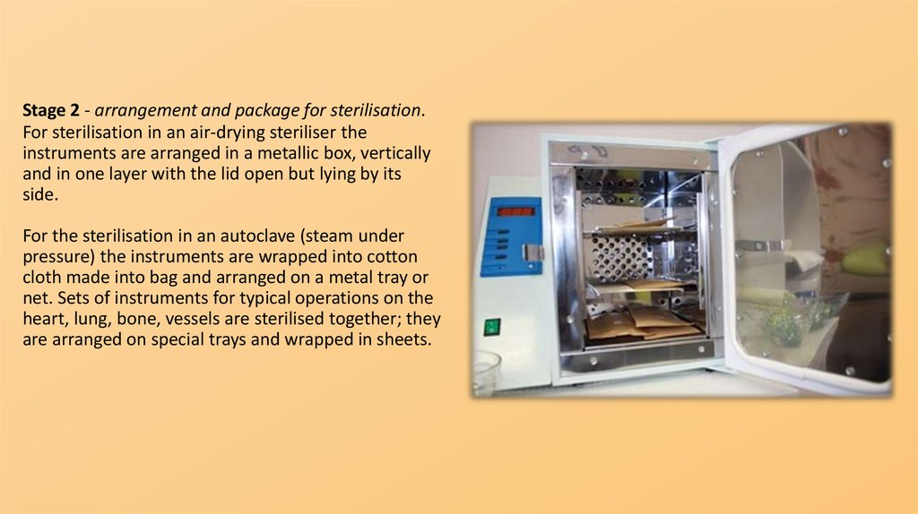
Stage 2 - arrangement and package for sterilisation.For sterilisation in an air-drying steriliser the
instruments are arranged in a metallic box, vertically
and in one layer with the lid open but lying by its
side.
For the sterilisation in an autoclave (steam under
pressure) the instruments are wrapped into cotton
cloth made into bag and arranged on a metal tray or
net. Sets of instruments for typical operations on the
heart, lung, bone, vessels are sterilised together; they
are arranged on special trays and wrapped in sheets.
13.
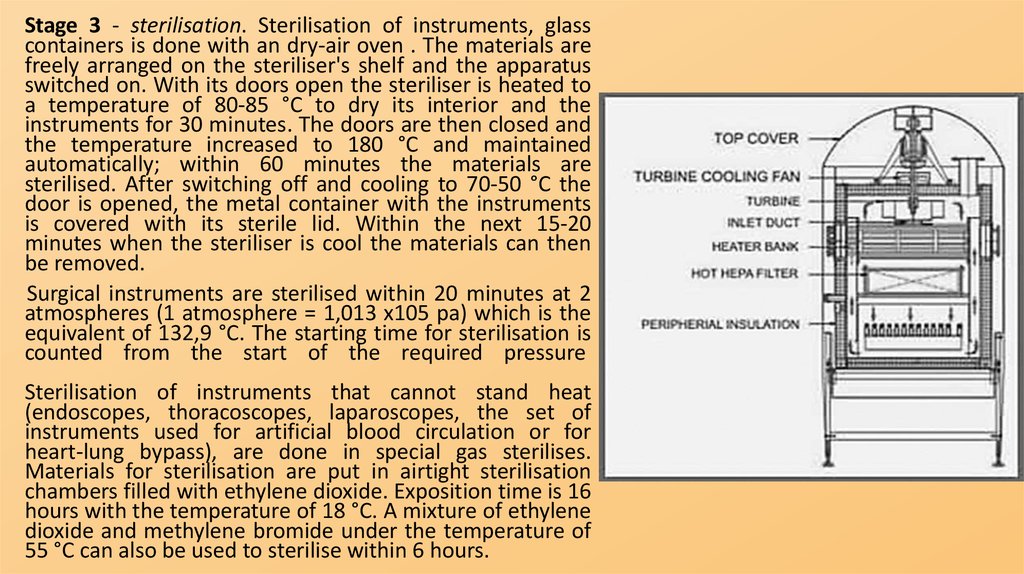
Stage 3 - sterilisation. Sterilisation of instruments, glasscontainers is done with an dry-air oven . The materials are
freely arranged on the steriliser's shelf and the apparatus
switched on. With its doors open the steriliser is heated to
a temperature of 80-85 °C to dry its interior and the
instruments for 30 minutes. The doors are then closed and
the temperature increased to 180 °C and maintained
automatically; within 60 minutes the materials are
sterilised. After switching off and cooling to 70-50 °C the
door is opened, the metal container with the instruments
is covered with its sterile lid. Within the next 15-20
minutes when the steriliser is cool the materials can then
be removed.
Surgical instruments are sterilised within 20 minutes at 2
atmospheres (1 atmosphere = 1,013 x105 pa) which is the
equivalent of 132,9 °C. The starting time for sterilisation is
counted from the start of the required pressure
Sterilisation of instruments that cannot stand heat
(endoscopes, thoracoscopes, laparoscopes, the set of
instruments used for artificial blood circulation or for
heart-lung bypass), are done in special gas sterilises.
Materials for sterilisation are put in airtight sterilisation
chambers filled with ethylene dioxide. Exposition time is 16
hours with the temperature of 18 °C. A mixture of ethylene
dioxide and methylene bromide under the temperature of
55 °C can also be used to sterilise within 6 hours.
14.
Stage 4 - Keeping the sterilised materials. Sterile materials are kept in special containers. Sterile and nonsterile items may not be kept at the same container. Materials can stay sterile in a dressing box, which hasnot yet been opened for 48 hours. If before packing in the dressing box the materials were wrapped in
(towels, sheets or napkins) as is the case with rubber drains), then they can stay sterile for 3 days. In cases
of centralised sterilisation syringes can be sterile for 25 days.
15. Sterilisation of dressing materials, operating sheets and suturing materials
STERILISATION OF DRESSING MATERIALS,OPERATING SHEETS AND SUTURING MATERIALS
Stage 1 - presterilisation. Dressing materials include gauze balls, towels, pack, and swabs. They
have to have the following characteristics:
1) they should be biologically and chemically inert and void of any negative effects on wound
healing;
2) they should have good hygroscopic, or water absorbing, properties;
3) they should have a few free threads from outside; this will prevent pieces of thread from falling
into the wound as these can act as foreign bodies in the wound;
4) they should be soft, elastic and not traumatise the wound;
5) they should be easy to sterilise without loosing its qualities;
6) they should be cheap, considering its wide use. Annually, 200 metres of gauze and 225 pieces
of bandage are normally spent per a surgical bed.
16.
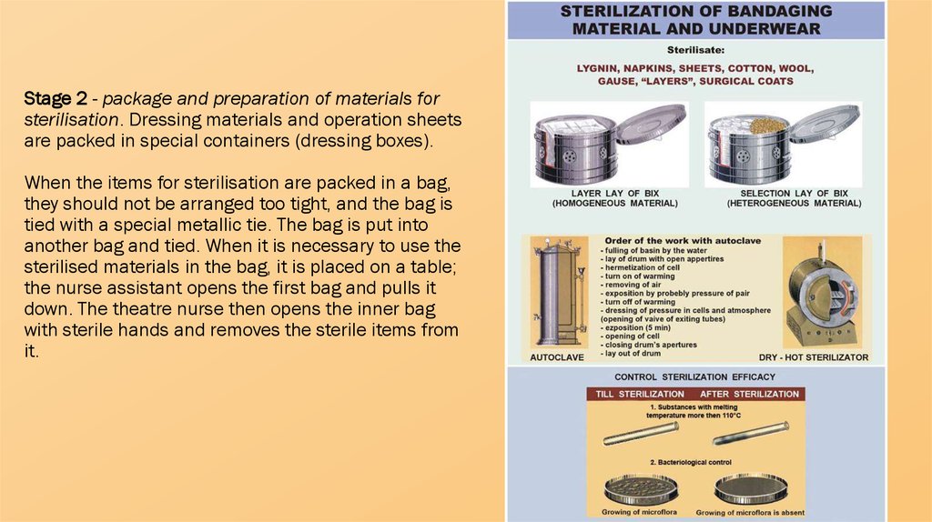
Stage 2 - package and preparation of materials forsterilisation. Dressing materials and operation sheets
are packed in special containers (dressing boxes).
When the items for sterilisation are packed in a bag,
they should not be arranged too tight, and the bag is
tied with a special metallic tie. The bag is put into
another bag and tied. When it is necessary to use the
sterilised materials in the bag, it is placed on a table;
the nurse assistant opens the first bag and pulls it
down. The theatre nurse then opens the inner bag
with sterile hands and removes the sterile items from
it.
17.
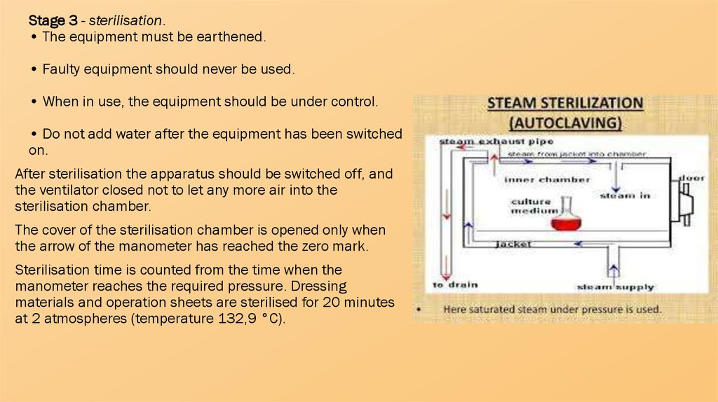
Stage 3 - sterilisation.• The equipment must be earthened.
• Faulty equipment should never be used.
• When in use, the equipment should be under control.
• Do not add water after the equipment has been switched
on.
After sterilisation the apparatus should be switched off, and
the ventilator closed not to let any more air into the
sterilisation chamber.
The cover of the sterilisation chamber is opened only when
the arrow of the manometer has reached the zero mark.
Sterilisation time is counted from the time when the
manometer reaches the required pressure. Dressing
materials and operation sheets are sterilised for 20 minutes
at 2 atmospheres (temperature 132,9 °C).
18.
Stage 4 - keeping the sterilised materials. Aftersterilisation ends the sterilisation chamber is
emptied, dressing boxes are removed, all openings
are immediately closed and brought to a special table
for sterile materials. Dressing boxes are kept locked
in a special room. With an intact dressing box
dressing materials and sheets can stay sterile for 48
hours after sterilisation has completed. Dressing
materials and sheets sterilised in the bag can stay
sterile for only 24 hours.
19. Control of sterility
CONTROL OF STERILITYDirect methods
• Inoculation of medium with a swab of the dressing material.
To inoculate medium with a swab, open the dressing box in the operating theatre, using a sterile
instrument. Soak a piece of sterile gauze in normal saline which is passed several times on the material to
be tested, then drop the piece of gauze into a sterile test tube and send it to the microbiological laboratory
Bacteriological tests.
A test tube that contains reference non-pathogenic cultured microorganisms known to die, if exposed to a
certain temperature, is used. Place the test tube inside the dressing box and send it to the laboratory after
sterilisation is over. Absence of bacterial growth implies that the items are sterile.
The swabs should be taken from once every 10 days.
Indirect methods
Control of sterility of materials is done each time they have been sterilised. Compounds with known specific
melting points are used for this purpose: benzoic acid (120 °C), resorcinol (119 °C), antipyrin (110 °C). These
compounds are kept in ampoules. One or two ampoules are placed in between the layers of materials to be
sterilised. Melting of the powdered compound into a liquid mass implies that the temperature in the box was
at least as high as the melting point of the compound. Thermometry is the most objective indirect methods of
sterility control. In each dressing box 1 or 2 thermometers are placed in between the layers of materials to be
sterilised. The readings will indicate the maximum temperatures but not the exposition time.
20. Suturing material
SUTURING MATERIALNatural resolvable threads are made of catgut. To lengthen the resolution time of catgut,
metallic compounds are impregnated into them (chromic and silver catguts). The examples of
synthetic resolvablesutures are dexon, vicryl and oxylon.
Non-resolvable natural sutures include sutures made of natural silk, cotton, yarn; their
synthetic equivalents are dacron, nylon, ftolon, silk, kapron, etc. Suturing material should meet
the main requirements as follows:
• have smooth level surface without causing additional damage to the tissues;
• have good manipulating qualities - slip easily through tissues;
• be elastic (sufficient elasticity prevents tissues from being pressed on and necrotized when
they subsequently become oedematous);
• be firm at the knots;
• be non-hygroscopic and not swell up;
• be biologically compatible with bodily tissues and not be allergic to the body.
21.
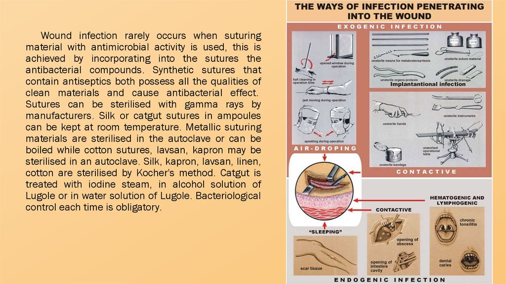
Wound infection rarely occurs when suturingmaterial with antimicrobial activity is used, this is
achieved by incorporating into the sutures the
antibacterial compounds. Synthetic sutures that
contain antiseptics both possess all the qualities of
clean materials and cause antibacterial effect.
Sutures can be sterilised with gamma rays by
manufacturers. Silk or catgut sutures in ampoules
can be kept at room temperature. Metallic suturing
materials are sterilised in the autoclave or can be
boiled while cotton sutures, lavsan, kapron may be
sterilised in an autoclave. Silk, kapron, lavsan, linen,
cotton are sterilised by Kocher's method. Catgut is
treated with iodine steam, in alcohol solution of
Lugole or in water solution of Lugole. Bacteriological
control each time is obligatory.
22. Preparation of the hands for operation
PREPARATION OF THE HANDS FOR OPERATIONScrubbing of the hands is a very important way of preventing
infection.The nails should always be trimmed and short. Whenever
very dirty work is to be done manually, gloves should be it is better
to worn. Taking good care of the hands should be regarded as a
step in the preparation for operation.
• Chlorhexidine (0,5% alcohol solution)
The hands (the finger up to the midforearm) are smeared with
gauze swabs soaked in the solution of chlorhexidine for about three
minutes; prior to this the hands are washed with soap for a minute.
• AHD solution and Eurosept
These solutions contain the antiseptics such as ethanol,
chlorhexidine, and polyiolic fatty acid ether. The hands are first
washed with soap and running water for a minute.
A few millilitres of the solution are then poured onto the hands twice
and rubbed for 2-3 minutes each.
The hands can also be cleansed by rubbing the hands with 96%
ethyl alcohol for 10 minutes (Brun's method).
23. Cleaning the operative field
CLEANING THE OPERATIVE FIELDPreparation of the place of the expected incision (operative
field or site) starts on the day preceding the operation, which
includes hygienic baths and a change of underwear. On the
day of operation, the skin of the expected place of incision is
dry-shaved and cleaned with alcohol.
Immediately before the operation, on the operating table, the
operative field is abundantly smeared with 5% alcohol
solution of iodine. The operation site itself is isolated with
sterile towels and again smeared with 5% alcohol solution of
iodine. Before suturing, the skin is smeared with 5% alcohol
solution of iodine and repeated after the suturing. This is
known as Grossich-Filonchikov's method.
In a patient allergic to iodine the skin can be prepared with
brilliant green (Bakkal's method). On the operating table, the
operation site can be can be prepared with derivatives of
iodine such as iodonate, povidon-iodine, betadin.
24. List of used literature
LIST OF USED LITERATURE• V. K. Gostishcev General Surgery 2002