
 biology
Similar presentations:


 - one of the classes of arthropods")


Tsetse flies
1.
TSETSE FLIES[GLOSSINA]
z
NAME : JYOTI PRIYA SINGH
VIDUSHI VERMA
GROUP NO : 192B
2.
z3.
EXTERNAL ANATOMY OF GLOSSINAz
The word anatomy means the structure of the body, in this case, of the tsetse
fly.
CUTICLE
Like all other insects, the tsetse fly has a tough outer covering or cuticle. The
whole of the body is covered with cuticle, even the eyes. Mast parts are hard,
but some areas remain flexible, especially the base of the wing, the joints on
things and where the mouth parts join on to the head; these parts can therefore
be moved easily. The cuticle on the underside (ventral side) of the abdomen in
the tsetse fly is elastic, so that it can stretch when the abdomen takes up the
large blood meal
Movements of the legs are controlled by muscles attached to the inside of the
cuticle of the legs; rapid movement of the wings for flying is controlled by very
large muscles in the thorax.
4.
zEXTERNAL APPEARANCE
The tsetse flies are nearly always some shade of brown or grey-brown; sometimes
there is a slight pink or sandy-red tinge. Several species are very dark. The body
usually has darker and lighter patches, making the insect difficult to see when it is
settled on bark, rock or soil. At rest, the tsetse normally appears quite slim
because the wings are placed one over the other on the back (Figure 1.3), not
projecting outwards at an angle to the body as in house flies or most blowflies.
Immediately after a blood meal the tsetse abdomen is large, rounded and red.
The body is made up of three main parts: the head, the thorax (to which are
attached the wings and legs) and the abdomen. These parts will now be described
in greater detail
5.
Compoundz eyes On the head is a pair of large compound eyes. Each of these eyes
is composed of thousands of small units, called ommatidia (singular: ommatidium).
The part of the ommatidium that forms the surface of the eye is the lens. The lenses
near the midline of the head are slightly larger than those at the sides of the head.
The compound eyes of some species are said to be able to detect moving objects
at 137 metres (150 yd). They are very good for nearer vision and a small movement
near the insect can make it fly off. The compound eyes are dark brown in the living
fly.
Simple eyes At the top of the head are three simple
eyes or ocelli (singular: ocellus); these are also sensitive to light, but their exact
function is un certain.
Antennae There are two antennae placed at the front of the head in a depression
between the two compound eyes. Each antenna has three segments of which the
third is the largest, and bears the arista.
6.
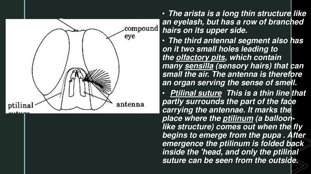
z• The arista is a long thin structure like
an eyelash, but has a row of branched
hairs on its upper side.
• The third antennal segment also has
on it two small holes leading to
the olfactory pits, which contain
many sensilla (sensory hairs) that can
small the air. The antenna is therefore
an organ serving the sense of smell.
• Ptilinal suture This is a thin line that
partly surrounds the part of the face
carrying the antennae. It marks the
place where the ptilinum (a balloonlike structure) comes out when the fly
begins to emerge from the pupa . After
emergence the ptilinum is folded back
inside the 'head, and only the ptilinal
suture can be seen from the outside.
7.
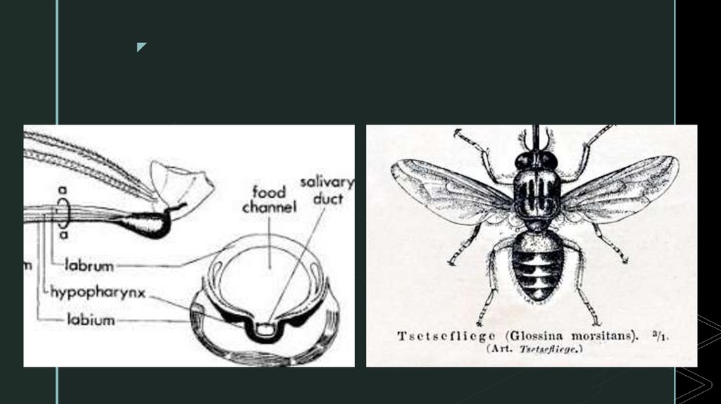
MouthpartsThe mouthparts are very important to the life of the fly. They are long
z
and narrow and can pierce the skin of an animal, so that blood can be sucked up
into the fly; at the same time saliva can be passed down the mouthparts into the
animal being fed on.
When the fly is not feeding, all the mouthparts are held so that they point
forwards from beneath the head.
A pair of maxillary palps help to protect the more delicate proboscis or
haustellum which lies between them when it is not in use.
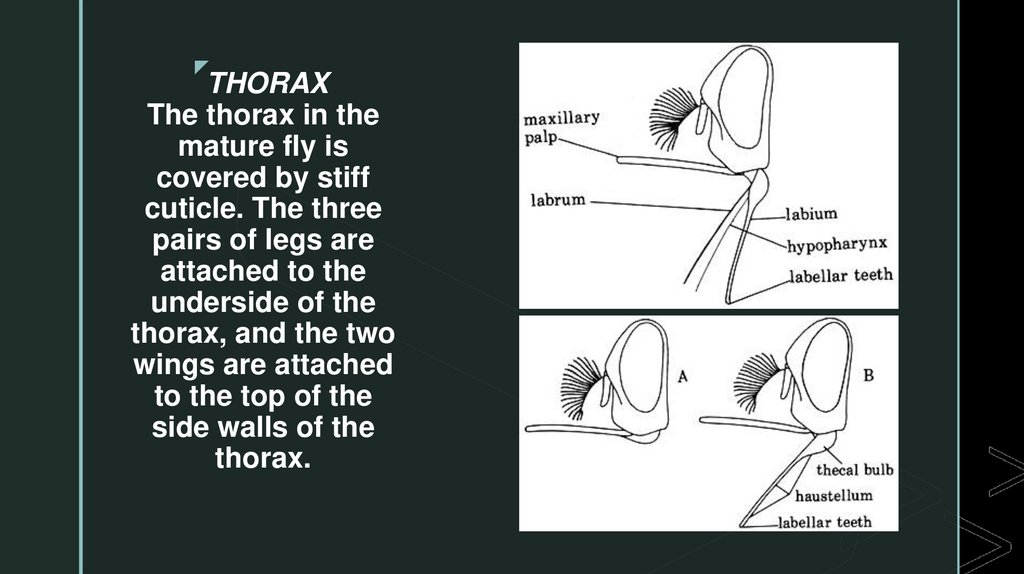
The proboscis is very narrow (Figure 1.6) • It is made up of three parts,
the labium, the labrum and the hypopharynx
8.
zLabium The labium is the thickest of these very thin structures. At the free end
it has a large number of very small teeth (labellar teeth). The teeth can cut their
way through the skin of an animal, so that the fly may suck blood. The other
end of the labium, where it is attached to the head of the fly, is swollen. This
part (the thecal bulb) contains the muscles that cause the teeth to move.
Labrum The labrum forms a tube through which blood is sucked up from the
animal being bitten. The tube is called the food canal.
Hypopharynx The hypopharynx is an extremely narrow tube through which
saliva is pumped into the host animal as the fly feeds
9.
zTHORAX
The thorax in the
mature fly is
covered by stiff
cuticle. The three
pairs of legs are
attached to the
underside of the
thorax, and the two
wings are attached
to the top of the
side walls of the
thorax.
10.
zABDOMEN
In the resting fly, the abdomen is covered over by the wings. It has seven visible
segments, or parts, and in the male there is, in addition, an extra structure
(hypopygium) folded beneath the last two segments . On the dorsal (upper) side of the
abdomen there are strong plates (each plate is called a tergite), one for each segment;
but the ventral side is made of highly elastic cuticle, which can stretch to allow the
abdomen to carry the enormous blood meal, and in the case of the female, the large
larva. Remains of the blood meal can often be seen within the abdomen if examined
from the ventral surface.
There are seven pairs of spiracles (breathing holes) along the sides of the abdomen.
The anus is at the posterior end of the abdomen.
The male genitalia The word genitalia means parts used for mating. When the male
tsetse fly is looked at from the ventral side, a rounded structure can be seen at the
posterior end of the abdomen. This is the hypopygium. Just in front of the
hypopygium is a plate bearing dark hairs called hectors
11.
DIGESTIVE SYSTEMz
Salivary glands and saliva The tsetse fly has two salivary glands.
The main part of each gland lies in the anterior part of the abdomen
and it sends forward to the head a very narrow tube that joins with
the one from the other side, before entering the hypopharynx .
When the fly feeds, saliva is sent forward from the glands and so
down the length of the hypopharynx. It mixes with the blood meal as
this is sucked up from the host's body. Saliva contains
an anticoagulant, a substance that helps to prevent the blood meal
from clotting in the mouthparts and anterior part of the alimentary
canal.
12.
Labellarzteeth
There are hundreds of very small sharp labellar teeth at the end of the labium. The
teeth help to cut into the skin of the host. As the proboscis moves through the
skin, the teeth cut the walls of small blood vessels (capillaries) and release the
blood from them, forming a pool of blood under the skin (pool feeding).
Pharynx
The released blood is drawn up the food canal by the action of the muscles of
the pharynx. When the fly is feeding, strong muscles in the head contract to make
the space inside the pharynx larger, and this pulls blood up into
the pharynx. When the muscles relax, the pharynx returns to its usual size, and
the blood meal is sent on to the next part of the alimentary canal, the oesophagus.
13.
NERVOUS SYSTEMz
The senses and the behaviour of the tsetse fly are coordinated by the nervous
system. The fly is able to see, small and feel with the aid of its sense organs.
These send messages along nerves to the larger masses of nervous tissue in the
head ('brain') and thorax (ganglion), which coordinate the sensory messages
coming in, and send out other messages along other nerves to the muscles of the
body, so that the fly moves (behaves) in an appropriate manner.
ENDOCRINE SYSTEM
Another messag sending system is the endocrine system. There are small glands
in different parts of the body which release chemical substances (hormones)into
the haemolymph, causing an appropriate reaction elsewhere in the body.
Processes such as pupation are under the control of the endocrine system.
14.
REPRODUCTIVE SYSTEMz
Glossina reproduces by the female fly producing eggs which hatch into larvae inside the body;
these later emerge from the body fully grown. In order that the female fly may develop these
eggs into larvae, the male fly has to make sperm and transfer it to the female (see 3.1).
Male reproduction system The main parts of the male reproduction system are:
a pair of testes
a pair of accessory glands
a sperm pump
various ducts joining these other parts together.
The main function of the male reproduction system is to produce sperm and transfer these to the
female, in order to fertilise the eggs.
Testes A testis is a coiled tube in which sperm is made and stored. The main part of the testis
has a covering of orange or brown material, which makes it easy to see in dissections.
15.
zLARVAL STAGES
As with other flies, the larva in Glossinapasses through several stages or instars,
as it grows. There are three larval instars in Glossina up to the time when the fully
grown larva is dropped by the female fly: the first, second and third instars. The
larva has a mouth at the anterior end, and two posterior spiracles. The unusual
feature of the Glossina life history is that the larva spends practically all its time,
and does all its feeding, within the body of the female fly
16.
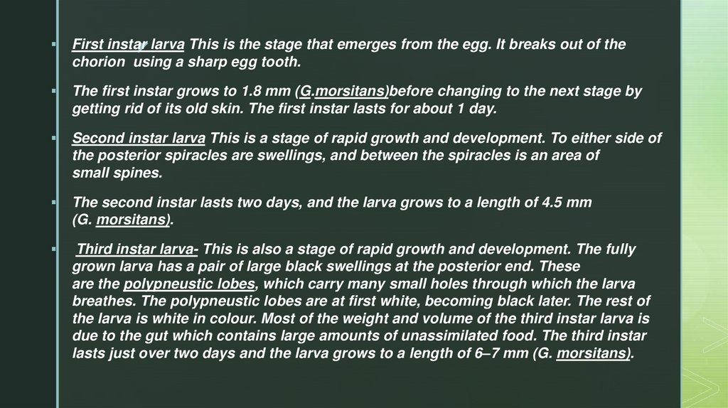
First instarz larva This is the stage that emerges from the egg. It breaks out of the
chorion using a sharp egg tooth.
The first instar grows to 1.8 mm (G.morsitans)before changing to the next stage by
getting rid of its old skin. The first instar lasts for about 1 day.
Second instar larva This is a stage of rapid growth and development. To either side of
the posterior spiracles are swellings, and between the spiracles is an area of
small spines.
The second instar lasts two days, and the larva grows to a length of 4.5 mm
(G. morsitans).
Third instar larva- This is also a stage of rapid growth and development. The fully
grown larva has a pair of large black swellings at the posterior end. These
are the polypneustic lobes, which carry many small holes through which the larva
breathes. The polypneustic lobes are at first white, becoming black later. The rest of
the larva is white in colour. Most of the weight and volume of the third instar larva is
due to the gut which contains large amounts of unassimilated food. The third instar
lasts just over two days and the larva grows to a length of 6–7 mm (G. morsitans).
17.
z18.
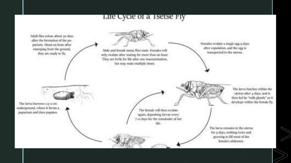
Female tsetsez mate just once. After 7 - 9 days she produces a single egg which develops
into a larva within her uterus. About nine days later, the mother produces a larva which
burrows into the ground where it pupates. The mother continues to produce a single larva at
roughly nine day intervals for her entire life.
The adult fly emerges from the pupa in the ground after about 30 days. Over a period of 1214 days it matures, mates and, if it is a female, deposits its first larva. Thus 50 days elapse
between the emergence of one female fly and the subsequent emergence of the first of its
progeny.
This life cycle, with a slow reproductive rate and substantial parental investment in the care
of young, is a relatively unusual example of an insect with a so-called 'K-type' life history.
This slow rate of reproduction means that tsetse populations can be eradicated by killing just
2-3% of the female population per day.
For a more detailed description of the life cycle and general biology of tsetse flies, see
Stephen Leak's excellent book