


 medicine
Similar presentations:


 isoimmunization")
Forensic medicine
1.
ZAPOROZHIAL STATE MEDICAL UNIVERSITYTHE DEPARTMENT OF PATHOLOGICAL ANATOMY and FORENSIC
MEDICINE
2.
MATERIAL EVIDENCEObjects which are served as crime
instruments or are saved on itself tracks of
crime or were the objects of criminal acts
of accused are material evidences.
They are explored by medico-legal experts,
forensic chemists and specialist in crime
detection.
3.
MATERIAL EVIDENCEAxe, rolling-pin, knives, screw-driver, scissors, iron, revolver
4.
MATERIAL EVIDENCEElectrical cable, rope, penknife, billiards ball with blood
stains, bullets
5.
MATERIAL EVIDENCECloth with tear and blood stains , coat with blood stains
6.
MATERIAL EVIDENCESketch of knife, fragment of skin with wounds, skull with the
fracture
7.
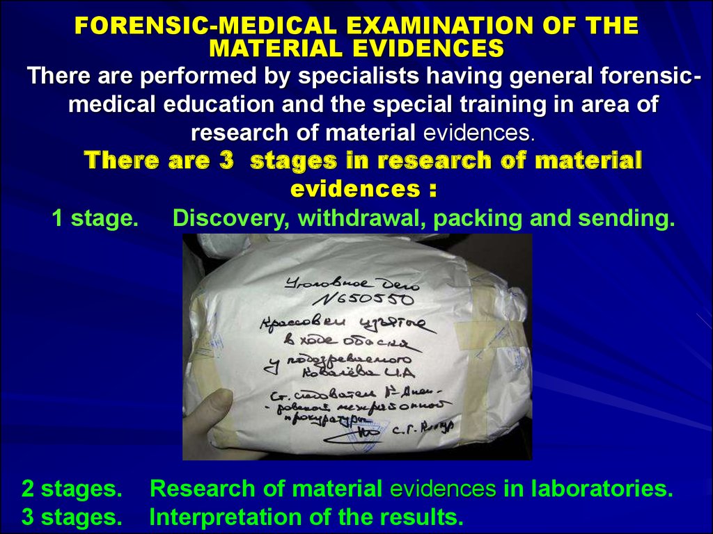
FORENSIC-MEDICAL EXAMINATION OF THEMATERIAL EVIDENCES
There are performed by specialists having general forensicmedical education and the special training in area of
research of material evidences.
There are 3 stages in research of material
evidences :
1 stage. Discovery, withdrawal, packing and sending.
2 stages.
3 stages.
Research of material evidences in laboratories.
Interpretation of the results.
8.
FORENSIC-MEDICAL EXAMINATION OF THE MATERIALEVIDENCES IN A FORENSIC-MEDICAL IMMUNOLOGICAL
LABORATORY
Forensic Immunological Examinations are carried out to
establish presence, kind and group-belonging of objects of
biological origin.
9.
FORENSIC-MEDICAL EXAMINATION OF THE MATERIALEVIDENCES IN A FORENSIC-MEDICAL CRIMINALISTICS
LABORATORY
Medico-Criminalistic Examinations are carried out to
establish the instruments of the trauma, their differentiation
and identification, personal identification, definition of the
nature and element structure of microobjects, traces,
reconstruction of the situation in which damages were
caused.
10.
FORENSIC-MEDICAL EXAMINATION OF BLOOD IN AFORENSIC-MEDICAL TOXICOLOGICAL LABORATORY
Forensic Toxicological Examinations are carried out to reveal
and determine chemical substances in objects of biological
origin and other proofs.
11.
The objects are taken by a medico-legal expert at autopsyand at examination of victim can be sent in a laboratory: pull
out hairs with bulbs, cut off nails
12.
The objects taken by a medico-legal expert at autopsy andat examination of victim can be sent in a laboratory: blood,
bile
13.
INVESTIGATION OF THE SCENE OF CRIME(BLLOD STAINS ON THE FLOOR AND WALL)
14.
BLOOD AS TRACE EVIDENCESometimes, something contaminated with other materials
come to tremendous help in medico-legal and other
forensic investigations.
For example, when a weapon is found stained with blood
of the victim of assault, then it becomes very much
reasonable to suspect that, that particular weapon might
have been used to injure the victim.
Blood of the victim on the weapon here acts as trace
evidence to link the weapon with the assault on the basis of
which further investigation proceeds.
Blood itself is a very important entity in medico-legal
practices, which alone or along with other trace evidence
play key role to unfold different criminal problems
15.
MEDICO-LEGAL QUESTIONS1. Whether the stain is due to blood or some
other material?
2. If it is due to blood, then whether it is of
human origin or it belongs to some other animal?
3. What is the source of the bleeding.
a) Is it from arterial or venous source?
b) Does it belong to the victim or the accused?
c) Is it from an injury or due to haemoptysis,
menstruation or miscarriage?
16.
MEDICO-LEGAL QUESTIONS4. What is the sex of the person?
5. What is the group of blood?
6. Is it blood of the adult person or
newborn child?
17.
All blood stains should be sent to the Forensicmedical immunology laboratory.The stained article is allowed to dry at room
temperature.
Extra heat should not be used as this will cause
deterioration of the stain.
If the stained clothes are not dried, putrefaction
sets in and it becomes difficult or impossible to
know whether the blood is of human or animal
origin.
18.
COLLECTION OF BLOOD STAINS1. A clean piece of white filter paper may be used, allowing
blood to soak into it, then drying it at room temperature.
2. If the object is porous, a portion of unstained area should
also be taken if this is practical.
3. If the object is non-porous and particularly if it is metallic,
stains can be removed by scraping and placed in small
glass containers.
4. Stains on clothing may be scraped off or a fragment of
the material cut.
19.
COLLECTION OF BLOOD STAINSTaking of blood on gauze, example of clean gauze
20.
EXAMINATION OF BLOOD STAINSSTAINS OF CLOTHING
In the case of clothing, type of garment, its
colour and consistence should be noted
and if the garment is torn, the position of
the tears should be noted.
Both the outer and inner surfaces of the
garments should be examined.
21.
EXAMINATION OF BLOOD STAINSSTAINS OF CLOTHING
The position of all stains should be given
correctly by a description of the stain in
its relation to the manner in which a
garment is usually worn, e.g., a stain on
the trousers should be described as being
above, behind, or to the outer side of the
knee.
Stains may also be described in relation to
the pockets, the buttons or the seams of a
garment.
22.
EXAMINATION OF BLOOD STAINSSTAINS OF CLOTHING
The size and the shape of the stain should
be noted.
If the stain is in the form of a smear, its
general direction should be noted.
Blood stains are extremely resistant to
washing by water.
Dried blood on a dead body or article is very
resistant for quite a long time even though
the body has been totally submerged.
23.
EXAMINATION OF BLOOD STAINSI. General examination:
• Scene of the crime.
• Part of the body from which stain is derived.
• Age of Blood Stains.
• Sex and Age of Person.
• Living or Dead Body.
• Source of Blood.
II. Chemical examination:
• Phenolphtalein Test
• Ortho-tolidine Test.
24.
EXAMINATION OF BLOOD STAINSIII. Microscopical and microchemical
examination:
• Red Corpuscles.
• Haemin Crystal Test (Teichmann's Test).
• Haemochromogen Crystal Test (Takayama
Test).
IV. Spectroscopic examination
V. Serological examination
25.
GENERAL EXAMINATION OF BLOOD STAINSScene of the crime:
1. When much blood is present it suggests serious injury
during life, but if a large vessel is cut, bleeding can occur
after death.
2. The collection of a pool of blood near the body during life
indicates that the deceased fell unconscious and
remained immobile after the injury.
3. A trail of blood stains will indicate that the victim was
wounded at some distance from the place at which the
body is found.
26.
GENERAL EXAMINATION OF BLOOD STAINSScene of the crime:
4. It can happen when the victim is attacked while running
or in case of suicide.
5. Blood coming from the arteries of a living person will be
scattered in fine spray over the surface upon which it has
fallen.
6. Venous bleeding is a slow steady flow, causing a pool if
the victim is at rest, and separate widely spaced drops, if
the victim walks about.
27.
GENERAL EXAMINATION OF BLOOD STAINSScene of the crime:
The direction of the fall of blood on to a surface may be
recognized:
- if it drops vertically on to flat surface, the stains are
circular.
- If the height does not exceed a few cm, the drop appears
as a round spot.
- If it has travelled thirty cm. or more, it shows prickly
edges, the projections growing finer and larger in number
with the increase in length.
28.
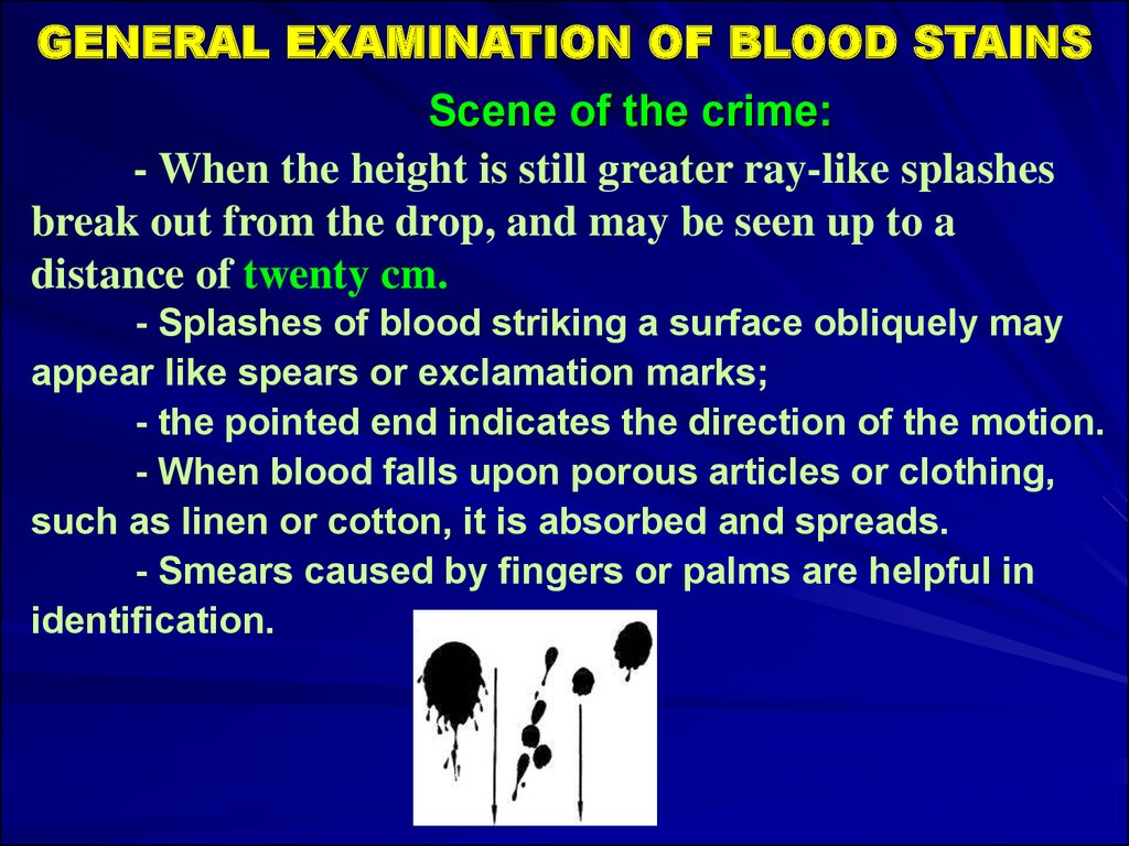
GENERAL EXAMINATION OF BLOOD STAINSScene of the crime:
- When the height is still greater ray-like splashes
break out from the drop, and may be seen up to a
distance of twenty cm.
- Splashes of blood striking a surface obliquely may
appear like spears or exclamation marks;
- the pointed end indicates the direction of the motion.
- When blood falls upon porous articles or clothing,
such as linen or cotton, it is absorbed and spreads.
- Smears caused by fingers or palms are helpful in
identification.
29.
GENERAL EXAMINATION OF BLOOD STAINSPart of the body from which stain is derived:
Menstrual blood is usually found on female
garments, diapers or pieces of cloth.
It is dark and fluid, has a disagreeable smell and
the reaction is acid.
On microscopic examination it shows endometrial
and vaginal epithelial cells and number of
microorganisms. It contains fibrinolysins.
If the blood is from the nose, mucus and hair from
the nose may be found.
30.
GENERAL EXAMINATION OF BLOOD STAINSPart of the body from which stain is derived:
- Vomited blood is of chocolate colour and acid in
reaction due to the action of gastric juice.
- Blood due to haemoptysis is bright-red and
frothy, with alkaline reaction.
- In blood due to rape, semen and pubic hair may
be found.
- Blood stains due to the boils and sores show a
smeared appearance without definite drops of
blood, and may contain pus cells and bacteria.
31.
MICROSCOPIC AND SPECTROSCOPICEXAMINATION OF BLOOD
MICROSCOPICAL EXAMINATION
Red Corpuscles: Intact red cells are seen only
when the stains are fresh, or when a fragment of
clot is available.
The red cells become unrecognizable when dried.
Red blood cells are circular, biconcave, nonnucleated discs in all mammals except camels.
In camels they are oval and biconvex but nonnucleated.
In birds, fishes, amphibian and reptiles they are
oval, biconvex and nucleated.
32.
MICROSCOPIC AND SPECTROSCOPICEXAMINATION OF BLOOD
SPECTROSCOPIC EXAMINATION
It is the most delicate and reliable test for
detecting the presence of blood in both recent and
old stains.
The blood stain is dissolved in water, normal
saline, or dilute ammonia, and is placed in a small
glass test tube, which is then put between the
spectroscope and the source of the light.
33.
MICROSCOPIC AND SPECTROSCOPICEXAMINATION OF BLOOD
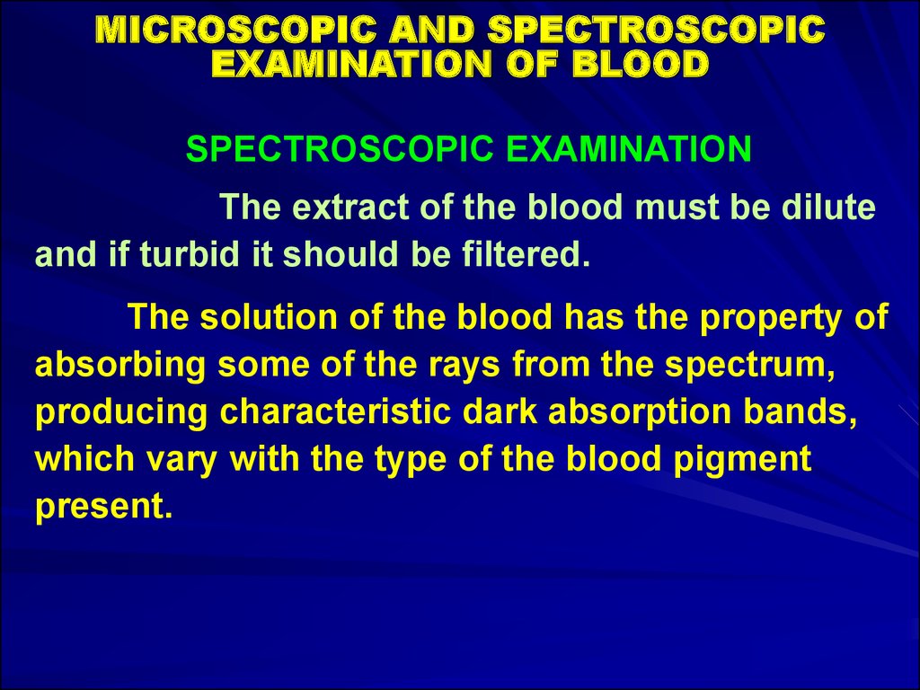
SPECTROSCOPIC EXAMINATION
The extract of the blood must be dilute
and if turbid it should be filtered.
The solution of the blood has the property of
absorbing some of the rays from the spectrum,
producing characteristic dark absorption bands,
which vary with the type of the blood pigment
present.
34.
SPECTROSCOPIC EXAMINATIONOxyhaemoglobin
Reduced haemoglobin
Carbon monoxide haemoglobin
Methaemoglobin
Alcaline haematin
Haemochromogen
Haematoporphyrin
35.
SEROLOGICAL EXAMINATION1. Precipitin Test:
Blood serum contains protein in colloidal
suspension, and when human serum is injected
into an animal, the animal becomes immunized
against these proteins, and antibodies develop in
its blood.
If human serum is then brought into contact with
this animal serum, the antibodies in the animal
serum react with the proteins in the human
serum, and a visible precipitate forms.
36.
SEROLOGICAL EXAMINATION1. Precipitin Test:
The antibodies causing this reaction are known as
precipitins, and the animal serum is known as an antihuman precipitin serum.
A rabbit or a fowl is injected with human blood every third
day for 3-5 injections. After this the animal is killed, and
the antiserum is collected.
A suitable antiserum should react immediately or within a
minute on the 1:1,000 dilution.
37.
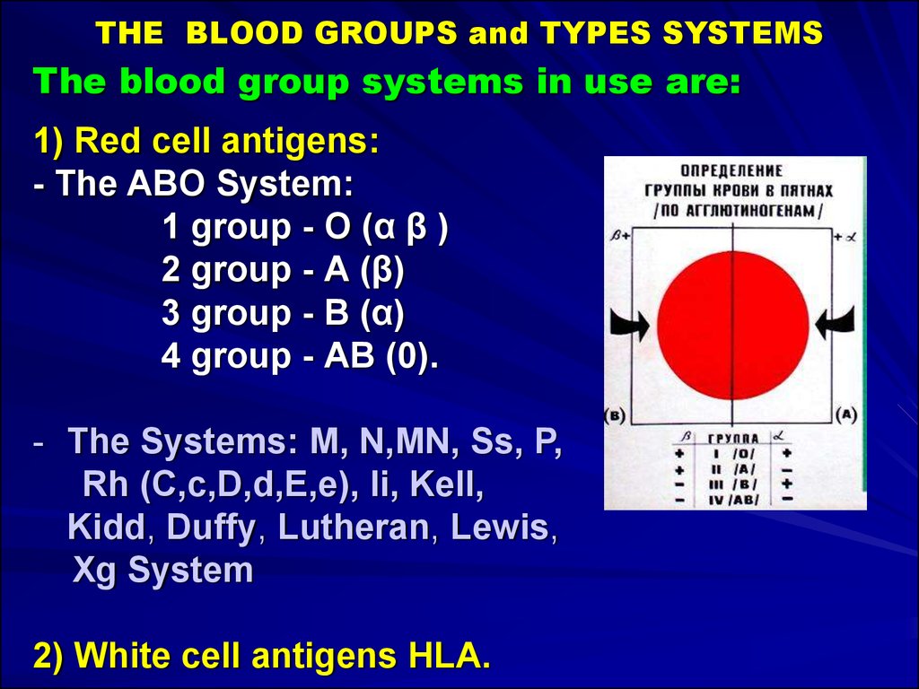
THE BLOOD GROUPS and TYPES SYSTEMSThe blood group systems in use are:
1) Red cell antigens:
- The ABO System:
1 group - О (α β )
2 group - А (β)
3 group - В (α)
4 group - АВ (0).
- The Systems: M, N,MN, Ss, P,
Rh (C,c,D,d,E,e), li, Kell,
Kidd, Duffy, Lutheran, Lewis,
Xg System
2) White cell antigens HLA.
38.
THE BLOOD GROUPS and TYPES SYSTEMSThe blood group systems in use are:
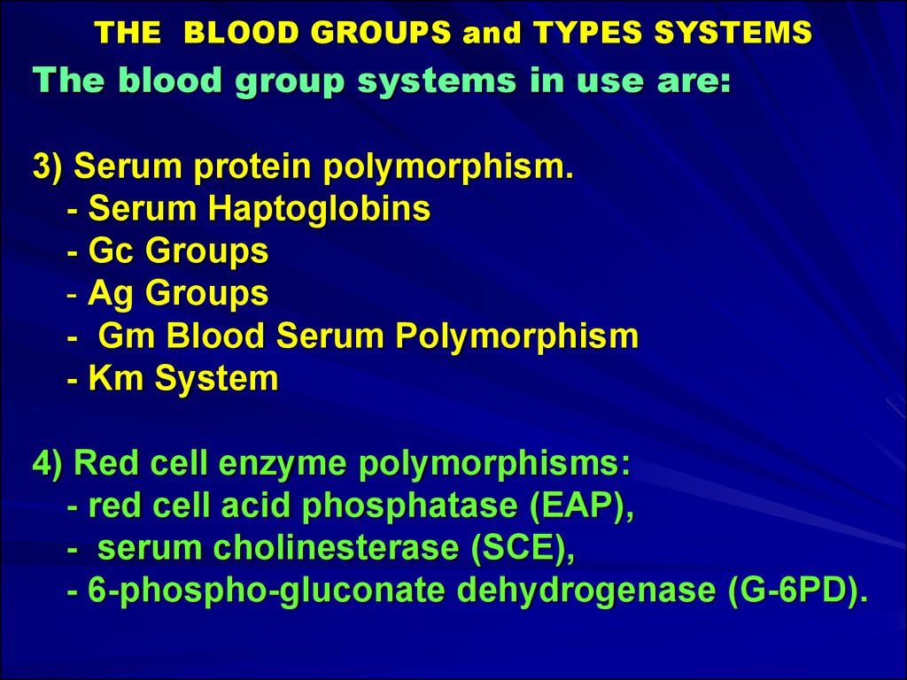
3) Serum protein polymorphism.
- Serum Haptoglobins
- Gc Groups
- Ag Groups
- Gm Blood Serum Polymorphism
- Km System
4) Red cell enzyme polymorphisms:
- red cell acid phosphatase (EAP),
- serum cholinesterase (SCE),
- 6-phospho-gluconate dehydrogenase (G-6PD).
39.
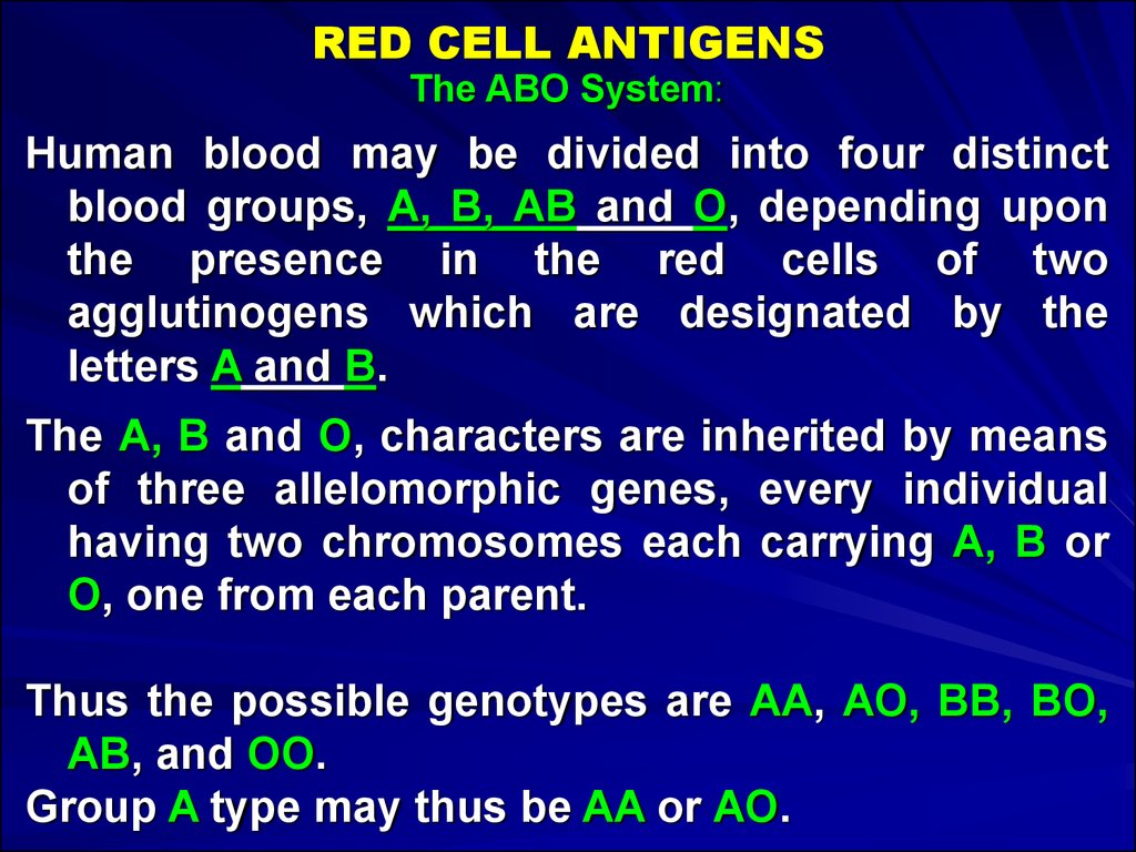
RED CELL ANTIGENSThe ABO System:
Human blood may be divided into four distinct
blood groups, А, В, АВ and O, depending upon
the presence in the red cells of two
agglutinogens which are designated by the
letters A and B.
The А, В and O, characters are inherited by means
of three allelomorphic genes, every individual
having two chromosomes each carrying А, В or
О, one from each parent.
Thus the possible genotypes are AA, АО, BB, BO,
AB, and OO.
Group A type may thus be AA or АО.
40.
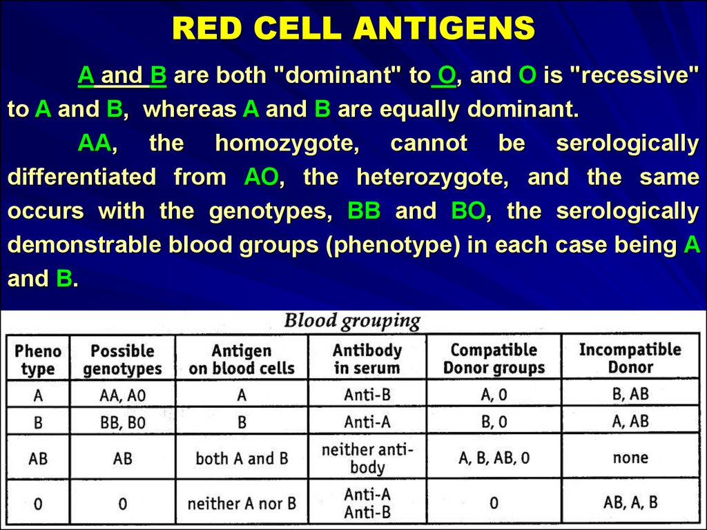
RED CELL ANTIGENSA and В are both "dominant" to O, and О is "recessive"
to A and B, whereas A and В are equally dominant.
AA, the homozygote, cannot be serologically
differentiated from АО, the heterozygote, and the same
occurs with the genotypes, BB and BO, the serologically
demonstrable blood groups (phenotype) in each case being A
and B.
41.
RED CELL ANTIGENSThe other phenotypes are AB and О.
А has two subgroups A1 and A2.
These are also found in group AB giving rise
to subgroups A1B and A2B.
Subgroups A3, A4 and A5 are weak and very
rare.
Anti-A or α and Anti-B or β agglutinins are
normally developed in the serum against whichever
agglutinogens are absent from the red blood cells.
There is no positive method of identifying the
'silent' gene O.
42.
RED CELL ANTIGENSThe MNSs System:
Two further agglutinogens M and N, which are
quite distinct and independent of the
agglutinogens A and B, occur in human blood.
The M and N factors are inherited as Mendelian
dominants. They are present at birth.
They form three groups, M, N and MN.
Anti-M and Anti-N agglutinins are not normally
developed in human sera but, they may be
present rarely.
The agglutinogens, M and N are feebly antigenic.
43.
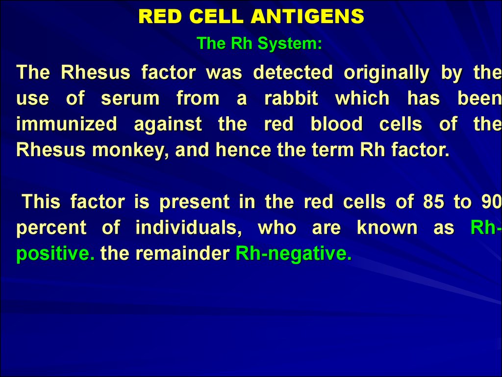
RED CELL ANTIGENSThe Rh System:
The Rhesus factor was detected originally by the
use of serum from a rabbit which has been
immunized against the red blood cells of the
Rhesus monkey, and hence the term Rh factor.
This factor is present in the red cells of 85 to 90
percent of individuals, who are known as Rhpositive. the remainder Rh-negative.
44.
RED CELL ANTIGENSThe Rh System:
A complex of antigens is involved, six in number,
named Cc, Dd. and Ее, each of which is capable of
producing antibodies, although they are all not
equally powerful in this respect.
The Rh antigens can be detected in a foetus after
six weeks of pregnancy.
45.
BLOOD GROUPS AND HEREDITYThe ABO, MN, and Rh factors
are inherited according to
Mendelian principles.
The rules of inheritance of ABO
system are:
1) Agglutinogen A or В cannot
appear in the child unless it is
present in one or both
parents.
2) Agglutionogen A1 or A2
cannot appear in the blood of
the child unless it is present
in one or both parents.
46.
BLOOD GROUPS AND HEREDITY3) The combination of A1B
parent with A2 child and
vice versa cannot occur.
4) An О parent cannot have
an AB child and an AB
parent cannot have О
child.
5) Parents of АО and АО
genotype may have a OO
child.
6) Parents of AA or АО
genotype may have A
child.
47.
BLOOD GROUPS AND HEREDITYThe rules of inheritance for
MN system are:
1) Agglutinogens M and N,
cannot appear in the
blood of a child unless
present in one or both
parents.
2) A type M parent cannot
produce a type N child,
and conversely an N
parent cannot produce M
child.
48.
BLOOD GROUPS AND HEREDITY3) In matings where both parents
are homozygous type M or N,
the children are always of the
same type as the parents.
4) In matings where one parent is
type M and other type N, all
children are type MN.
5) In matings where one parent is
homozygous (M or N) the
children are of parental types in
fifty to fifty ratio.
6) In matings where the parents
are both MN, children of all
three types are possible.
49.
BLOOD GROUPS AND HEREDITYRules of inheritance of Rh groups are:
1) Rh-negative parents cannot produce an Rhpositive child.
2) Rh-positive and mixed parents can have Rhpositive and Rh-negative children.
50.
DNA FINGERPRINTINGEach human nucleus contains about a metre of DNA, but
only ten percent is used for genetic coding, the rest
being redundant or silent segments (stutters; hyper
variable regions (HVR); minisatellites).
Of these redundant segment, there may be two hundred to
fourteen thousand repeats of each identical sequence on
each DNA strand.
These segments of nucleotides are called "repetitive DNA".
51.
DNA FINGERPRINTINGThe length, constitution, and number of the repetitive
sequences are different for each person, but are unique
for an individual (except unovular twins), and are stably
inherited in a Mandelian fashion. This method is as
unique as fingerprints to an individual.
Nucleated cells are the source of DNA for extraction from
blood, semen, vaginal epithelial cells, tooth pulp, bone
marrow, hair roots, muscle, skin, mucous membranes,
etc.
Because every person’s DNA sequence is different, the
fragments in DNA specimen from one individual to
another are different in number and length, from those in
a DNA specimen from another individual.
52.
MEDICO-LEGAL IMPORTANCE DNAFINGERPRINTING
1)
The blood on a weapon can be matched against
the blood of the victim.
2) Hair roots found on a weapon can be matched
against the blood of the victim and accused.
3) Seminal fluid recovered from the vagina of a
victim can be matched against the blood of an
accused.
53.
MEDICO-LEGAL IMPORTANCE DNA FINGERPRINTING4) It can exonerate a falsely implicated person in a
crime.
5) Paternity can be established positively.
6) It can be applied for tracing pedigrees and for
establishing family relationship.
7) Identification of victims of accident, mass
disasters, mutilated bodies can be made by
matching prints with prints of parents or close
relatives.
54.
EXAMINATION OF SEMINAL FLUIDSeminal stains have to be detected in cases of rape
or attempted rape, sexual murder of the female,
sodomy and bestiality. Fertility of the fluid has to
be proved in civil cases, e.g., disputed paternity.
Semen is greyish- yellow, thick, jelly-like and sticky when
fresh.
The quantity of seminal fluid in a single emission is two to
five ml. and contains about 60 to 150 million sperms per
ml. of which 90 % are motile at the time of ejaculation.
55.
EXAMINATION OF SEMINAL FLUIDThe fluid is alkaline with a pH of 7.4.
The stains are usually found on the clothing but
may be found on the person of either the victim
or the accused.
They may also be found on bed clothes, on floor or
on the grass where the offence was committed.
Seminal stains have to be differentiated from those
due to starch, pus.
56.
MEDICO-LEGAL ASPECTS OF EXAMINATIONOF SEMINAL FLUID.
Examination of seminal fluid is important on the
many accounts:
Civil importance:
- Compensation on the ground acquired sterility
- Disputed paternity
- Legitimacy
- Artificial insemination and other.
Criminal importance: in relation to sex offence cases
- Concerning commission of sex offence
- Identification of the offender.
57.
EXAMINATION OF SEMINAL FLUIDCOLLECTION OF MATERIAL:
1. Dried or drying seminal fluid on the perineum or thighs is
collected with a wet throat swab.
2. Fluid from the vagina is collected with a pipette or throat
swab inserted with or without the aid of a speculum.
3. The pubic hair should be removed and placed in a small
container.
4. A portion of cloth containing the stain is cut out, dried
and preserved.
5. Stains on smooth, impervious surface should be gently
scraped off with the point of a knife into a glass
container.
58.
EXAMINATION OF SEMINAL FLUID1) Physical Examination.
2) Chemical Examination:
a) Florens Test,
b) Barberios Test,
c) The Acid Phosphatase Test,
d) Creatine Phosphokinase.
3) Microscopic Examination
59.
EXAMINATION OF SEMINAL FLUID4) Precipitin Test:
The principle is the same as that for blood.
Spermatozoa of rat (1); rabbit (2); horse (3); human (4,5)
5) Group of Seminal Fluid:
The specific aglutinable substances A and B are
present in the semen of secretors.
As such, the group of the individual may be determined.
60.
EXAMINATION OF HAIRQuestions are decided:
1. Are there the hair?
61.
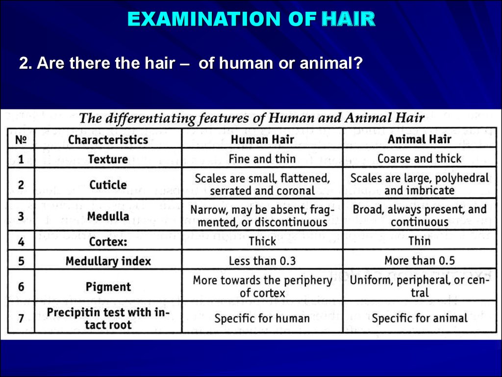
EXAMINATION OF HAIR2. Are there the hair – of human or animal?
62.
EXAMINATION OF HAIR2. Are there the hair – of human or animal?
63.
EXAMINATION OF HAIR3. What region of body is a hair from? (head, beard,
moustache, pubis)
64.
EXAMINATION OF HAIR4. Did hair pull out or did it fall out?
5. Had hair additionally physical and chemical influences?
6. Can hair belong to the certain person?
65.
EXAMINATION OF SALIVA, FAECIES, VAGINALSECRETION
SALIVA:
It contains enzymes like ptyalin, glucose 6-phosphate
dehydrogenase, various proteins, lipids. chlorides,
thiocyanate ions, etc. The stains are identified from the
presence of amylase and buccal epithelial cells. ABO
grouping and species origin can be carried out.
FAECES:
The stains can be identified from odour, presence of
undigested muscle and vegetable fibres and stercobilin.
VAGINAL SECRETION:
It consists of white coagulated material consisting of shed
vaginal epithelium and Doderlein's bacilli.