
 medicine
Similar presentations:


")
Forensic or legal medicine
1.
ZAPOROZHIAL STATE MEDICAL UNIVERSITYTHE DEPARTMENT OF PATHOLOGICAL ANATOMY and FORENSIC
MEDICINE
2.
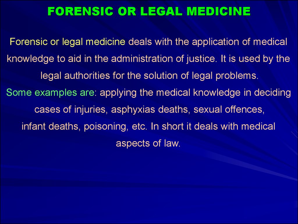
FORENSIC OR LEGAL MEDICINEForensic or legal medicine deals with the application of medical
knowledge to aid in the administration of justice. It is used by the
legal authorities for the solution of legal problems.
Some examples are: applying the medical knowledge in deciding
cases of injuries, asphyxias deaths, sexual offences,
infant deaths, poisoning, etc. In short it deals with medical
aspects of law.
3.
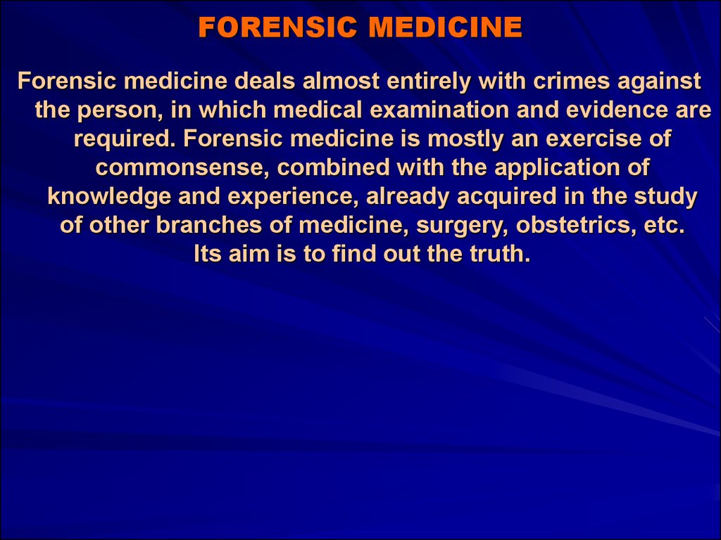
FORENSIC MEDICINEForensic medicine deals almost entirely with crimes against
the person, in which medical examination and evidence are
required. Forensic medicine is mostly an exercise of
commonsense, combined with the application of
knowledge and experience, already acquired in the study
of other branches of medicine, surgery, obstetrics, etc.
Its aim is to find out the truth.
4.
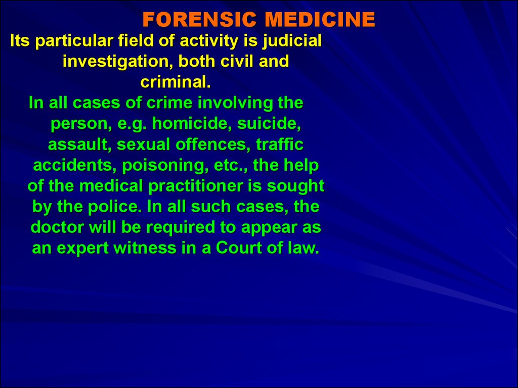
FORENSIC MEDICINEIts particular field of activity is judicial
investigation, both civil and
criminal.
In all cases of crime involving the
person, e.g. homicide, suicide,
assault, sexual offences, traffic
accidents, poisoning, etc., the help
of the medical practitioner is sought
by the police. In all such cases, the
doctor will be required to appear as
an expert witness in a Court of law.
5.
FORENSIC MEDICINEThe medical expert should be very careful when he is
examining living people.
He should not encourage an accused person to talk about the
crime with which he is charged, or about the events that led to
his arrest.
If, during a medical examination, a person says anything that
might incriminate himself, it should be neither recorded not
repeated.
6.
FORENSIC MEDICINEThree things are needed for success:
1) the power of observation,
2) a wide range of exact knowledge,
3) the power of deduction.
A good command of language, clear presentation, and ability in
expressing a relatively firm opinion are necessary for the
success of the forensic pathologist.
The forensic pathologist must be alert to where evidence should
be looked for, and how it should be interpreted.
7.
FORENSIC MEDICINEForensic medicine is not an exact science. Unexpected
results are produced due to biological variations.
In every case, there is an element of uncertainty, and
absolute proof is a rarity in any medical problem.
Doctors should bear in mind the essential difference between
probability and proof.
The medical witness should not be dogmatic about his
opinion.
8.
FORENSIC MEDICINEThe doctor should be ready to defend every finding
and conclusion on the report on clinical and
scientific grounds.
He should be aware of professional and scientific
viewpoints which might differ from his, and
should be familiar with the latest scientific
literature in relation to the subject involved.
9.
FORENSIC MEDICINEFor the purpose of illustrating and clarifying his
testimony, the medical expert may employ
photographs, maps, diagrams, charts, X-rays,
skeletons, models, etc., when they are properly
verified.
10.
FORENSIC-MEDICINE EXAMINATIONForensic-medical examination is performed only when there
is a written resolution or direction from the investigative
or judicial organs.
Objects of forensic-medical examination:
1. Dead body.
2. Living person (a victim , is suspected and other persons).
11.
FORENSIC-MEDICINE EXAMINATIONObjects of forensic-medical examination:
3. Material evidences.
4. Materials of crime cases.
12.
FORENSIC-MEDICINE EXAMINATIONExamination or research of these objects is produced in
Bureau of forensic-medical examination or Institute of
Forensic Medicine which consists of the following
departments:
1. Forensic-medical mortuary (morgue).
2. Department for examination of victims, suspected and
other living persons.
3. Forensic-medical laboratories ( histological
immunological, cytological, chemical, criminalisticals).
13.
INVESTIGATION OF THE SCENE OF DEATHI. The first research of dead body an expert conducts on
the scene of death.
The basic rules for investigation of any scene of crime are:
1) verify that a crime has been committed,
2) look for signs of how it was committed,
3) recover and preserve evidence that might lead to the
arrest and conviction of the guilty.
14.
INVESTIGATION OF THE SCENE OF DEATHMedico-legal Masquerades:
Many cases of homicide go undetected because of the lack
of suspicion and improper or inadequate investigation.
All cases of death should be regarded as unnatural,
until proved otherwise.
15.
INVESTIGATION OF THE SCENE OF DEATHAccidental deaths and suicides can occur under
circumstances which suggest homicide.
In a suicide case, alterations may be made at the scene
because of stigma. In a homicide case, the scene may
be altered or rigged to suggest that death resulted from
suicide or accident.
16.
INVESTIGATION OF THE SCENE OF DEATHThe doctor must look for any possible inconsistencies
between the apparent death scene and his actual
scientific findings. In a case of hanging, the manner in
which a ligature is applied to the neck, or the mode of
suspension of a body may be determining features in
the circumstances.
17.
INVESTIGATION OF THE SCENE OF DEATHIn such cases, the real cause of death can be established
by complete autopsy and police investigation.
The investigating officer should obtain information about
the circumstances of death and the background of the
deceased.
18.
INVESTIGATION OF THE SCENE OF DEATHThe answers to the following questions have to be found:
1) Who is the victim? (identification).
2) When the death and injuries occurred? (time of death
and injuries).
3) Where the death occurred? (scene and circumstances
of death).
4) What injuries are present? (description of injuries).
19.
INVESTIGATION OF THE SCENE OF DEATHThe answers to the following questions have to be found:
5) Which injuries are significant? (major, minor, true,
artifacts, post-mortem injuries).
6) Why and how injuries were produced? (mechanism and
manner of death, i.e., accidental, suicidal or homicidal).
If the death is violent, determine the means or agent
causing death, e.g., knife, firearm, poison, etc. and if
homicide assist in identifying the person responsible
for death.
20.
CONDUCT AND DUTIES OF THE DOCTOR AT THE SCENE OFCRIME
Complete and accurate
recording of the scene
as it was found is very
important.
This can be done by
accurate diagrams,
notes and photography.
The scene may show
evidence of a struggle,
and on the body vital
trace evidence may be
present.
21.
CONDUCT AND DUTIES OF THE DOCTOR AT THESCENE OF CRIME
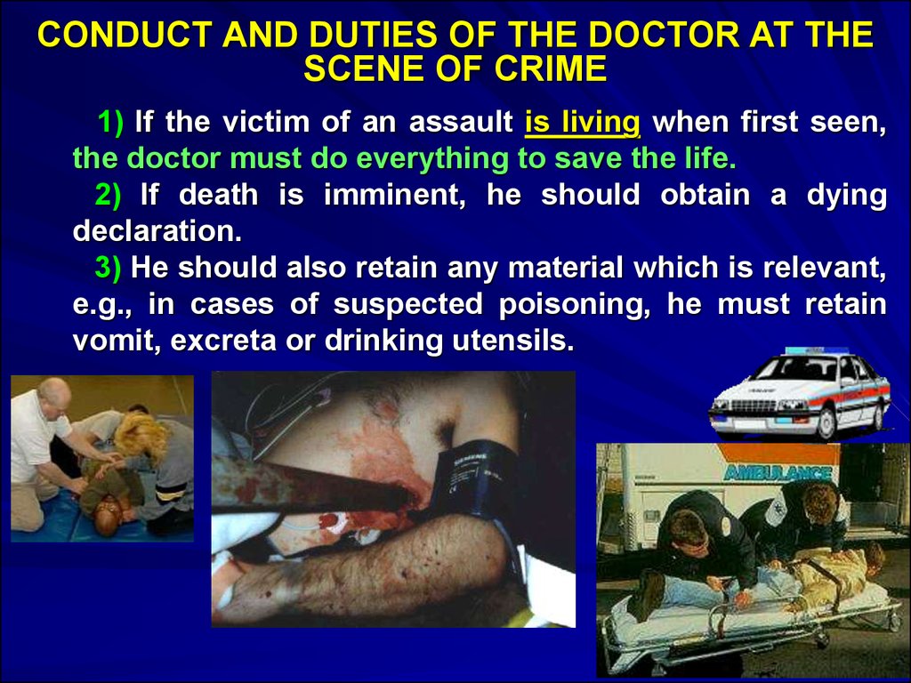
1) If the victim of an assault is living when first seen,
the doctor must do everything to save the life.
2) If death is imminent, he should obtain a dying
declaration.
3) He should also retain any material which is relevant,
e.g., in cases of suspected poisoning, he must retain
vomit, excreta or drinking utensils.
22.
CONDUCT AND DUTIES OF THE DOCTOR AT THESCENE OF CRIME
4) He must make sure that death has occurred.
5) If he suspects foul play, the police should be informed.
6) He must obtain all possible information regarding the
crime.
7) He must identify the body, which should also be
identified by the relatives and the police.
8) He must enquire whether the body has been moved at all
before he first saw it.
23.
CONDUCT AND DUTIES OF THE DOCTOR AT THESCENE OF CRIME
9) He should ask the investigating officer before moving
anything.
Photograph the scene from several angles.
He should follow but not lead the police around the
scene.
10) He should not give opinion without proper thought.
11) He should make adequate notes:
a) Date, time, address or location,
b) Name and sex of deceased,
24.
CONDUCT AND DUTIES OF THE DOCTOR AT THESCENE OF CRIME
c) A list of all persons present,
d) General observation about the scene; any evidence of
struggle, such as overturned furniture or trampled
ground. Note surroundings of the body, such as walls,
flooring, fixtures, furniture, doors and windows.
e) Temperature of the surroundings, and the rectal
temperature of the deceased should be taken,
25.
CONDUCT AND DUTIES OF THE DOCTOR AT THESCENE OF CRIME
f) Make a sketch noting such points or importance as
direction and position of blood (pools or splashes),
position of the body and any weapons. If the weapon is
in the hand of the deceased, note whether it is loosely
held or tightly grasped.
The distribution of blood stains and their shape which may
point to the site of injury should be noted.
26.
CONDUCT AND DUTIES OF THE DOCTOR AT THESCENE OF CRIME
Note the amount of bleeding at the scene. Describe the
clothing and note any tears, cuts, missing buttons, etc.
Examine the hands and forearms for defense wounds.
Make note of injuries and record them on body
diagrams,
g) Position and appearance of the body, rigor mortis,
postmortem lividity, etc., which assist in estimating the
time of death,
27.
CONDUCT AND DUTIES OF THE DOCTOR AT THESCENE OF CRIME
h) Free hair, fibers or other foreign matter which is
likely to be dislodged when the body is moved, should
be searched and removed with adhesive tape,
i) If there are any bite marks, they should be swabbed
with a cotton wool swab moistened with saline,
j) The pubic hair should be combed in situ in cases of
sexual assault, and loose hair collected.
28.
CONDUCT AND DUTIES OF THE DOCTOR AT THESCENE OF CRIME
k) The objects on premises, e.g., dates on mail and
newspapers, condition of food on table, etc., to
determine the time of death.
l) Photograph any ligature before removal, cut if
necessary leaving the knot or knots intact.
29.
CONDUCT AND DUTIES OF THE DOCTOR AT THESCENE OF CRIME
m) If a weapon is found, handle it with care to preserve
fingerprints, blood stains, hair, fibers, etc.
n) Leave firearms in the condition they are found.
o) Note position of each bullet and casing. Bullets
should be marked for identification.
30.
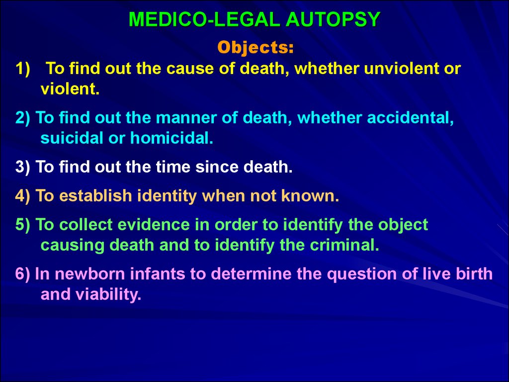
MEDICO-LEGAL AUTOPSYObjects:
1) To find out the cause of death, whether unviolent or
violent.
2) To find out the manner of death, whether accidental,
suicidal or homicidal.
3) To find out the time since death.
4) To establish identity when not known.
5) To collect evidence in order to identify the object
causing death and to identify the criminal.
6) In newborn infants to determine the question of live birth
and viability.
31.
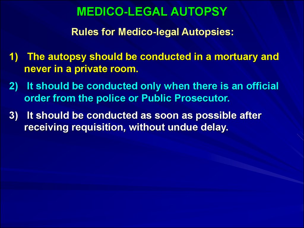
MEDICO-LEGAL AUTOPSYRules for Medico-legal Autopsies:
1) The autopsy should be conducted in a mortuary and
never in a private room.
2) It should be conducted only when there is an official
order from the police or Public Prosecutor.
3) It should be conducted as soon as possible after
receiving requisition, without undue delay.
32.
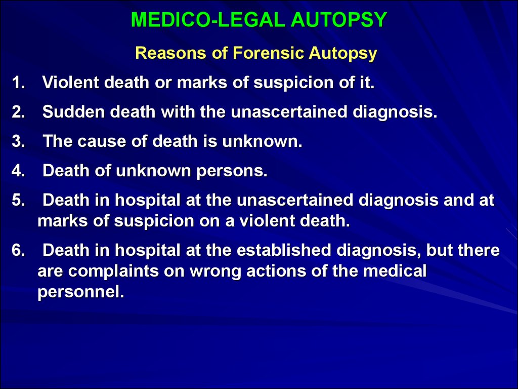
MEDICO-LEGAL AUTOPSYReasons of Forensic Autopsy
1. Violent death or marks of suspicion of it.
2. Sudden death with the unascertained diagnosis.
3. The cause of death is unknown.
4. Death of unknown persons.
5. Death in hospital at the unascertained diagnosis and at
marks of suspicion on a violent death.
6. Death in hospital at the established diagnosis, but there
are complaints on wrong actions of the medical
personnel.
33.
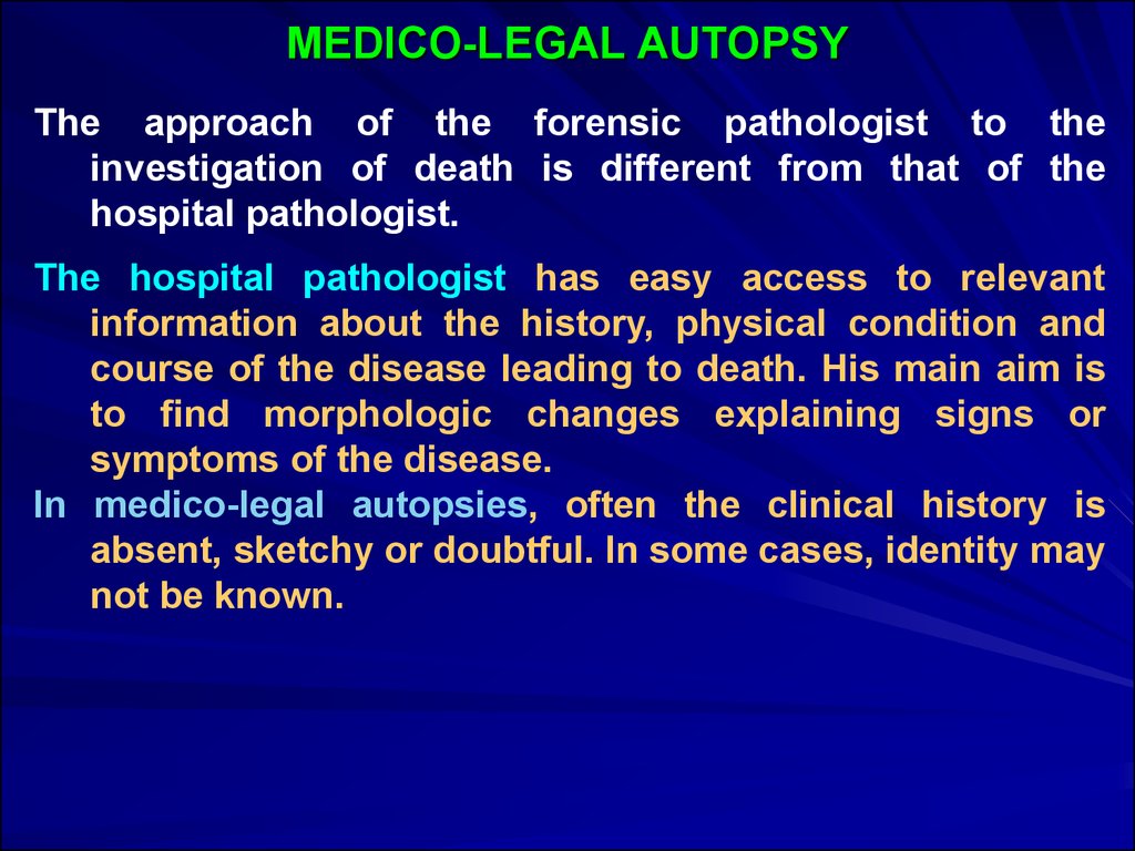
MEDICO-LEGAL AUTOPSYThe approach of the forensic pathologist to the
investigation of death is different from that of the
hospital pathologist.
The hospital pathologist has easy access to relevant
information about the history, physical condition and
course of the disease leading to death. His main aim is
to find morphologic changes explaining signs or
symptoms of the disease.
In medico-legal autopsies, often the clinical history is
absent, sketchy or doubtful. In some cases, identity may
not be known.
34.
MEDICO-LEGAL AUTOPSYForensic pathologist has to determine time of death and
age of injuries.
He has to carry out careful external examination including
clothing, in the determination of the pattern of injuries
and their relationship to the object or weapon causing
them.
He has also to determine the manner and mechanism of
death. The autopsy should be carried out by the doctor,
and not left to the mortuary attendant.
35.
DOCUMENTS OF FORENSIC-MEDICALEXAMINATION
In all cases of forensic-medical examination is made:
1. Conclusion of forensic-medical examination
Structure of conclusion of forensic-medical
examination
1. Introduction.
2. Research part.
3. Conclusions.
36.
ThanatologyThanatos is Greek god of death
Thanatology deals with death in all its aspects.
Three modes of death are:
Coma (Brain)
Asphyxia (Lungs)
Syncope (Heart)
"Tripod of life" are Brain, Lungs and Heart
37.
ThanatologyPOSTMORTEM CHANGES
Signs of death appear in the following order:
1.Immediate (somatic death):
a. Insensibility and loss of voluntary power
earliest sign at death.
b. Cessation of respiration.
c. Cessation of circulation.
38.
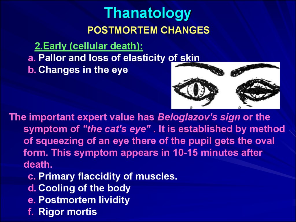
ThanatologyPOSTMORTEM CHANGES
2.Early (cellular death):
a. Pallor and loss of elasticity of skin
b. Changes in the eye
The important expert value has Beloglazov's sign or the
symptom of "the cat's eye" . It is established by method
of squeezing of an eye there of the pupil gets the oval
form. This symptom appears in 10-15 minutes after
death.
c. Primary flaccidity of muscles.
d. Cooling of the body
e. Postmortem lividity
f. Rigor mortis
39.
ThanatologyPOSTMORTEM CHANGES
3. Late (decomposition and decay):
a. Putrefaction
b. Adipocere formation
c. Mummification
40.
ThanatologyPOSTMORTEM CHANGES
LIVOR MORTIS (Postmortem Staining)
Synonyms of postmortem staining are cadaveric or
postmortem lividity, hypostasis, or livor mortis.
Cadaveric lividity is an early sign of death.
Postmortem hypostasis is bluish purple or purplish red
discoloration, which appears under the skin (rete
mucosum) of the dependent part of the body after death
due to capillo-venous distention.
Postmortem lividity begins shortly after death, but it may
not be visible for about half to one hour after death.
41.
ThanatologyPOSTMORTEM CHANGES
LIVOR MORTIS (Postmortem Staining)
- It is usually well developed within 4 hours and reaches a
maximum between 6 and 12 hours.
- The postmortem staining gets fixed in 6-7-8-10 hours.
- Postmortem hypostasis persists till it merges with
discoloration of putrefaction.
- It is more marked in asphyxia and is less marked in death
from hemorrhage, anemia, lobar pneumonia and
wasting disease.
42.
ThanatologyPOSTMORTEM CHANGES
LIVOR MORTIS (Postmortem Staining)
Location of Postmortem hypostasis
In case of Hanging: Hypostasis is more marked in lower
limbs, external genetalia, lower part of arms and
forearms.
In case of Drowning: If the body is constantly moving,
postmortem staining may not develop.
In a body lying on its back, it first appears in the neck
and then spreads over the entire back except parts
directly pressed on i.e. shoulder-blades, buttocks,
calves and heel.
43.
ThanatologyPOSTMORTEM CHANGES
LIVOR MORTIS (Postmortem Staining)
Location of Postmortem hypostasis
Distribution of Livores mortis depends on position of the body
after death
44.
ThanatologyPOSTMORTEM CHANGES
LIVOR MORTIS (Postmortem Staining)
The hypostatic areas have a distinct colour in certain cases
of poisoning - e.g.
- In carbon monoxide poisoning - cherry red.
- In hydrocyanic poisoning - bright red,
- In poisoning by nitrites, potassium chlorate,
nitrobenzene, aniline (causing methaemoglobinaemia)
the colour is red brown or brown.
- In Pottasium Cyanide Poisoning - Deep Blue.
- In Clostridum perfringens infection - Bronze colour.
- Hypothermia – Bright pink
- Opium - Black color
45.
ThanatologyPOSTMORTEM CHANGES: LIVOR MORTIS
46.
ThanatologyPOSTMORTEM CHANGES: LIVOR MORTIS
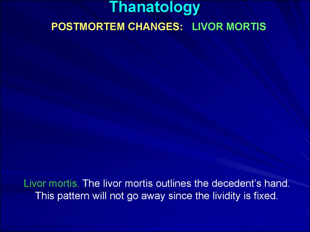
Livor mortis. The livor mortis outlines the decedent’s hand.
This pattern will not go away since the lividity is fixed.
47.
ThanatologyPOSTMORTEM CHANGES: LIVOR MORTIS
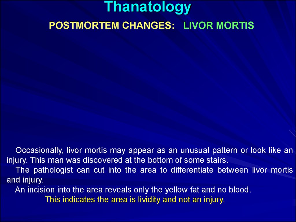
Occasionally, livor mortis may appear as an unusual pattern or look like an
injury. This man was discovered at the bottom of some stairs.
The pathologist can cut into the area to differentiate between livor mortis
and injury.
An incision into the area reveals only the yellow fat and no blood.
This indicates the area is lividity and not an injury.
48.
ThanatologyPOSTMORTEM CHANGES: Rigor Mortis
Definition
This is stage of stiffening of muscles with shortening of the
fibers. Individual cell death takes place at this stage.
Mechanism
When the ATP is reduced to 85% of the normal, the
overlapping portions of myosin and action filaments
combine as rigid links of actomyosin , which is viscous and
inextensible and causes hardness and rigidity of muscle
rigor.
49.
ThanatologyPOSTMORTEM CHANGES: Rigor Mortis
The Order of Appearance of Rigor Mortis
- All muscles of the body, both voluntary and involuntary
are affected.
- It first appears in involuntary muscles - the myocardium
becomes rigid in an hour.
- In Voluntary muscle in
Order of onset of rigor mortis is-eyelids-thorax-lower
limbs.
Order of disappearance of rigor mortis is-eyelidsthorax-lower extremities.
When rigor is fully developed, the entire body is stiff,
the muscles shortened, hard and opaque.
50.
ThanatologyPOSTMORTEM CHANGES: Rigor Mortis
Time of Onset
In India, it begins 1 to 2 hours after death and
takes further 1 to 2 hours to develop
In, temperature countries, it begins in 3 to 6 hours
and takes further 2 to 3 hours to develop.
Duration of Rigor Mortis
Rigor mortis follows "RULE OF 12" 12 hours
to set in, 12 hours to remain, 12 hours to pass off.
It takes roughly 36 hours for rigor mortis to vanish.
51.
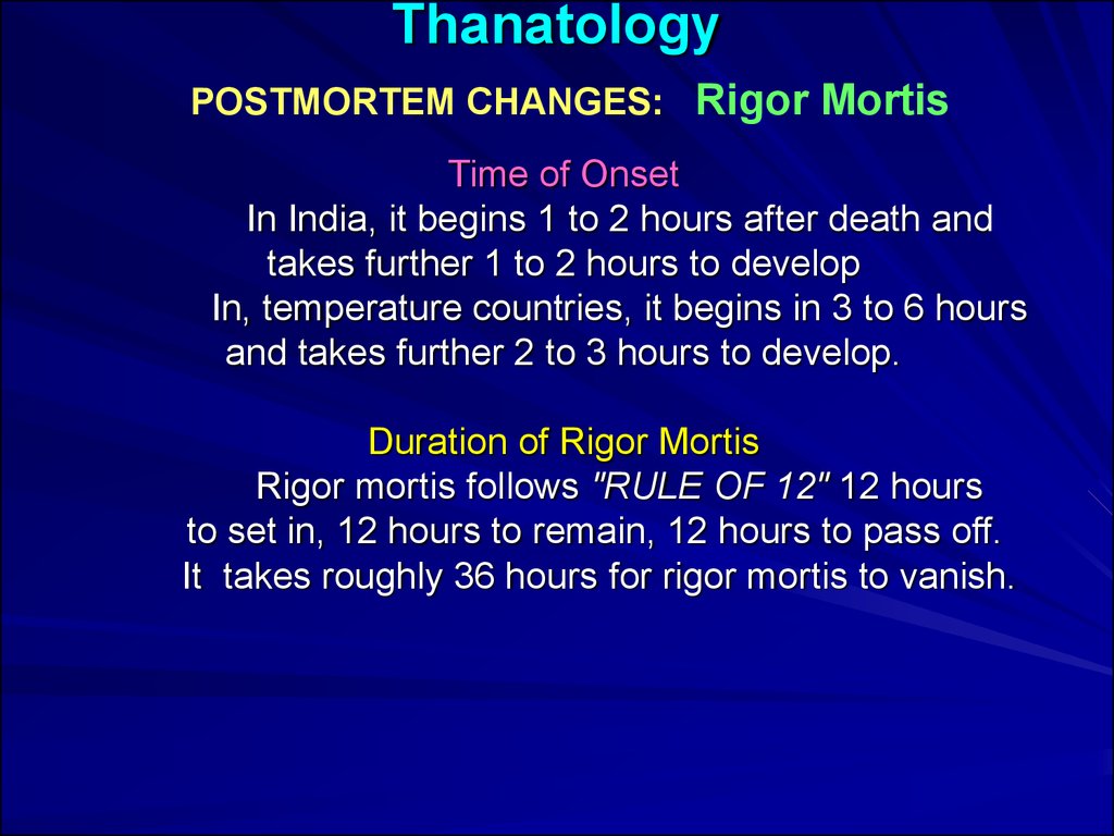
ThanatologyPOSTMORTEM CHANGES: Rigor Mortis
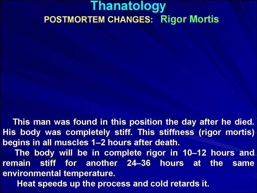
This man was found in this position the day after he died.
His body was completely stiff. This stiffness (rigor mortis)
begins in all muscles 1–2 hours after death.
The body will be in complete rigor in 10–12 hours and
remain stiff for another 24–36 hours at the same
environmental temperature.
Heat speeds up the process and cold retards it.
52.
ThanatologyPOSTMORTEM CHANGES: Rigor Mortis
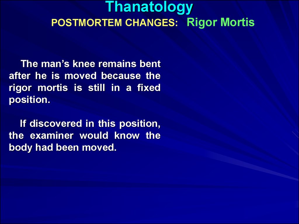
The man’s knee remains bent
after he is moved because the
rigor mortis is still in a fixed
position.
If discovered in this position,
the examiner would know the
body had been moved.
53.
ThanatologyPOSTMORTEM CHANGES: Putrefaction
Putrefaction is the final stage following death, produced
mainly by the action of bacterial enzymes, mostly anaerobic
organism derived from the bowel.
Enzymes Lecithinase produced by CI. Welchii is most
important.
The characteristic features of putrefaction are:
- Changes in the colour of the tissue.
- Collection of gases in the tissues.
- Liquefaction of tissues.
54.
ThanatologyPOSTMORTEM CHANGES: Putrefaction
The first external sign of putrefaction
in a body lying in air is a greenish discoloration of skin
over caecum i.e. right iliac fossa, then all stomach.
Sulphmethhaemoglobin causes greenish discoloration.
The color appears in 12 to 18 hours if
the environmental temperature is high, and 2 to 3 days in
temperature about 18-20°.
55.
ThanatologyPOSTMORTEM CHANGES: Putrefaction
The marbled appearance is prominent in 36 to 48
hours.
Most bodies turn green during the progression
of decomposition. This one did not. The body is swollen
(bloated) from bacterial gas formation and there is skin slippage
and subcutaneous marbling (the outlines of the blood
vessels under the skin).
56.
ThanatologyPOSTMORTEM CHANGES: Putrefaction
Due to the presence of gases in the abdomen, the
diaphragm is forced upwards resulting in expulsion of
blood stained froth/gastric contents from mouth and
nostrils—POSTMORTEM PURGE.
Internal pressure occurs on
the internal organs
when gas develops.
Pressure pushes bloody
fluid out the nose
and mouth (purging). This
should not be confused with
trauma to the nose and
mouth.
57.
ThanatologyPOSTMORTEM CHANGES: Putrefaction
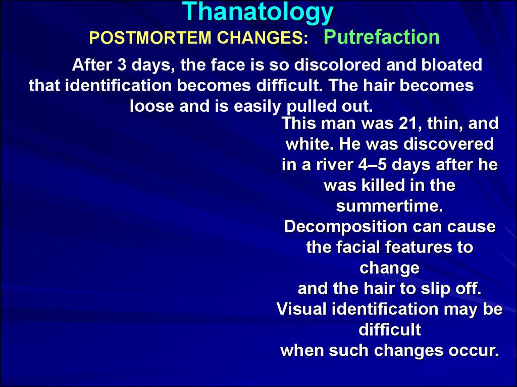
After 3 days, the face is so discolored and bloated
that identification becomes difficult. The hair becomes
loose and is easily pulled out.
This man was 21, thin, and
white. He was discovered
in a river 4–5 days after he
was killed in the
summertime.
Decomposition can cause
the facial features to
change
and the hair to slip off.
Visual identification may be
difficult
when such changes occur.
58.
ThanatologyPOSTMORTEM CHANGES: Putrefaction
The organs show putrefactive changes in the following
order:
1. Larynx and trachea
2. Stomach, intestines, spleen
3. Liver and lungs
4. Brain
5.Uterus,prostate
6. Skin, muscle tendon
7. Bone
In putrefaction, liver has a honey combed or foamy
appearance.
Prostate and Non-gravid uterus resist putrefaction for a
very long time.
Putrefaction begins above 10°C and optimum 'between 21
°C and 38°C.
59.
ThanatologyPOSTMORTEM CHANGES: Mummification
- It is modification of putrefaction, occur when the
environmental condition is hot and dry.
- Mummification occurs when body is buried in dry
sandy shallow graves.
- Desiccation or Dehydration or Drying and shrinkage
of the cadaver occur due to evaporation of water but the
natural appearance and features of the body are
preserved.
- A mummified body is odorless.
- The time required for complete mummification of a
body varies from three months to a year or two.
60.
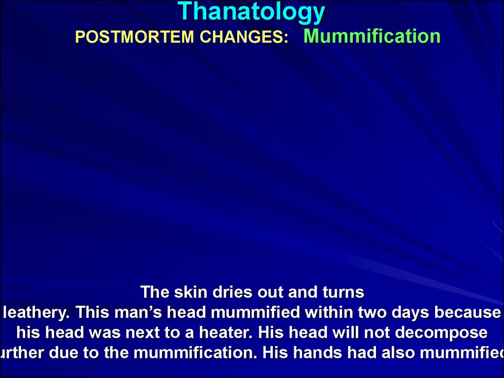
ThanatologyPOSTMORTEM CHANGES: Mummification
The skin dries out and turns
leathery. This man’s head mummified within two days because
his head was next to a heater. His head will not decompose
urther due to the mummification. His hands had also mummified
61.
ThanatologyExhumation
- Exhumation is the digging out of an already buried body
from the grave.
- There is no time limit for exhumation .
- The body is exhumed only when there is a written
order from the first class magistrate.
- It should be conducted in natural light in early
morning.
- Average number of sample of earth taken is 6-7.
- Disinfectants should not be sprinkled on the body.
- In suspected mineral poisoning, hair, nails, and long
bone e.g. femur should be preserved for chemical
analysis.