


 medicine
Similar presentations:


")


Anemia in children
1. Anemia in children
2.
3.
4.
an
d
r
e
l
a
t
e
d
n
u
t
r
i
t
i
o
n
a
l
a
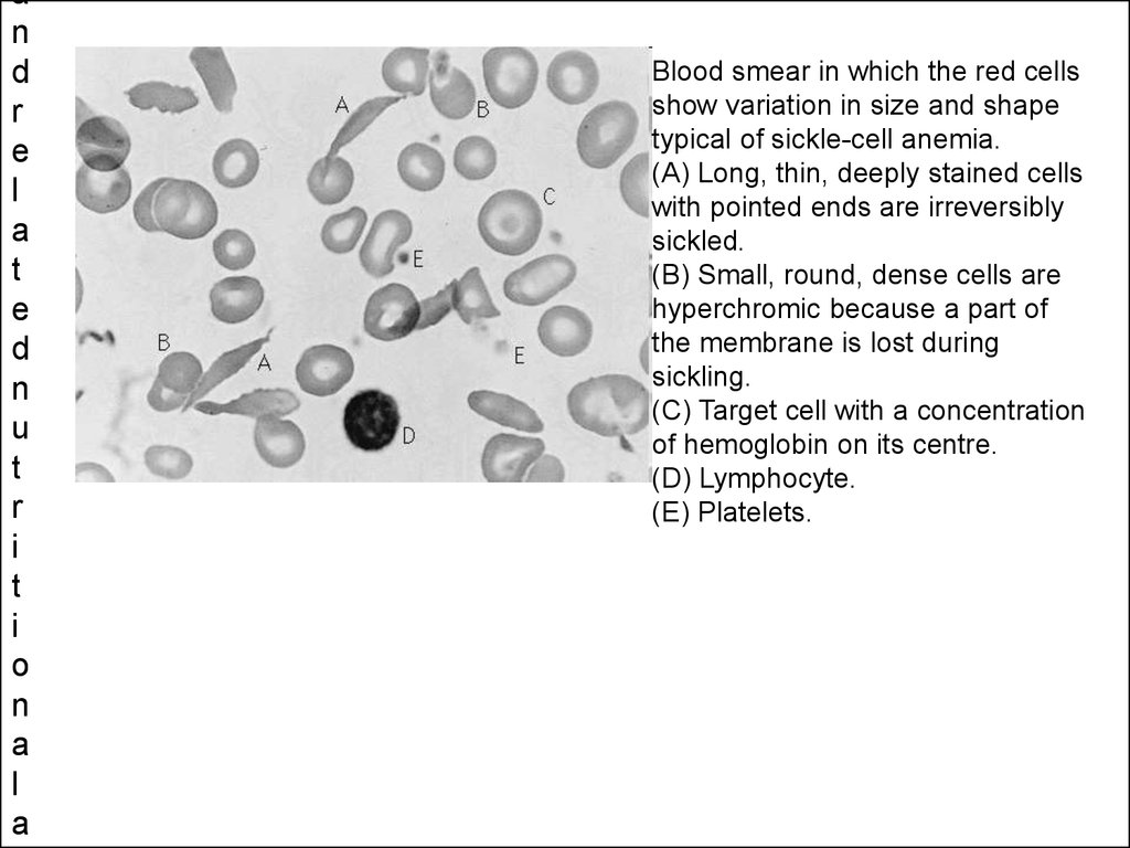
Blood smear in which the red cells
show variation in size and shape
typical of sickle-cell anemia.
(A) Long, thin, deeply stained cells
with pointed ends are irreversibly
sickled.
(B) Small, round, dense cells are
hyperchromic because a part of
the membrane is lost during
sickling.
(C) Target cell with a concentration
of hemoglobin on its centre.
(D) Lymphocyte.
(E) Platelets.
5. AVERAGE NORMAL BLOOD VALUES AT DIFFERENT AGE GROUPS
AGEHb
(gm
%)
RBC
(m/L)
HCT
%
MCV
MCH
(cu.mm) (pg)
MCHC
%
Retic
%
1 day
18.0
5.14
61
119
36.0
31.6
32
4 weeks 14.2
4.0
43
106
35.5
33.5
0.6
1 year
11.6
4.6
35
77
25.0
33.0
0.9
10-12
years
13.0
4.8
39
80
27.0
33.0
1.0
AdultMen
16.0
5.4
47
87
29.0
34.0
1.0
6. CLASSIFICATION AND AETIOLOGY OF ANEMIA :
There are four basic causes of anemia - loss, destruction,sequestration and hypoproduction.
Anemia can be further classified by
RBC size - micro, normo, and macrocytic
anemia.
RBC shape - e.g. Sickle cell.
Etiology
Blood loss : Acute
Chronic
Decreased iron assimilation : Nutritional
deficiency
Hypoplastic or aplastic anemia
Bone marrow infiltration like leukemia &
other malignancies, myelodysplastic
syndrome
Dyserythropoietic anemia
Increased physiologic requirement
Extracorpscular like alloimmune &
isoimmune
hemolytic
anemia,
microangiopathic anemias, infections,
hypersplenism, Intracorpsular defect
like :
Red
cell
membranopathy
i.e.
congenital spherocytosis, elliptocytosis
Hemoglobinopathy like HbS, C,D,E etc.
Thalassemia syndrome
RBC enzymopathies like G6PD
deficiency, PK deficiency etc.
7.
8.
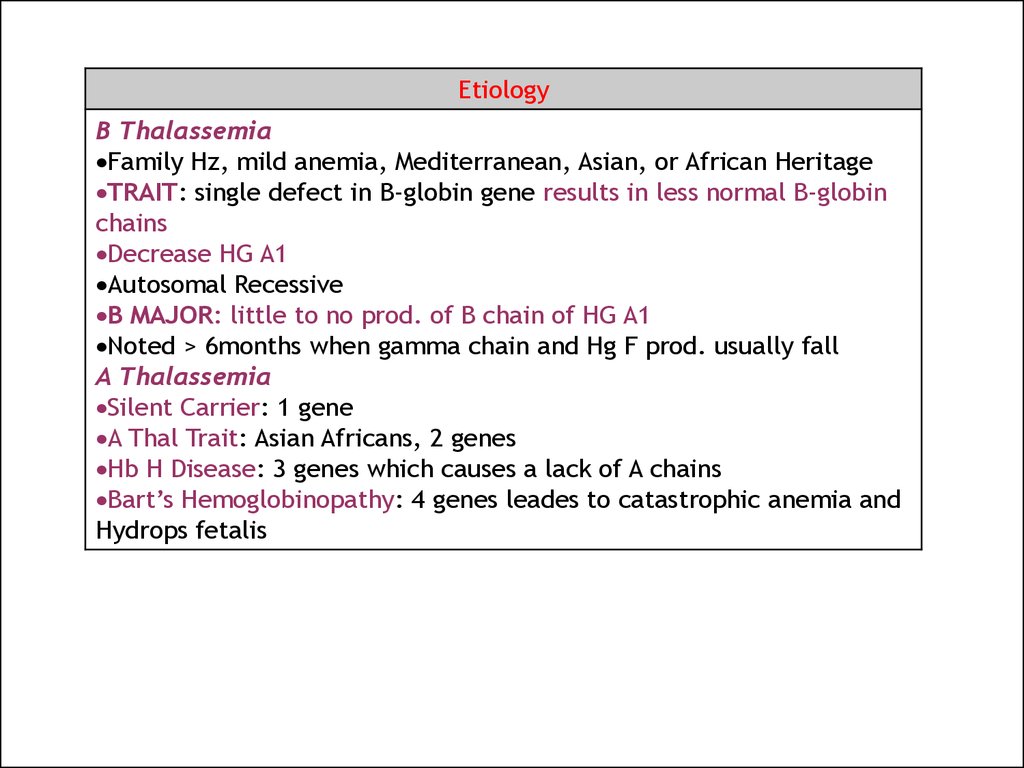
EtiologyB Thalassemia
Family Hz, mild anemia, Mediterranean, Asian, or African Heritage
TRAIT: single defect in B-globin gene results in less normal B-globin
chains
Decrease HG A1
Autosomal Recessive
B MAJOR: little to no prod. of B chain of HG A1
Noted > 6months when gamma chain and Hg F prod. usually fall
A Thalassemia
Silent Carrier: 1 gene
A Thal Trait: Asian Africans, 2 genes
Hb H Disease: 3 genes which causes a lack of A chains
Bart’s Hemoglobinopathy: 4 genes leades to catastrophic anemia and
Hydrops fetalis
9. HEREDITARY SPHEROCYTOSIS
RBC membrane defect with SPECTRINRBC destroyed prematurely in spleen
MOST COMMON HEREDITARY RED CELL DISORDER!!!!!!
Autosomal Dominant
Increased RBC turnover leads to cholelithiasis and cholecystitis
Susceptible to aplastic crisis from PARVOvirus
Physical shows pallor, jaundice, splenomegaly
Lab findings include reticulocytosis, increased MCHC (decreased in
IDA), spherocytes in smear.
Antiglobulin test rules out an immunce cause for the HA
Osmotic fragility test
Tx includes careful management of situation, esp. aplastic crisis
Splenectomy: spherocytes remain but RBC destruction stops! (only after 5 yrs old)
Immunize prior to procedure with Hib, Pneumovax, and N. meningitis vaccine b/c
increase rish for encapsulated organisms.
Also penicillin prophy
10. AREGENERATIVE ANEMIAS
Parvo B19
Fifth’s Disease with affinity for red cell precursors causing marrow aplasia
Causes Hydrops Fetalis
Diamond-Blackfan Anemia
Relative insensitivity to EPO (idiopathic)
Develops insidiously in 1st year of life and no recovery!
Short stature, abnormal facies, abnormal thumbs
Macrocytosis (any anemic child with Macrocytosis is very serious)
Tx includes transfusions, steroids for life
Increase risk of myelogenous leukemia
Transient Erythroblastopenia of Childhood
2nd yr of life and is idiopathic
VERY low Hbn but no symptoms
Recover with no intervention at all!
Normochromic/normocytic anemia
11. SICKLE CELL DISEASE
Etiology:Valine for glutamic Acid in 6th position
of Beta chain Hb
• Most common in African descent
• Only appears after 6 months when B
chains have fully developed into Hb
A1.
• Defect on Chrom 11 Neonatal
screening!!!
12. G6PD DEFICIENCY
• Central enzyme in PPP pathway• Makes NADPH which forms reduced
Glutathione that removes radicals
• X-Linked
• A form is common in AA and mild
• B form in Meds and very serious
• Canton form in oriental and rare but most
serious
13.
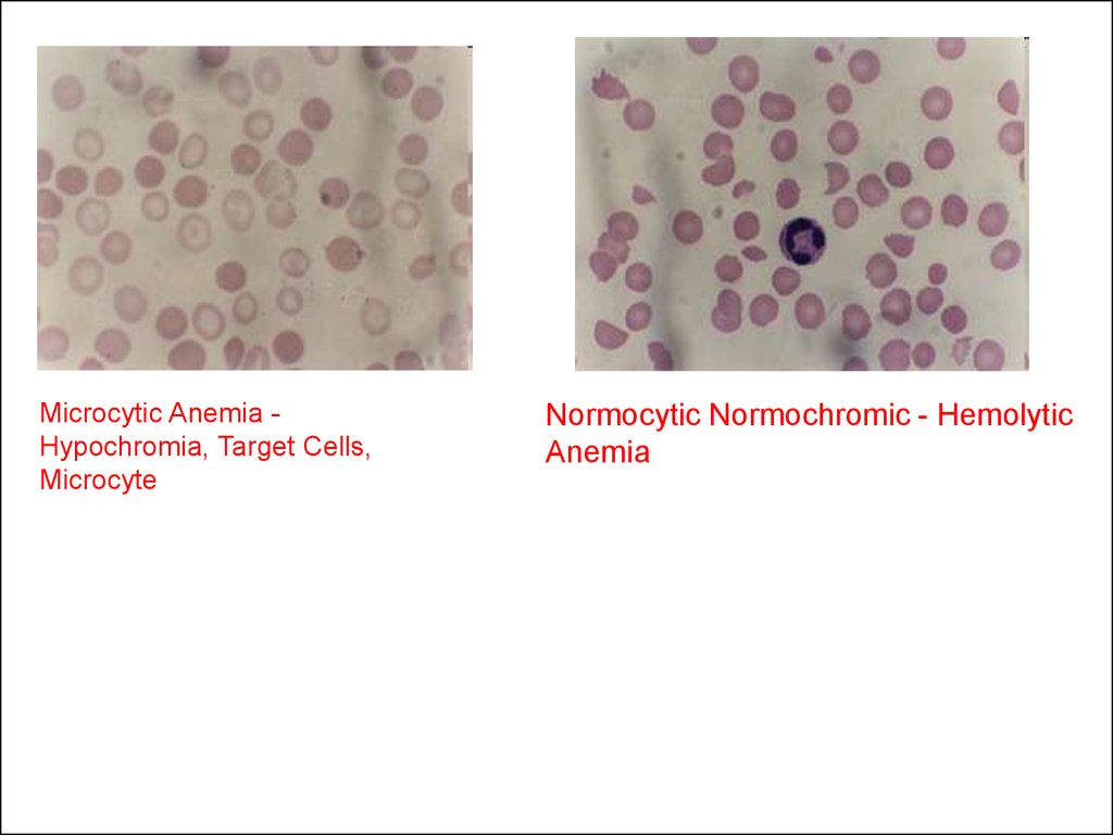
Microcytic Anemia Hypochromia, Target Cells,Microcyte
Normocytic Normochromic - Hemolytic
Anemia
14.
Megaloblastic Anemia, Bonemarrow smear, May-Giemsa
stain, x1000
Pernicious Anemia, Bone marrow smear,
May-Giemsa stain, x1000