


 medicine
Similar presentations:


Neurobiology and neurophysiology of schizophrenia
1.
Neurobiology andNeurophysiology of
schizophrenia
Scientific advisers: Antokhina RI,
Antokhin EY.
By: Yagnik Kharat
407i
2.
INTRODUCTION•Schizophrenia is a chronic mental illness affecting approximately 1 percent of the
population
•several neurotransmitter systems appear to play a role, particularly in the expression
of positive as well as negative psychotic symptoms
•dopamine system is the most compelling.
• Шизофрения - хроническое психическое заболевание, которым
страдает примерно 1 процент населения.
• несколько систем нейротрансмиттеров, по-видимому, играют роль,
особенно в выражении как положительных, так и отрицательных
психотических симптомов
• дофаминовая система является наиболее привлекательной.
3.
•Other neurotransmitters have also been implicated, includingglutamate, serotonin, and γaminobutyric acid (GABA).
• Также были задействованы другие нейротрансмиттеры, включая
глутамат, серотонин и гамма-аминомасляную кислоту (ГАМК).
4.
Epidemiology of schizophreniaЭпидемиология шизофрении
-Prevalence - 0.7% of the population in
any country in the world
-The highest incidence in the age
period 20-29 years
-Men: Women = 1: 1 (1.4: 1)
-Распространенность - 0,7%
населения в любой стране мира.
-Наибольшая заболеваемость в
возрастном периоде 20-29 лет.
-Мужчины: Женщины = 1: 1 (1,4: 1)
5.
6.
7.
8.
9.
10.
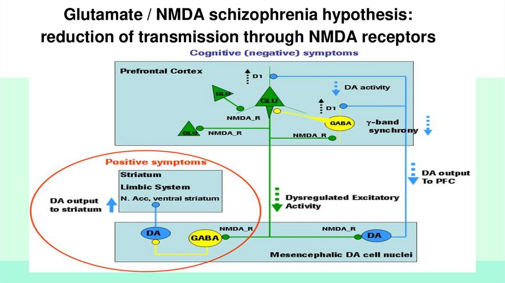
Glutamate / NMDA schizophrenia hypothesis:reduction of transmission through NMDA receptors
11.
FIGURE 12.4-3 A, Schematic diagram of the mesial temporal lobe at the level of the body of the hippocampusand posterior entorhinal cortex, in coronal section. B, The illustrated connections of the coronal section are
described in this table. (Drawn by Kyle Christensen.)
12.
STRUCTURAL AND FUNCTIONAL NEUROIMAGINGСТРУКТУРНО-ФУНКЦИОНАЛЬНОЕ НЕЙРО ИЗОБРАЖЕНИЕ
13.
FIGURE 12.4-2 PET scans using H2O15 of two monozygotic twins, one with (right) and one without (left)schizophrenia. Top and bottom scans show two levels through the dorsolateral prefrontal cortex. At the time
of scanning, subjects are performing a cognitive task that typically requires prefrontal cortical function. The
affected twin blood flow to the dorsolateral prefrontal cortex is markedly reduced compared to the unaffected
twin. (Courtesy of R. Berman and D.
РИСУНОК 12.4-2. ПЭТ-сканирование с
использованием H2O15 двух монозиготных
близнецов, один с (справа), а другой без
(слева) шизофренией. Верхнее и нижнее
сканирование показывает два уровня
дорсолатеральной префронтальной коры.
Во время сканирования субъекты
выполняют когнитивную задачу, которая
обычно требует префронтальной корковой
функции. Кровоток пораженного близнеца
к дорсолатеральной префронтальной коре
заметно снижен по сравнению с
непораженным близнецом. (Предоставлено
Р. Берманом и Д.
14.
FIGURE: MRI scans (coronal sections) of two sets of discordantmonozygotic twins (A and B = set 1; C and D = set 2). For each pair,
one has schizophrenia (A and C) while the other does not (B and D).
For both pairs, the affected twin has larger ventricles than the
unaffected twin, even though ventricular size appears to be within the
normal range for the affected twin (C). (Courtesy of D. Weinberger
and E. F. Torrey.)
МРТ (коронарные срезы) двух наборов дискордантных
монозиготных близнецов (A и B = набор 1; C и D = набор 2). В
каждой паре один болен шизофренией (A и C), а другой - нет (B и
D). Для обеих пар пораженный близнец имеет желудочки
большего размера, чем здоровый близнец, хотя размер
желудочка, по-видимому, находится в пределах нормы для
пораженного близнеца (С). (С любезного разрешения Д.
Вайнбергера и Э. Ф. Торри.)
15.
The total decrease in the volume of the brain of schizophrenic patients incomparison with healthy ones is only 3%
Суммарное уменьшение объема головного мозга больных шизофренией по сравнению со
здоровыми составляет всего 3%.
16.
17.
18.
THANK YOU!СПАСИБО!