


 medicine
Similar presentations:


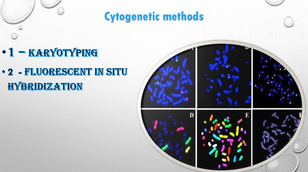
Cytogenetic methods
1.
MEDICAL ACADEMY NAMED AFTER S.I.GEORGIEVSKY OFVERENADSKY CFU
NAME : RISHABH JAIN AND RIRITES MIRASE
GROUP : LA3 -204(2)
TOPIC :CYTOGENETIC METHOD
TEACHER NAME :MAM SVETLANA SMIRNOVA
2.
CYTOGENETIC3.
4.
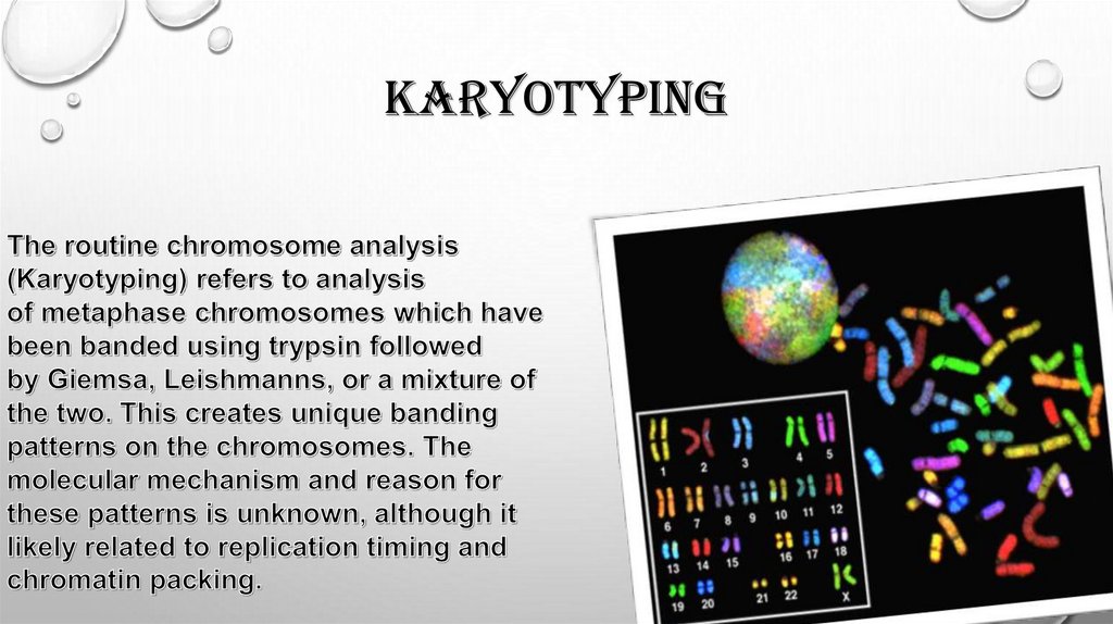
HISTORY AND EVOLUTION OF CYTOGENETICShas been a key part of biology since 1842, when Swiss botanist
Karl Nägeli first discovered chromosomes in pollen. In the decades
since, the science has been defined as the study of chromosomes,
including their behavior, mechanics, and role in inheritance. Ever
since Nägeli’s discovery, methods for examining chromosomes have
become more and more effective, further illuminating their roles in
cell biology and human and animal health in ways undreamed-of
when chromosomes were first discovered. Their behavior in animal
(salamander) cells was described by Walther Flemming, the
discoverer of mitosis, in 1882. The name was coined by another
German anatomist, von Waldeyer in 1888.
5.
Cytogenetic methods6.
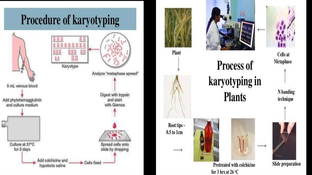
karyotyping7.
8.
staining9.
10.
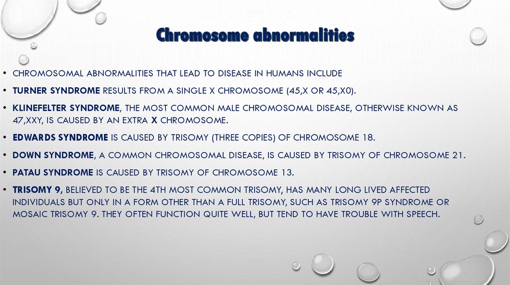
• CHROMOSOMAL ABNORMALITIES THAT LEAD TO DISEASE IN HUMANS INCLUDE• TURNER SYNDROME RESULTS FROM A SINGLE X CHROMOSOME (45,X OR 45,X0).
• KLINEFELTER SYNDROME, THE MOST COMMON MALE CHROMOSOMAL DISEASE, OTHERWISE KNOWN AS
47,XXY, IS CAUSED BY AN EXTRA X CHROMOSOME.
• EDWARDS SYNDROME IS CAUSED BY TRISOMY (THREE COPIES) OF CHROMOSOME 18.
• DOWN SYNDROME, A COMMON CHROMOSOMAL DISEASE, IS CAUSED BY TRISOMY OF CHROMOSOME 21.
• PATAU SYNDROME IS CAUSED BY TRISOMY OF CHROMOSOME 13.
• TRISOMY 9, BELIEVED TO BE THE 4TH MOST COMMON TRISOMY, HAS MANY LONG LIVED AFFECTED
INDIVIDUALS BUT ONLY IN A FORM OTHER THAN A FULL TRISOMY, SUCH AS TRISOMY 9P SYNDROME OR
MOSAIC TRISOMY 9. THEY OFTEN FUNCTION QUITE WELL, BUT TEND TO HAVE TROUBLE WITH SPEECH.
11.
SOME DISORDERS ARISE FROM LOSS OF JUST A PIECE OFONE CHROMOSOME, INCLUDING
• Cri du chat (cry of the cat), from a truncated short arm on
chromosome 5. The name comes from the babies'
distinctive cry, caused by abnormal formation of the larynx.
• 1p36 Deletion syndrome, from the loss of part of the short
arm of chromosome 1.
• Angelman syndrome – 50% of cases have a segment of
the long arm of chromosome 15 missing; a deletion of the
maternal genes, example of imprintingdisorder.
• Prader-Willi syndrome – 50% of cases have a segment of
the long arm of chromosome 15 missing; a deletion of the
paternal genes, example of imprinting disorder.
12.
13.
FLUORESCENCE IN SITU HYBRIDIZATION (FISH) IS A LABORATORY TECHNIQUEFOR DETECTING AND LOCATING A SPECIFIC DNA SEQUENCE ON A
CHROMOSOME. THE TECHNIQUE RELIES ON EXPOSING CHROMOSOMES TO A
SMALL DNA SEQUENCE CALLED A PROBE THAT HAS A FLUORESCENT MOLECULE
ATTACHED TO IT. THE PROBE SEQUENCE BINDS TO ITS CORRESPONDING
SEQUENCE ON THE CHROMOSOME.
14.
LINKAGE MAPPING USING MOLECULAR MARKERS• THE NEXT STEP IN GENE ID IS TO GENETICALLY MAP ITS POSITION WITH RESPECT TO KNOWN GENETIC
MARKERS IN THE GENOME. THIS METHOD CAN BE PERFORMED BY BREEDING STUDIES IN SIMPLE EXPERIMENTAL
ORGANISMS IN WHICH GENETIC MARKERS CONFER READILY DETECTABLE PHENOTYPES. HOWEVER SUCH
PHENOTYPIC MARKERS ARE UNCOMMON IN HUMANS, AND INSTEAD DNA-BASED MOLECULAR MARKERS ARE
USED. MOLECULAR MARKERS CAUSED BY DNA POLYMORPHISMS (SEQUENCE DIFFERENCES) OCCUR AT A
FREQUENCY OF ABOUT 1/1,000 NUCLEOTIDES. POLYMORPHISMS ARE USED AS LANDMARKS IN LOCATING THE
POSITION OF A DISEASE GENE. IN SOME CASES, POLYMORPHISMS CHANGE THE LOCATIONS OF RESTRICTION
SITES. THIS RESULTS IN RESTRICTION FRAGMENT LENGTH POLYMORPHISMS (RLFPS) WHICH CAN BE USED IN
LINKAGE STUDIES. OTHER DNA POLYMORPHISMS DO NOT AFFECT RESTRICTION SITES. THESE MOLECULAR
MARKERS--CALLED SINGLE NUCLEOTIDE POLYMORPHISMS (SNPS) AND SIMPLE SEQUENCE REPEATS (SSRS)--CAN
BE IDENTIFIED AND STUDIED BY PCR AMPLIFICATION AND SEQUENCING OF GENOMIC DNA.
15.
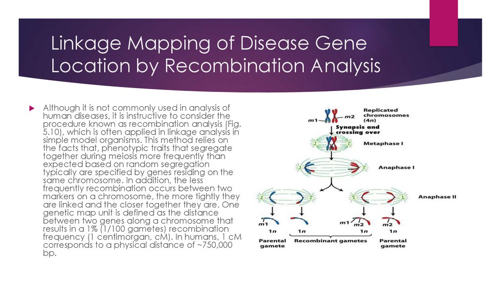
Linkage Mapping of Disease GeneLocation by Recombination Analysis
Although it is not commonly used in analysis of
human diseases, it is instructive to consider the
procedure known as recombination analysis (Fig.
5.10), which is often applied in linkage analysis in
simple model organisms. This method relies on
the facts that, phenotypic traits that segregate
together during meiosis more frequently than
expected based on random segregation
typically are specified by genes residing on the
same chromosome. In addition, the less
frequently recombination occurs between two
markers on a chromosome, the more tightly they
are linked and the closer together they are. One
genetic map unit is defined as the distance
between two genes along a chromosome that
results in a 1% (1/100 gametes) recombination
frequency (1 centimorgan, cM). In humans, 1 cM
corresponds to a physical distance of ~750,000
bp.
16.
Final Steps of Mutant Gene IsolationOften mutations can be mapped only to 1
cM regions of human DNA using the
methods discussed above (Fig. 5.38).
Regions of this length can contain dozens
of genes. The final identification of the
disease gene typically involves sequencing
and mapping of all SNPs, etc. in a long
region of DNA. The responsible gene is likely
to be located in regions where SNPs
associated with the disease consistently are
found in a number of affected individuals.
The mutation itself eventually is identified
by DNA sequencing. The analysis of gene
expression by Northern blotting and in situ
hybridization in affected tissues also may
help in identifying a disease gene in cases
where grossly defective mRNA transcripts
are produced from a gene.
17.
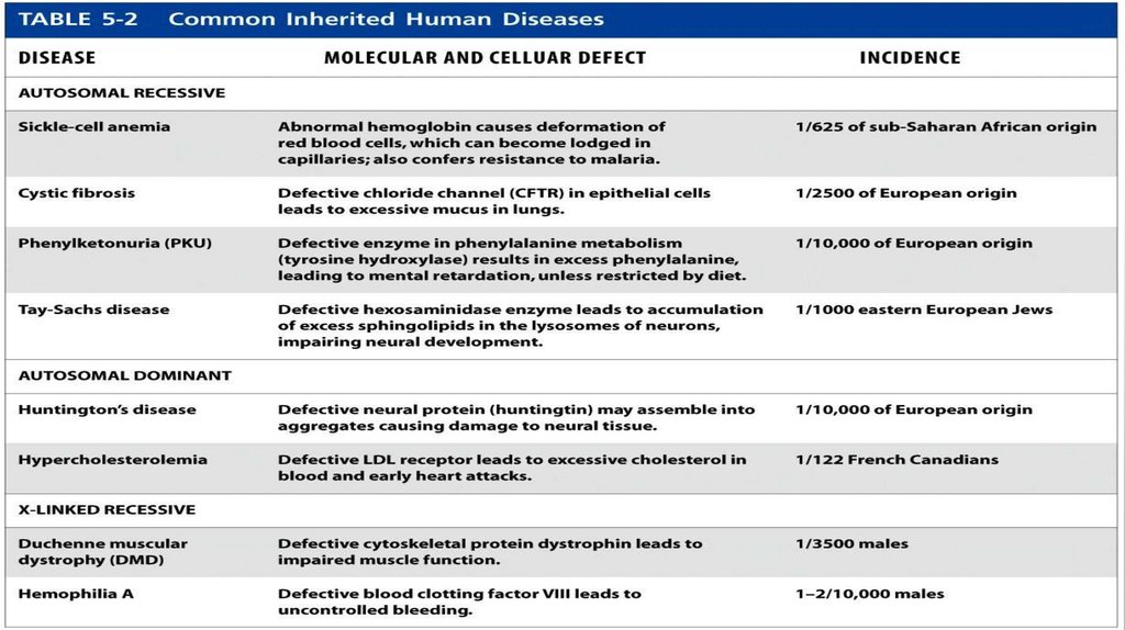
Common Hereditary Human DiseasesMost inherited diseases are caused by
preexisting mutant alleles that have
been passed down from one generation
to the next. Examples of common
autosomal recessive, autosomal
dominant, and X-linked recessive human
diseases of monogenic origin are listed in
Table 5.2. At least some fraction of
diseases such as cancers, diabetes,
obesity, and heart disease are hereditary
and polygenic in origin. The molecular
bases of these diseases are even harder
to solve than those of monogenic
diseases.